In this nursing care plan and management guide, learn how to provide care for patients with with impaired balance of gas exchange. Get to know the nursing assessment, interventions, goals, and nursing diagnosis specific to inadequate ventilation/perfusion by referring to this comprehensive guide.
Table of Contents
- What is an impaired gas exchange?
- Nursing Care Plans and Management
- Nursing Problem Priorities
- Nursing Assessment
- Nursing Diagnosis
- Nursing Goals
- Nursing Interventions and Actions
- Recommended Resources
- See also
- References and Sources
What is an impaired gas exchange?
Gas is exchanged between the alveoli and the pulmonary capillaries via diffusion. Diffusion of oxygen and carbon dioxide occurs passively, according to their concentration differences across the alveolar-capillary barrier. These concentration differences must be maintained by ventilation (airflow) of the alveoli and perfusion (blood flow) of the pulmonary capillaries.
A balance between the two exists typically, but certain conditions can alter this balance causing gas exchange impairment. Dead space is the volume of a breath that does not participate in gas exchange. It is ventilation without perfusion. A dead space results in a high ventilation/perfusion (V/Q) ratio, decreasing alveolar ventilation and reducing PaO2 for functional alveoli. Hypoxemia results from reduced PaO2 (Powers & Dharmoon, 2023).
Conditions that cause changes or collapse of the alveoli (e.g., atelectasis, pneumonia, pulmonary edema, and acute respiratory distress syndrome) impair ventilation. High altitudes, hypoventilation, and altered oxygen-carrying capacity of the blood from reduced hemoglobin are other factors that affect gas exchange. The total pulmonary blood flow in older adults is lower than in young subjects. Obesity in COPD and the impact of excessive fat mass on lung function put clients at greater risk for hypoxia. Smokers and clients suffering from pulmonary problems, prolonged periods of immobility, and chest or upper abdominal incisions are also at risk for this condition.
Nursing Care Plans and Management
When creating a nursing care plan and management for a client with impaired gas exchange, the primary goal is to optimize oxygenation and ensure adequate ventilation. Key components to consider include assessment and monitoring, positioning and airway management, medication and treatment, fluid and nutrition management, client education and support, and collaboration with and referrals to healthcare professionals.
Nursing Problem Priorities
The following are the nursing priorities for clients with impaired gas exchange:
- Inadequate oxygen perfusion. This is a priority because gas exchange directly affects oxygenation. The focus should be on optimizing oxygen delivery, monitoring oxygen saturation levels, and administering oxygen therapy.
- Alteration in breathing patterns. Impaired breathing patterns can further compromise gas exchange. Signs of respiratory distress must be monitored and deep breathing exercises must be initiated.
- Risk for respiratory failure. Clients with severely impaired gas exchange may be at great risk for respiratory failure. Monitoring for worsening gas exchange may reveal results such as increased respiratory rate, decreased oxygen saturation, or altered mental status.
- Relief from fear or anxiety. Providing emotional support and using therapeutic communication techniques effectively may alleviate anxiety.
- Client and caregiver education. Clients and their families may lack understanding of the disease process and its management. Providing education about the underlying causes, treatment strategies, and the importance of follow-up care can make a big difference in the client’s condition.
Nursing Assessment
The major signs and symptoms of respiratory disease include dyspnea, cough, sputum production, chest pain, wheezing, and hemoptysis. When evaluating symptoms, the nurse should also consider nonpulmonary diseases, as they may also occur with a variety of other illnesses. Use these subjective and objective data to help guide you through nursing assessment. Alternatively, you can check out the assessment guide below.
- Hypoxemia. Low blood oxygen level
- Abnormal breathing pattern. Deviation from the normal or expected rate, depth, and rhythm of respirations.
- Abnormal arterial blood gases. Refers to laboratory information about the client’s respiratory status, acid-base balance, and oxygenation.
- Restlessness. A common manifestation of agitation or unease due to inadequate oxygen perfusion.
- Cyanosis. Bluish coloring of the skin
- Dyspnea. The subjective feeling of labored or difficult breathing.
- Cough. A reflex that protects the lungs from the accumulation of secretions or the inhalation of foreign bodies.
- Nasal flaring. A visible sign that allows the healthcare professional to observe the client’s respiratory effort.
- Hypercapnia. Elevated levels of carbon dioxide in the bloodstream.
- Hypoxia. A deficiency of oxygen in the tissues and cells of the body.
- Orthopnea. Shortness of breath when lying flat.
- Tachypnea. Abnormally rapid respirations.
- Use of accessory muscles. A group of muscles that can assist in breathing when there is increased demand for ventilation.
Nursing Diagnosis
Following a thorough assessment, a nursing diagnosis is formulated to specifically address the challenges associated with impairment of gas exchange based on the nurse’s clinical judgement and understanding of the patient’s unique health condition. While nursing diagnoses serve as a framework for organizing care, their usefulness may vary in different clinical situations. In real-life clinical settings, it is important to note that the use of specific nursing diagnostic labels may not be as prominent or commonly utilized as other components of the care plan. It is ultimately the nurse’s clinical expertise and judgment that shape the care plan to meet the unique needs of each patient, prioritizing their health concerns and priorities. However, if you still find value in utilizing nursing diagnosis labels, here are some examples to consider:
- Impaired Gas Exchange related to decreased lung expansion due to restrictive lung disease, leading to insufficient oxygenation of blood as evidenced by decreased breath sounds, use of accessory muscles to breathe, and abnormal chest X-ray findings.
- Impaired Gas Exchange related to pulmonary fluid accumulation (e.g., as seen in heart failure or acute respiratory distress syndrome (ARDS), affecting oxygen and carbon dioxide exchange) as evidenced by crackles on auscultation, dyspnea at rest, and hypoxemia on arterial blood gas analysis.
- Impaired Gas Exchange related to altered blood flow to the lungs due to pulmonary embolism, compromising oxygen uptake and carbon dioxide elimination as evidenced by sudden onset of shortness of breath, chest pain, and hypoxia despite adequate ventilation.
- Impaired Gas Exchange related to reduced alveolar surface area available for gas exchange, (e.g., emphysema), impacting oxygenation levels as evidenced by prolonged expiratory phase, barrel chest, and decreased oxygen saturation levels on pulse oximetry.
- Impaired Gas Exchange related to increased metabolic demands (e.g., fever or sepsis requiring enhanced oxygen delivery and carbon dioxide removal) as evidenced by tachypnea, altered mental status, and increased lactate levels.
- Impaired Gas Exchange related to airway obstruction (e.g., asthma or bronchiolitis), as evidenced by wheezing on auscultation, increased peak expiratory flow rate variability, and difficulty speaking in full sentences.
- Impaired Gas Exchange related to impaired hemoglobin function (e.g., anemia or carbon monoxide poisoning) as evidenced by pallor, fatigue, and carboxyhemoglobin levels above normal in cases of carbon monoxide exposure.
Nursing Goals
The following are the common goals and expected outcomes for impaired balance of gas exchange.
- The client maintains optimal gas exchange as evidenced by usual mental status, unlabored respirations at 12 to 20 per minute, oximetry results within normal range, blood gases within normal range, and baseline HR for the client.
- The client maintains clear lung fields and remains free of signs of respiratory distress.
- The client verbalizes understanding of oxygen and other therapeutic interventions.
- The client participates in procedures to optimize oxygenation and in the management regimen within the level of capability/condition.
- The client manifests resolution or absence of symptoms of respiratory distress.
Nursing Interventions and Actions
The following are the therapeutic nursing interventions for managing clients with an impaired balance of gas exchange.
1. Improving oxygen perfusion
Assessment of oxygen saturation
Monitor oxygen saturation continuously, using a pulse oximeter.
Pulse oximetry is a useful tool to detect changes in oxygenation. An oxygen saturation of <90% (normal: 95% to 100%) or a partial pressure of oxygen of <80 (normal: 80 to 100) indicates significant oxygenation problems. Values less than 90% indicate that the tissues are not receiving enough oxygen.
Monitor for signs and symptoms of atelectasis: bronchial or tubular breath sounds, crackles, diminished chest excursion, limited diaphragm excursion, and tracheal shift to the affected side.
Low ventilation-perfusion states may be called shunt-producing disorders. When perfusion exceeds ventilation, a shunt exists. Blood bypasses the alveoli without gas exchange occurs. This is seen with obstruction of the distal airways, such as pneumonia, atelectasis, tumor, or a mucus plug.
Observe for nail beds and cyanosis in the skin; especially note the color of the tongue and oral mucous membranes.
Central cyanosis of the tongue and oral mucosa indicates severe hypoxia and is a medical emergency (Pahal et al., 2021). Peripheral cyanosis in extremities may or may not be serious. The presence or absence of cyanosis is determined by the amount of unoxygenated hemoglobin in the blood. Cyanosis appears when there is at least 5g/dL of unoxygenated hemoglobin. A client with a hemoglobin level of 15 g/dL does not demonstrate cyanosis until 5 g/dL of that hemoglobin becomes unoxygenated.
Monitor the client’s behavior and mental status for the onset of restlessness, agitation, confusion, and (in the late stages) extreme lethargy.
Changes in behavior and mental status can be early signs of impaired gas exchange. When oxygen is severely compromised, organ function will start to deteriorate. Neurologic manifestations include restlessness, headache, and confusion with moderate hypoxia. In severe cases, altered mentation and coma can occur, and if not corrected quickly may lead to death (Bhutta et al., 2022).
Observe for signs and symptoms of pulmonary infarction: bronchial breath sounds, consolidation, cough, fever, hemoptysis, pleural effusion, pleuritic pain, and pleural friction rub.
Increased dead space and reflex bronchoconstriction in areas adjacent to the infarct result in hypoxia (ventilation without perfusion). The alveoli do not have an adequate blood supply for gas exchange to occur. This is also characteristic of a variety of other disorders, such as pulmonary emboli and cardiogenic shock.
Note blood gas (ABG) results as available and note changes.
Increasing PaCO2 and decreasing PaO2 are signs of respiratory acidosis and hypoxemia. As the client’s condition deteriorates, the respiratory rate will decrease, and PaCO2 will increase. Some clients, such as those with COPD, have a significant decrease in pulmonary reserves, and additional physiological stress may result in acute respiratory failure. ABG studies aid in assessing the ability of the lungs to provide adequate oxygen and remove carbon dioxide, which reflects ventilation.
Monitor the effects of position changes on oxygenation (ABG and pulse oximetry.
Putting the most compromised lung areas in the dependent position (where perfusion is greatest) potentiates ventilation and perfusion imbalances. The lungs and chest wall structures expand together and share identical volumes. Regional compliance of the lung and chest wall varies in response to differences in the anatomic shape of these structures, the local effects of gravity, and the heterogenous mechanical properties of the diseased lung (Guerin et al., 2020).
Check on hemoglobin levels.
Low levels reduce the uptake of oxygen at the alveolar-capillary membrane and oxygen delivery to the tissues. Anemic hypoxia occurs when the oxygen-carrying ability of the blood decreases, and thus, this defect is specifically associated with the blood. This implies that fewer hemoglobin molecules are available for binding oxygen (Pittman, 2011).
Assess venous oxygen saturation (SvO2) as indicated.
Venous blood gas (VBG) studies provide additional data on oxygen delivery and consumption. VBG levels reflect the balance between the amount of oxygen used by tissues and organs and the amount of oxygen returning to the right side of the heart in the blood. VBG analysis is recommended to guide goal-directed therapy in postoperative clients at risk for hemodynamic instability or in clients with septic shock.
Assess for signs of obstructive sleep apnea (OSA).
Clients with OSA are often not previously diagnosed prior to hospitalization. The nurse may notice the client snores, has pauses in breathing while snoring, or awakens not feeling rested. These signs may indicate the client is unable to maintain an open airway while sleeping, resulting in periods of apnea and hypoxia (Hoyord, 2019).
Arrange for nocturnal trend oximetry.
This provides information about oxyhemoglobin saturation over a period (usually overnight). This test is primarily used to assess the adequacy or need for oxygen supplementation at night (Bhutta et al., 2022).
Assist the client during the six-minute walk test.
This test provides information on oxyhemoglobin saturation response to exercise as well as the total distance a client can walk in six minutes on a ground level. This information can be used to titrate oxygen supplementation as well as evaluate the response to therapy (Bhutta et al., 2022).
Optimal client positioning
Regularly check the client’s position so that they do not slump down in bed.
Slumped positioning causes the abdomen to compress the diaphragm and limits full lung expansion. According to a study, in clients breathing spontaneously, for any given volume above the functional residual capacity, the elastic pressures of the chest wall and respiratory system are positive in the supine position as compared to the upright position and greater in the former than in the latter. The main determinant of the changes in chest wall elastic properties with a change in position is abdominal pressure (Mezidi & Guerin, 2018).
If the client has unilateral lung disease, position them correctly to promote ventilation-perfusion.
Gravity and hydrostatic pressure cause the dependent lung to become better ventilated and perfused, which increases oxygenation. The good side should be down when the client is positioned on the side (e.g., a lung with pulmonary embolus or atelectasis should be up). When placed in the non-dependent position, increased transpulmonary pressure may help to expand the diseased, poorly compliant lung unit. However, when conditions like lung hemorrhage and an abscess are present, the affected lung should be placed downward to prevent drainage to the healthy lung (Lanks, 2022).
Turn the client every two hours. Monitor mixed venous oxygen saturation closely after turning. If it drops below 10% or fails to return to baseline promptly, turn the client back into a supine position, and evaluate oxygen status.
Turning is important to prevent complications of immobility, but in critically ill clients with low hemoglobin levels or decreased cardiac output, turning on either side can result in desaturation. In the supine position, functional residual capacity decreases from the sitting position in normal subjects breathing spontaneously. This was also found in healthy persons mechanically ventilated under general anesthesia (Mezidi & Guerin, 2018).
Consider positioning the client prone with the upper thorax and pelvis supported, allowing the abdomen to protrude. Monitor oxygen saturation, and turn back if desaturation occurs. Do not put in a prone position if the client has multisystem trauma.
The partial pressure of arterial oxygen has been shown to increase in the prone position, possibly because of greater diaphragm contraction and increased ventral lung regions’ function. Prone positioning improves hypoxemia significantly. In acute respiratory distress (ARDS) clients, the change from a supine to a prone position generates a more even distribution of the gas-tissue ratios along the dependent-nondependent axis and a more homogenous distribution of lung stress and strain (Guerin et al., 2020).
Support the client with pillows or cushions as needed for comfort and proper alignment. Use positioning aids for immobile clients.
Proper alignment prevents muscle fatigue and respiratory effort, allowing the client to breathe more efficiently, thus enhancing oxygenation. Positioning aids, such as wedges or rolled blankets, can be used to provide additional support and maintain proper positioning. These aids help align the body, prevent slouching or slumping, and promote optimal lung expansion, allowing for improved oxygenation.
Instruct the client in positioning during chest physiotherapy.
Chest physiotherapy includes postural drainage. By promoting clearance of secretions postural drainage allows the force of gravity to assist in the removal of bronchial secretions. The secretions drain from the affected bronchioles into the bronchi and trachea and are removed by coughing or suctioning. Several positions are used so that the force of gravity helps move secretions from the smaller bronchial airways to the main bronchi and trachea. Each position contributes to the effective drainage of a different lobe of the lungs; lower and middle lobe bronchi drain more effectively when the head is down, whereas the upper lobe bronchi drain more effectively when the head is up.
Consider the use of special positioning devices.
Continuous lateral rotation beds or prone positioning devices may be necessary to optimize oxygenation. The rotation of a client on a bed is hypothesized to improve drainage of secretions within the lung and lower airways, to increase functional residual capacity by providing an increased critical opening pressure to the independent lung. Continuous lateral rotation involves a programmable bed that turns on its longitudinal axes, intermittently or continuously (Goldhill et al., 2020).
Oxygen therapy
Maintain an oxygen administration device as ordered, attempting to maintain oxygen saturation at 90% or greater.
Supplemental oxygen may be required to maintain PaO2 at an acceptable level. Oxygen transport to tissues depends on factors such as cardiac output, arterial oxygen content, concentration of hemoglobin, and metabolic requirements. These factors must be kept in mind when oxygen therapy is considered.
Avoid a high concentration of oxygen in clients with COPD unless ordered.
Hypoxia stimulates the drive to breathe in the client who chronically retains carbon dioxide. When administering oxygen, close monitoring is imperative to prevent unsafe increases in the client’s PaO2, resulting in apnea. It is also important to monitor the respiratory rate and the oxygen saturation as measured by pulse oximetry in order to maintain an oxygen saturation between 90% and 93% on the lowest liter flow of oxygen.
If the client is permitted to eat, provide oxygen to the client but differently (changing from a mask to a nasal cannula).
More oxygen will be consumed during the activity. The original oxygen delivery system should be returned immediately after every meal. The nasal cannula allows the client to move about in bed, talk, cough, and eat without interrupting oxygen flow. Caution the client using the nasal cannula that may cause irritation and drying of the nasal and pharyngeal mucosa.
Administer humidified oxygen through an appropriate device (e.g., nasal cannula or face mask per provider’s order); watch for the onset of hypoventilation as evidenced by increased somnolence after initiating or increasing oxygen therapy.
A client with chronic lung disease may need a hypoxic drive to breathe and hypoventilate during oxygen therapy. Oxygen toxicity occurs when too high a concentration of oxygen (greater than 50% is given for an extended period (generally longer than 24 hours). If left untreated, this can severely damage the alveolar-capillary membrane leading to pulmonary edema and progressing to cell death.
For clients who should be ambulatory, provide extension tubing or a portable oxygen apparatus.
These measures may improve exercise tolerance by maintaining adequate oxygen levels during activity. The use of oxygen concentrators is another means of providing varying amounts of oxygen, especially in the home setting. These devices are relatively portable, easy to operate, and cost-effective but require more maintenance than tanks of liquid systems. These models can deliver oxygen flows from one to ten liters/minute.
Schedule nursing care to provide rest and minimize fatigue.
The hypoxic client has limited reserves; inappropriate activity can increase hypoxia. For example, clients diagnosed with COPD have decreased exercise tolerance during specific periods of the day, especially in the morning on arising, because bronchial secretions have collected in the lungs during the night while the client was lying down. The client may have difficulty bathing or dressing and may become fatigued. The nurse may help the client reduce these limitations by planning self-care activities and determining the best times for these activities.
Assess for indicators of the effectiveness of oxygen therapy.
Oxygen is a medication, and except in emergency situations, it is given only when prescribed by a provider with prescriptive authority. As with other medications, the nurse administers oxygen with caution and carefully assesses its effects on each client. The nurse should also assess for indicators of inadequate oxygenation, such as confusion, restlessness progressing to lethargy, diaphoresis, pallor, tachycardia, tachypnea, and hypertension.
Monitor for signs of oxygen toxicity.
Oxygen toxicity may occur when a too high concentration of oxygen (greater than 50%) is given for an extended period (generally longer than 24 hours). Clinical manifestations of oxygen toxicity causing lung damage are similar to acute respiratory distress syndrome. Signs and symptoms of oxygen toxicity include substernal discomfort, paresthesias, dyspnea, restlessness, fatigue, malaise, progressive respiratory difficulty, refractory hypoxemia, alveolar atelectasis, and alveolar infiltrates evident from chest X-rays.
Consider gerontologic factors when assessing oxygen delivery efficacy and effectiveness.
The respiratory system changes throughout the aging process, and it is important for nurses to be aware of these changes when assessing older adults who are receiving oxygen therapy. Older adults may display increased chest rigidity and respiratory rate and decreased PaO2 and lung expansion. The older adult is at risk for aspiration and infection related to these changes.
Treatment for hypercapnia
Monitor for signs of hypercapnia.
Hypercapnia is the buildup of carbon dioxide in the bloodstream. Signs of hypercapnia include headaches, dizziness, lethargy, and reduced ability to follow instructions, disorientation, and coma. Sudden hypercapnia can cause an increased pulse and respiratory rate and increased blood pressure. An elevated PaCO2, greater than 60 mm Hg, causes cerebrovascular vasodilation, and increased cerebral blood flow (Guerra, 2022).
Monitor ABG or VBG analysis results.
An arterial or venous blood gas is possibly the most valuable laboratory test as it allows for the evaluation of pH status, serum CO2, and serum HCO3. additionally, an anion gap can be calculated to assist in determining if acidosis is metabolic or respiratory in nature (Guerra, 2022).
Monitor the client’s respiratory rate, depth, and effort.
Frequent monitoring of the client’s respiratory parameters allows for early identification of any changes in breathing patterns or effort. In hypercapnia, it is important to assess for signs of respiratory distress, such as increased respiratory rate, shallow breaths, or the use of accessory muscles. Timely recognition of these changes can guide appropriate interventions and prevent further impairment of gas exchange.
Provide instructions in performing incentive spirometry.
Spirometry evaluates the general functioning of the lung. Forced expiratory volume over one second and forced vital capacity measurements are used to determine whether a restrictive or obstructive process is the etiology of hypoventilation. If air trapping is is suspected, this may indicate COPD or asthmatic pathologies most commonly (Guerra, 2022).
Instruct the client to maintain an upright position or elevate the head of the bed.
Keeping the client in an upright position or elevating the head of the bed promotes optimal lung expansion and ventilation. This position allows the diaphragm to function effectively and reduces the pressure on the chest, facilitating improved gas exchange and decreasing the level of CO2 in the blood.
Encourage the client to perform deep breathing exercises.
The breathing pattern of most clients diagnosed with COPD is shallow, rapid, and insufficient; the more severe the disease, the more inefficient the breathing pattern. With practice, this type of upper chest breathing can be changed to diaphragmatic breathing, which reduces respiratory rate, increases alveolar ventilation, and sometimes helps expel as much air as possible. Pursed-lip breathing helps slow expiration, prevents the collapse of small airways, and helps the client control the rate and depth of respiration.
Administer bronchodilators as prescribed.
Bronchodilators help relax the airway’s smooth muscles, dilate the bronchioles, and improve airflow. By reducing airway resistance, these medications facilitate better ventilation and gas exchange, addressing impaired gas exchange associated with hypercapnia.
Assist in noninvasive ventilatory support as indicated.
Noninvasive ventilation refers to techniques that provide ventilatory support without endotracheal intubation. The most frequently used is positive-pressure ventilation delivered through a tight-fitting mask. The beneficial effects of NIV include reducing the need for endotracheal intubation, decreasing the rate of complications, and lowering the cost of care. NIV can also be used as a long-term treatment for chronic hypercapnic respiratory failure in COPD (Csoma et al., 2022).
Assist in endotracheal ventilation and mechanical ventilation as appropriate.
BiPAP, CPAP, and intubation with mechanical ventilation are supportive measures that aim to optimize oxygenation while removing CO2 from the body. Mechanical ventilation is the most invasive option, but it allows better control of both respiratory rate and tidal volume in addition to FiO2 and pressure support (Guerra, 2022).
2. Promoting effective breathing patterns
Effective breathing ensures adequate ventilation and oxygenation, allowing for effective gas exchange in the lungs. When the gas exchange is compromised, as in conditions such as COPD, pneumonia, or asthma, it is essential to implement interventions that support and enhance respiratory function.
Assessment of respirations and pulmonary function
Assess respiratory rate, depth, and effort, including the use of accessory muscles, nasal flaring, and abnormal breathing patterns.
The presentation of hypoxia can be acute or chronic; acutely the hypoxia may present with dyspnea and tachypnea. Stridor can be heard once the client arrives in cases of upper airway obstruction. The chronic presentation includes dyspnea on exertion as the most common complaint (Bhutta et al., 2022). Increased respiratory rate, use of accessory muscles, nasal flaring, abdominal breathing, and a look of panic in the client’s eyes may be seen with hypoxia.
Assess the lungs for areas of decreased ventilation and auscultate the presence of adventitious sounds.
Any irregularity of breath sounds may disclose the cause of impaired gas exchange. The presence of crackles and wheezes may alert the nurse to airway obstruction, leading to or exacerbating existing hypoxia. Diminished breath sounds are linked with poor ventilation. The nurse may need to listen to two full inspirations and expirations at each anatomic location for a valid interpretation of the sound heard.
Monitor for alteration in blood pressure (BP) and heart rate (HR).
BP, HR, and respiratory rate all increase with initial hypoxia and hypercapnia. However, when both conditions become severe, BP and HR decrease and dysrhythmias may occur. Sufficiently severe hypoxia can result in tachycardia to provide sufficient oxygen to the tissues (Bhutta et al., 2022).
Assess for the presence of chest pain.
Chest pain or discomfort may occur with pneumonia, pulmonary infarction, or pleurisy, or as a late symptom of bronchogenic carcinoma. The nurse should assess the quality, intensity, and radiation of pain and identify and explore precipitating factors and their relationship to the client’s position. In addition, the nurse must assess the relationship of pain to the inspiratory and expiratory phases of respiration.
Assess for risk factors that may contribute to the client’s ineffective breathing pattern.
Many lung disorders are related to or exacerbated by tobacco smoke; therefore smoking history (including exposure to secondhand smoke) must be obtained. Socioeconomic differences rooted in race and ethnicity may predispose certain groups to greater burdens related to lung disease. Adults living below the poverty level are more likely to experience severe asthma exacerbations, hospitalizations, and death.
Breathing and coughing techniques
Assess the client’s ability to cough out secretions. Take note of the quantity, color, and consistency of the sputum.
Retained secretions weaken gas exchange. Cough is a reflex that protects the lungs from the accumulation of secretions or the inhalation of foreign bodies. The cough reflex may be impaired by weakness or paralysis of the respiratory muscles. Prolonged inactivity, the presence of a nasogastric tube, or depressed function of the brain’s medullary centers.
Client positioning
Position the client with the head of the bed elevated, in a semi-Fowler’s position (head of the bed at 45 degrees when supine) as tolerated.
Upright or semi-Fowler’s position allows increased thoracic capacity, total descent of the diaphragm, and increased lung expansion preventing the abdominal contents from crowding. In ARDs clients, an upright position (45 degrees trunk elevation and <45 degrees legs down) can improve end-expiratory lung volume in some clients. This improvement was associated with better oxygenation (Mezidi & Guerin, 2018).
If the client is obese or has ascites, consider positioning in the reverse Trendelenburg position at 45 degrees for periods as tolerated.
Trendelenburg’s position at 45 degrees results in increased tidal volumes and decreased respiratory rates. In clients with a body mass index greater than 35 kg/m², a sitting position with an angulation of 70 degrees was associated with a reduction of the expiratory flow limitation, auto-PEEP, and plateau pressure as compared to the supine position (Mezidi & Guerin, 2018).
If the client is acutely dyspneic, consider having them lean forward over a bedside table if tolerated.
Leaning forward can help decrease dyspnea, possibly because gastric pressure allows better contraction of the diaphragm. The orthopneic position, or the tripod position, is a sitting position where an individual leans slightly forward with their arms propped up on an overbed table or their knees. This position can be achieved by raising the head of the bed to 90 degrees and placing a table with pillows across the bed in front of the individual. Additional pillows, typically behind the lower back, can be used to increase comfort and support (Arps, 2017).
Pulmonary function testing
Consider the use of pulmonary function tests for the client.
PFTs are routinely used for clients with chronic respiratory disorders to aid diagnosis. They are performed to assess respiratory function and to determine the extent of dysfunction, response to therapy, and as screening tests in potentially hazardous industries. Such tests include the mechanics of breathing, diffusion, and gas exchange.
Instruct the client on how to measure their peak flow rate.
The client with respiratory symptoms usually undergoes a complete diagnostic evaluation, even if the results of PFTs are “normal”. Clients with respiratory disorders may be taught how to measure their peak flow rate (which refers to maximal expiratory flow) at home using a spirometer. This allows them to monitor the progress of therapy, to alter medications and other interventions as needed on the basis of caregiver guidelines.
Encourage slow deep breathing using an incentive spirometer as indicated.
This technique promotes deep inspiration, which increases oxygenation and prevents atelectasis. In the volume type, the client takes a deep breath through the mouthpiece, pauses at peak lung inflation, and then relaxes and exhales. In the flow type, the spirometer contains a number of movable balls that are pushed up by the force of the breath and held suspended in the air while the client inhales. The amount of air inhaled and the flow of the air are estimated by how long and how high the balls are suspended.
Administer medications as prescribed.
The type depends on the etiological factors of the problem (e.g., antibiotics for pneumonia, bronchodilators for COPD, anticoagulants, thrombolytics for pulmonary embolus, and analgesics for thoracic pain).
- Bronchodilators
These agents effectively relax smooth muscles and open airways. The administration of bronchodilators is primarily through inhalation devices to deliver the drug to the lung’s bronchioles. The best way to achieve maximum bioavailability is by fully exhaling, placing the inhaler in the mouth, and taking a full inhalation. After inhaling completely, it is followed by ten seconds of holding the breath to wait for the medication to dissipate into the lung space, then the client can slowly exhale (Almadhoun & Sharma, 2023). - Glucocorticoids
These agents relieve inflammation and also assist in opening air passages. The use of glucocorticoids in clients diagnosed with ARDS was associated with a significant reduction in hospital and ICU mortality and duration of mechanical ventilation. However, there is an increased risk of hyperglycemia (Zayed et al., 2020). - Mucolytics
Mucolytics decrease the thickness of pulmonary secretions so that they can be expectorated more easily. These agents can be administered orally, intravenously, and topically in a nebulized form. Though the topical route offered the advantage of activating the mucociliary clearance mechanism along with inducing a cough reflex, the oral route offers much better tolerability (Gupta & Wadha, 2023).
Instruct the client in proper positioning during PFTs.
Proper positioning ensures optimal ventilatory processes can be achieved during testing. The spirometry procedure is usually performed in a standard sitting position. However, spirometry measurement in the supine position may be indicated for clients with certain neuromuscular disorders (Ponce, 2022).
Assist in the measurement of lung volumes.
The lung volume measurement is important to detect changes in lung volume independent of effort, especially when functional vital capacity is reduced on spirometry. In the gas dilution method, the client breathes a gas mixture until equilibrium is achieved. The volume and mixture of gas exhaled after the equilibrium has been achieved permit the calculation of FRC. In body plethysmography, the subject sits inside a body box and breathes against a shutter valve. This method is considered the gold standard for lung volume measurement (Ponce, 2022).
Assess the client’s respiratory muscle pressure.
The respiratory muscle strength is assessed with maximal inspiratory pressure (MIP) and maximal expiratory pressure (MEP). The MIP reveals the strength of the diaphragm and other inspiratory muscles, whereas MEP indicates the strength of the abdominal and other expiratory muscles. An MEP of less than 60 cm H20 predicts a weak cough and difficulty clearing secretions (Ponce, 2022).
Emphasize the importance of informing the healthcare staff if the client is feeling ill or has a cardiopulmonary disorder before the testing.
PFTs are safe in general, and tehre are no complications. However, contraindications of PFTs include acute coronary syndrome, rupture of aneurysms, and dehiscence of the surgical wound. Clients with myocardial infarction, unstable heart disease, or stroke within the previous three months should not perform bronchoprovocation testing. If the client has an acute illness or symptom, they may likely have a suboptimal result (Ponce, 2022).
3. Reducing the risk of respiratory infection or failure
Impaired gas exchange can contribute to respiratory failure by preventing the efficient exchange of oxygen and carbon dioxide between the lungs and the bloodstream. Timely identification and management of the underlying cause are essential to prevent or treat respiratory failure and restore adequate gas exchange.
Identification of worsening respiratory symptoms
Monitor chest X-ray reports.
Chest X-ray studies reveal the etiological factors of impaired gas exchange. Normal pulmonary tissue is radiolucent because it consists mostly of air and gases; therefore, densities produced by fluid, tumors, foreign bodies, and other pathologic conditions can be detected by X-ray examination. In the absence of symptoms, a chest X-ray may reveal an extensive pathologic process in the lungs.
Closely monitor the client’s ABG results.
Acute respiratory failure is defined as a decrease in arterial oxygen tension (PaO2) to less than 60 mm Hg and an increase in arterial carbon dioxide tension (PaCO2) to greater than 50 mm Hg, with an arterial pH of less than 7.35.
Assess for symptoms of respiratory failure.
Clients with COPD are at risk for respiratory insufficiency and respiratory infections or COPD exacerbation, which in turn increase the risk of acute and chronic respiratory failure. Asterixis may be observed with severe hypercapnia. Tachycardia and a variety of arrhythmias may result from hypoxemia and acidosis. Dyspnea often accompanies respiratory failure (Kaynar & Sharma, 2020).
Monitor for the presence of nosocomial infections.
Nosocomial infections, such as pneumonia, urinary tract infections, and catheter-related sepsis, are frequent complications of respiratory failure. These usually occur with the use of mechanical devices. The incidence of nosocomial pneumonia is high and associated with significant mortality (Kaynar & Sharma, 2020).
Identify the PaO2:FiO2 ratio as indicated.
This ratio is another way to measure the degree of hypoxia. A normal PaO2/FiO2 ratio is about 300 to 500 mm Hg. A ratio of less than 300 indicates abnormal gas exchange, and values less than 200 mm hg indicate severe hypoxemia. The PaO2/FiO2 ratio is used mostly as a definition of ARDS severity (Bhutta et al., 2022).
For clients with mechanical ventilation and endotracheal intubation
Consider the need for intubation and mechanical ventilation.
Early intubation and mechanical ventilation are recommended to prevent full decompensation of the client. Mechanical ventilation provides supportive care to maintain adequate oxygenation and ventilation. If the client has evidence of respiratory failure or a compromised airway, endotracheal intubation and mechanical ventilation are indicated. This clinical evidence may be corroborated by a continuous decrease in oxygenation (PaO2), an increase in arterial carbon dioxide levels (PaCO2), and persistent acidosis (decreased pH).
Suction as necessary.
Suction clears secretions if the client is not capable of effectively clearing the airway. Airway obstruction blocks ventilation that impairs gas exchange. In clients who are mechanically ventilated, an in-line suction catheter may be used to allow rapid suction when needed and to minimize cross-contamination by airborne pathogens. In-line suctioning decreases hypoxemia, sustains PEEP, and can decrease client anxiety associated with suctioning.
Monitor the effects of sedation and analgesics on the client’s respiratory pattern; use judiciously.
Both analgesics and medications that cause sedation can depress respiration at times. However, these medications can be beneficial for decreasing the sympathetic nervous system discharge that accompanies hypoxia. The nurse must note changes in the client’s vital signs and evidence of hemodynamic instability and report them to the primary provider, because they may indicate that the mechanical ventilation is ineffective or the client may be receiving analgesics or sedation.
Instruct the client to limit exposure to persons with respiratory infections.
This is to reduce the potential spread of droplets between clients. Clients experiencing an impaired gas exchange often have weakened immune systems or respiratory defenses, making them more susceptible to respiratory infections. Being exposed to these clients with respiratory infections increases the likelihood of contracting the same infection, which can further worsen their already compromised respiratory function.
Place the client in a semi-Fowler position.
Once the client who has undergone tracheostomy for mechanical ventilation has stable vital signs, the nurse may put them in a semi-Fowler position to facilitate ventilation, promote drainage, minimize edema, and prevent strain on the suture lines.
Ensure that the cuff pressure on the endotracheal/tracheostomy tube is within normal parameters.
The cuff on an endotracheal or tracheostomy tube should be inflated if the client requires mechanical ventilation or is at risk for aspiration. The pressure within the cuff should be at the lowest possible pressure (20 to 25 mm Hg) which allows delivery of adequate tidal volumes and prevents pulmonary aspiration. Cuff pressure must be monitored by the nurse at least every eight hours by attaching a handheld pressure gauge to the pilot balloon of the tube.
Adjust ventilator settings as appropriate and as indicated.
The ventilator is adjusted so that the client is comfortable and breathes synchronously with the machine. If the volume ventilator is adjusted appropriately, the client’s ABG values will be satisfactory and there will be little or no cardiovascular compromise. The nurse, in collaboration with the respiratory therapist, always reviews the manufacturer’s instructions, which vary according to the equipment, before beginning mechanical ventilation.
Implement care bundle interventions for the client with mechanical ventilation.
Current best practices can include the implementation of specific evidence-based bundle interventions that, when used together, improve client outcomes. The five key elements of ventilator-associated pneumonia (VAP) bundle include elevation of the head of the bed (30 to 45 degrees), daily “sedation vacations” and assessment of readiness to extubate, peptic ulcer disease prophylaxis, deep venous thrombosis prophylaxis, and daily oral care with chlorhexidine (0.12% oral rinses).
Perform tracheostomy care routinely.
Clients with an endotracheal or tracheostomy tube do not have the normal defenses of the upper airway. These clients frequently have multiple additional body system disturbances that lead to immunocompromise. Tracheostomy care is performed at least every eight hours, and more frequently if needed, because of the increased risk of infection.
Perform oral hygiene frequently.
The nurse must administer oral hygiene frequently because the oral cavity is a primary source of contamination of the lungs in the client who is intubated. The presence of an NG tube can also increase the risk of aspiration, leading to nosocomial pneumonia.
4. Providing relief from anxiety
Air hunger, or the uncomfortable or unpleasant urge to breathe, can be provoked by a rise in respiratory drive or by a reduction in achieved ventilation. This can elicit very strong emotional responses of anxiety, fear, and frustration (Banzett et al., 2021).
Assessment of the level of anxiety
Assess for the level of anxiety in clients with dyspnea or air hunger.
Anxiety, a feeling of apprehension, nervousness, fear, or worry, is common in clients diagnosed with COPD. The Hospital Anxiety and Depression Scale (HADS) is a commonly used instrument, wherein significant improvement after intervention was found in most studies. Other studies used the Beck Anxiety Inventory, Anxiety Inventory for Respiratory disease scale, the Emotional State, the Revised Symptom Checklist 90, and the Spielberger State-Trait Anxiety Inventory (Yohannes et al., 2017).
Observe the client for cues that identify the level of anxiety.
Observe the client’s body language and behavior for signs of anxiety, such as restlessness, fidgeting, pacing, or tense facial expressions. Note for any verbal cues, such as expressions of fear, worry, or statements related to feeling anxious or overwhelmed. The client may be reluctant to admit the presence of anxiety but physical manifestations may be contrary to this.
Monitor changes in the client’s respiratory symptoms.
Dyspnea and anxiety often coexist, and respiratory distress can contribute to anxiety levels. Assessing the client’s respiratory status helps identify any physical factors exacerbating their anxiety, such as increased work of breathing or signs of respiratory distress like nasal flaring or intercostal retractions.
Monitor the client’s vital signs.
Monitoring vital signs, including the heart rate, blood pressure, and oxygen saturation levels provides objective data on the client’s physiological response to anxiety. Anxiety can lead to increased sympathetic nervous system activation, resulting in elevated heart rate or blood pressure. Changes in oxygen saturation levels during dyspnea episodes may indicate the presence of anxiety.
Pulmonary Rehabilitation
Introduce pulmonary rehabilitation to the client and their caregivers.
Pulmonary rehabilitation is widely accepted as a means to alleviate symptoms and optimize functional status. The primary goals of rehabilitation are to reduce symptoms, improve the quality of life, and increase physical and emotional participation in everyday activities. Breathing exercises, as well as retraining and exercise programs, are used to improve functional status. The majority of studies report that PR reduces anxiety and dyspnea in clients with COPD during short-term follow-up (Yohannes et al., 2017).
Assist the client during breathing exercises.
Nurses can be instrumental in educating clients and families as well as facilitating physical therapy for breathing retraining. Training in diaphragmatic breathing reduces the respiratory rate, increases alveolar ventilation, and sometimes helps expel as much air as possible during expiration. Pursed-lip breathing helps slow expiration, prevent the collapse of small airways, and control the rate and depth of respiration. It also promotes relaxation, which allows the client to gain control of dyspnea and reduce feelings of panic.
Arrange for the client to attend a pulmonary rehabilitation program weekly.
An intensive, 3-week PR program (six hours per day for five days per week) showed significant improvement in reducing dyspnea and anxiety symptoms and improving exercise capacity and quality of life. The improvement observed after rehabilitation on anxiety symptoms related to the magnitude of clients’ perceived dyspnea at rest. However, this program may not be applicable to moderate-to-severe clients with COPD (Yohannes et al., 2017).
Instruct the client about activity pacing.
Clients with COPD have decreased exercise tolerance during specific periods of the day, especially in the morning on arising, because bronchial secretions have collected in the lungs during the night while the client was lying down. The nurse can help the client reduce these limitations by planning self-care activities and determining the best times for bathing, dressing, and other daily activities.
Promote physical conditioning techniques.
Clients diagnosed with COPD of all grades may benefit from exercise training programs. Physical conditioning techniques include breathing exercises and general exercises intended to conserve energy and increase ventilation. Graded exercises and physical conditioning programs using treadmills, stationary bicycles, and measured-level walks can improve symptoms and increase work capacity and exercise tolerance.
Provide nutritional counseling as appropriate.
Nutritional status is reflected in the severity of symptoms, degrees of disability, and prognosis. A thorough assessment of caloric needs and counseling about meal planning and supplementation is part of the rehabilitation process. Continual monitoring of weight and interventions as necessary are important parts of the care of clients with COPD.
Promote coping measures to reduce anxiety and depression.
Any factor that interferes with normal breathing quite naturally induces anxiety, depression, and changes in behavior. Constant shortness of breath and fatigue may make the client irritable and apprehensive to the point of panic. The nurse should provide education and support to spouses or significant others and families because the caregiver role in end-stage COPD can be challenging.
Providing interventions for the relief of anxiety
Provide reassurance and reduce anxiety.
Anxiety increases dyspnea, respiratory rate, and work of breathing. Dyspnea can be a distressing symptom that often leads to heightened anxiety or panic in clients. By providing reassurance, the nurse alleviates the client’s distress, which can give a sense of calm. When a client feels reassured, they are also more likely to trust the nurse’s expertise and cooperate with the care plan wholeheartedly.
Support the family of a client with chronic illness.
Severely compromised respiratory functioning causes fear and anxiety in patients and their families. Reassurance from the nurse can be helpful. This trust from the family can also foster a positive therapeutic relationship and encourage the client and the family to communicate openly about their symptoms and concerns.
Encourage verbalization of feelings for both the client and their caregivers.
Dyspnea and anxiety can be distressing experiences, causing emotional turmoil and increasing psychological burden. Verbalizing feelings enables the clients to release their emotions, providing psychological relief. Verbalizing provides an outlet for them to share their experiences, fears, and worries and can help validate their emotions.
Provide therapeutic communication.
Providing therapeutic communication can help alleviate anxiety by creating a calming and supportive environment. Clear and empathetic communication can also help the client understand their condition and promote trust between the client and the nurse.
Promote relaxation techniques, such as deep breathing and guided imagery.
Relaxation techniques can help reduce anxiety and improve the client’s ability to cope with dyspnea. Deep breathing exercises can help the client regulate their breathing pattern, decrease respiratory distress, and increase oxygenation. Guided imagery provides a distraction and promotes a sense of calmness, aiding in anxiety relief.
Encourage the client to assume a position of comfort during dyspnea.
Finding a position that promotes comfort can help alleviate anxiety and improve the client’s breathing. Clients may find it helpful to sit upright, lean forward, or use pillows to support themselves. These positions can aid in reducing the work of breathing, opening up the chest, and improving ventilation.
Refer the client for cognitive behavioral therapy, as indicated.
CBT is a popular nonpharmacologic therapy that combines behavioral with cognitive therapy. CBT disentangles clients’ patterns of thoughts and beliefs that may induce excessive worry, anxiety, and depressed mood. Delivered in either a one-to-one or a group format session, CBT deals with current problems that the clients are experiencing and works collaboratively with the therapist and clients (Yohannes et al., 2017).
5. Client and caregiver education
Set short- or long-term client education goals with the client and their caregivers.
A major area of client education involves setting and accepting realistic short-term and long-range goals. If the COPD is mild, the objectives of treatment are to increase exercise tolerance and prevent further loss of pulmonary function. If the COPD is severe, the objectives are to preserve current pulmonary function and relieve symptoms as much as possible. It is important to plan and share the goals and expectations of treatment with the client.
Consider the client’s nutritional status.
Certain conditions affect lung expansion. Obesity may restrict the downward movement of the diaphragm, increasing the risk for atelectasis, hypoventilation, and respiratory infections. Labored breathing is present in severe obesity as a result of excessive weight on the chest wall. Malnutrition may also reduce respiratory mass and strength, affecting muscle function. Significant weight loss is often a major problem in clients diagnosed with COPD; most clients have difficulty gaining and maintaining weight. Continual monitoring of weight and interventions as necessary are important parts of the care of clients with COPD.
Evaluate the client’s hydration status.
Overhydration may impair gas exchange in clients with heart failure. On the other hand, insufficient hydration may reduce the ability to clear secretions in clients with pneumonia and COPD. Adequate hydration helps loosen respiratory secretions, making it easier for the client to expel them. Thick and sticky secretions can obstruct the air passages, leading to exacerbation of respiratory compromise and difficulty in gas exchange.
Assess the home environment for irritants that impair gas an exchange. Help the client adjust the home environment as necessary (e.g., installing an air filter to decrease dust).
Irritants in the environment decrease the client’s effectiveness in accessing oxygen during breathing. Each client reacts differently to external exposures, such as significant air pollution, high or low temperatures, high humidity, or strong smells, therefore, the nurse must assess the client’s actual and potential triggers that cause bronchospasm so that avoidance or a treatment plan can be established.
Encourage or assist with ambulation as indicated.
Ambulation facilitates lung expansion, and secretion clearance and stimulates deep breathing. Clients with COPD experience progressive activity and exercise intolerance that may lead to disability. Education should include pacing activities throughout the day or using supportive devices to decrease energy expenditure. The use of walking aids may also be recommended to improve activity levels and ambulation.
Help the client deep breathe, and perform controlled coughing. Have the client inhale deeply, hold breath for several seconds, and cough two to three times with mouth open while tightening the upper abdominal muscles as tolerated.
This technique can help increase sputum clearance and decrease cough spasms. Controlled coughing uses the diaphragmatic muscles, making the cough more forceful and effective. Diaphragmatic breathing reduces respiratory rate, increases alveolar ventilation, and sometimes helps expel as much air as possible during expiration. Pursed-lip breathing helps slow expiration, prevents collapse of small airways, and helps the client control the rate and depth of respiration.
For postoperative clients, assist with splinting the chest.
Splinting optimizes deep breathing and coughing efforts. Careful splinting of abdominal or thoracic incision sites helps the client overcome the fear that the exertion of coughing might open the incision. Analgesic agents are also given to permit more effective coughing.
Pace activities and schedule rest periods to prevent fatigue. Assist with ADLs.
Activities will increase oxygen consumption and should be planned, so the client does not become hypoxic. Allow the client to rest frequently, and space out interventions to decrease oxygen demand in clients whose reserves are likely limited.
Instruct family on complications of the disease and the importance of maintaining a medical regimen, including when to call a healthcare provider.
Knowledge of the family about the disease is critical to prevent further complications. The nurse instructs to report any signs of infection, such as fever, or change in sputum color, character, consistency, or amount. Any worsening of symptoms, such as increased tightness of the chest, increased dyspnea, or fatigue, also suggests infection and must be reported.
Provide information about self-management programs during discharge teaching.
The use of a Self-Management Program of Activity, Coping, and Education (SPACE), which includes a structured exercise program, showed a statistically significant reduction in dyspnea at six weeks. Another study showed significant improvement in clients who attended a health management program comprising an hour lecture on general information about medications, smoking cessation, counseling, encouragement for regular exercise, instruction on rehabilitation, and psychological counseling (Yohannes et al., 2017).
Refer the client to community-based or transitional care facilities or managers.
Referral for home, community-based, or transitional care is important to enable assessment of the client’s home environment and physical and psychological status, to evaluate the client’s adherence to a prescribed regimen, and to assess the client’s ability to cope with changes in lifestyle and physical status. Home visits provide an opportunity to reinforce the information and activities learned in the inpatient or outpatient pulmonary rehabilitation program and to have the client and family demonstrate the correct administration of medications and oxygen, and performance of exercises.
Recommended Resources
Recommended nursing diagnosis and nursing care plan books and resources.
Disclosure: Included below are affiliate links from Amazon at no additional cost from you. We may earn a small commission from your purchase. For more information, check out our privacy policy.
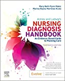
Ackley and Ladwig’s Nursing Diagnosis Handbook: An Evidence-Based Guide to Planning Care
We love this book because of its evidence-based approach to nursing interventions. This care plan handbook uses an easy, three-step system to guide you through client assessment, nursing diagnosis, and care planning. Includes step-by-step instructions showing how to implement care and evaluate outcomes, and help you build skills in diagnostic reasoning and critical thinking.

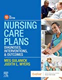
Nursing Care Plans – Nursing Diagnosis & Intervention (10th Edition)
Includes over two hundred care plans that reflect the most recent evidence-based guidelines. New to this edition are ICNP diagnoses, care plans on LGBTQ health issues, and on electrolytes and acid-base balance.

Nurse’s Pocket Guide: Diagnoses, Prioritized Interventions, and Rationales
Quick-reference tool includes all you need to identify the correct diagnoses for efficient patient care planning. The sixteenth edition includes the most recent nursing diagnoses and interventions and an alphabetized listing of nursing diagnoses covering more than 400 disorders.

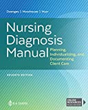
Nursing Diagnosis Manual: Planning, Individualizing, and Documenting Client Care
Identify interventions to plan, individualize, and document care for more than 800 diseases and disorders. Only in the Nursing Diagnosis Manual will you find for each diagnosis subjectively and objectively – sample clinical applications, prioritized action/interventions with rationales – a documentation section, and much more!

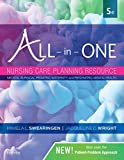
All-in-One Nursing Care Planning Resource – E-Book: Medical-Surgical, Pediatric, Maternity, and Psychiatric-Mental Health
Includes over 100 care plans for medical-surgical, maternity/OB, pediatrics, and psychiatric and mental health. Interprofessional “patient problems” focus familiarizes you with how to speak to patients.

See also
Other recommended site resources for this nursing care plan:
- Nursing Care Plans (NCP): Ultimate Guide and Database MUST READ!
Over 150+ nursing care plans for different diseases and conditions. Includes our easy-to-follow guide on how to create nursing care plans from scratch. - Nursing Diagnosis Guide and List: All You Need to Know to Master Diagnosing
Our comprehensive guide on how to create and write diagnostic labels. Includes detailed nursing care plan guides for common nursing diagnostic labels.
Other nursing care plans related to respiratory system disorders:
- Asthma
- Aspiration Risk & Aspiration Pneumonia
- Airway Clearance Therapy & Coughing
- Bronchiolitis
- Bronchopulmonary Dysplasia (BPD)
- Chronic Obstructive Pulmonary Disease (COPD)
- Cystic Fibrosis
- Hemothorax and Pneumothorax
- Influenza (Flu)
- Ineffective Breathing Pattern (Dyspnea)
- Impairment of Gas Exchange
- Lung Cancer
- Mechanical Ventilation
- Near-Drowning
- Pleural Effusion
- Pneumonia
- Pulmonary Embolism
- Pulmonary Tuberculosis
- Tracheostomy
References and Sources
Recommended sources, interesting articles, and references about management of patients with gas exchange problems to further your reading.
- Carlson‐Catalano, J., Lunney, M., Paradiso, C., Bruno, J., Luke, B. K., Martin, T., … & Pachter, S. (1998). Clinical validation of ineffective breathing pattern, ineffective airway clearance, and impaired gas exchange. Image: the Journal of Nursing Scholarship, 30(3), 243-248.
- Gosselink, R., & Stam, H. (Eds.). (2005). Lung Function Testing: European Respiratory Monograph (Vol. 31). European Respiratory Society.
- Pascoal, L. M., Lopes, M. V. D. O., Chaves, D. B. R., Beltrão, B. A., Silva, V. M. D., & Monteiro, F. P. M. (2015). Impaired gas exchange: accuracy of defining characteristics in children with acute respiratory infection1. Revista latino-americana de enfermagem, 23, 491-499.
- Pahal, P., & Goyal, A. (2021). Central and Peripheral Cyanosis. StatPearls [Internet].
- Sousa, V. E. C., Pascoal, L. M., de Matos, T. F. O., do Nascimento, R. V., Chaves, D. B. R., Guedes, N. G., & de Oliveira Lopes, M. V. (2015). Clinical indicators of impaired gas exchange in cardiac postoperative patients. International journal of nursing knowledge, 26(3), 141-146.
- Svedenkrans, J., Stoecklin, B., Jones, J. G., Doherty, D. A., & Pillow, J. J. (2019). Physiology and predictors of impaired gas exchange in infants with bronchopulmonary dysplasia. American journal of respiratory and critical care medicine, 200(4), 471-480.
- Yoost, B. L., & Crawford, L. R. (2019). Fundamentals of Nursing E-Book: Active Learning for Collaborative Practice. Elsevier Health Sciences.
- Almadhoun, K., & Sharma, S. (2023, April 28). Bronchodilators – StatPearls. NCBI.
- Banzett, R. B., Lansing, R. W., & Binks, A. P. (2021). Air Hunger: A Primal Sensation and a Primary Element of Dyspnea. Comprehensive Physiology, 11. 10.1002/cphy.c200001
- Bhutta, B. S., Alghoula, F., & Berim, I. (2022). Hypoxia – StatPearls. NCBI.
- Csoma, B., Vulpi, M. R., Dragonieri, S., Bentley, A., Felton, T., Lazar, Z., & Bikov, A. (2022). Hypercapnia in COPD: Causes, Consequences, and Therapy. Journal of Clinical Medicine, 11(11).
- Goldhill, D. R., Imhoff, M., McLean, B., & Waldmann, C. (2020). Rotational Bed Therapy to Prevent and Treat Respiratory Complications: A Review and Meta-Analysis. American Journal of Critical Care, 16(1).
- Guerin, C., Albert, R. K., Beitler, J., Gattinoni, L., Jaber, S., Marini, J. J., Munshi, L., Papazian, L., Pesenti, A., Vieillard-Baron, A., & Mancebo, J. (2020). Prone position in ARDS patients: why, when, how and for whom. Intensive Care Medicine, 46.
- Guerra, P. (2022). Hypercapnia – StatPearls. NCBI.
- Gupta, R., & Wadha, R. (2023, February 9). Mucolytic Medications – StatPearls. NCBI.
- Hinkle, J. L., & Cheever, K. H. (2018). Brunner & Suddarth’s Textbook of Medical-surgical Nursing. Wolters Kluwer.
- Hoyord, D. (2019). 11.2 Basic Concepts of Oxygenation – Nursing Skills. WI Technical Colleges Open Press.
- Kaynar, A. M., & Sharma, S. (2020, April 7). Respiratory Failure: Background, Pathophysiology, Etiology. Medscape Reference. Retrieved June 17, 2023, from
- Lanks, C. W. (2022). Match Me If You Can: The Relationship between Ventilation and Perfusion with Position Changes in Nonhomogenous Lung Injury. Annals of the American Thoracic Society, 19(2).
- Mezidi, M., & Guerin, C. (2018). Effects of patient positioning on respiratory mechanics in mechanically ventilated ICU patients. Annals of Translational Medicine, 6(19).
- Pittman, R. N. (2011). Regulation of Tissue Oxygenation. NCBI.
- Ponce, M. (2022). Pulmonary Function Tests – StatPearls. NCBI.
- Powers, K. A., & Dharmoon, A. S. (2023, January 23). Physiology, Pulmonary Ventilation and Perfusion – StatPearls. NCBI.
- Yohannes, A. M., Junkes-Cunha, M., & Smith, J. (2017). Management of Dyspnea and Anxiety in Chronic Obstructive Pulmonary Disease: A Critical Review. JAMDA, 18(12).
- Zayed, Y., Barbarawi, M., Ismail, E., Samji, V., Kerbage, J., Rizk, F., Salih, M., Bala, A., Obeid, M., Deliwala, S., Demian, S., Al-Sanouri, I., & Reddy, R. (2020). Use of glucocorticoids in patients with acute respiratory distress syndrome: a meta-analysis and trial sequential analysis. Journal of Intensive Care, 8(43).

God knowledge achieved on nursing care management
Well written, good review and easy to understand.
Good nursing care and clear to understand