Discover effective nursing care strategies in this comprehensive care plan and management guide for clients experiencing dyspnea or ineffective and abnormal breathing patterns. Gain valuable insights into the nursing assessment, interventions, goals, and nursing diagnosis specifically tailored to address the challenges associated with dyspnea.
Table of Contents
- What is dyspnea?
- Nursing Care Plans and Management
- Recommended Resources
- See also
- References and Sources
What is dyspnea?
When the abdominal wall excursion during inspiration, expiration or both do not maintain optimum ventilation for the individual, dyspnea or abnormal breathing pattern occurs. It is considered the state in which the rate, depth, timing, rhythm, or pattern of breathing is altered. When the breathing pattern is ineffective, the body will likely not get enough oxygen to the cells. Respiratory failure may be correlated with variations in respiratory rate, and abdominal and thoracic patterns.
Breathing pattern alteration may also transpire in several circumstances from heart failure, hypoxia, airway obstruction, diaphragmatic paralysis, infection, neuromuscular impairment, trauma or surgery resulting in musculoskeletal impairment, and pain, cognitive impairment and anxiety, diabetic ketoacidosis, uremia, thyroid dysfunction, peritonitis, drug overdose, AIDS, acute alcohol withdrawal, cardiac surgery, cholecystectomy, liver cirrhosis, craniocerebral trauma, disc surgery, lymphomas, renal dialysis, seizure disorders, spinal cord injuries, mechanical ventilatory assistance, and pleural inflammation.
Having a clear and effective airway is vital in inpatient care. Appropriate management for clients with oxygenation difficulties is to sustain or enhance pulmonary ventilation and oxygenation, promote comfort and ease of breathing, improve the ability to participate in physical activities and prevent risks associated with oxygenation problems such as skin and tissue breakdown, syncope, acid-base imbalances, and feelings of hopelessness and social isolation.
Nursing Care Plans and Management
The nursing care plan and management for dyspnea or ineffective breathing pattern typically involve assessing the underlying cause, providing immediate and appropriate interventions to alleviate the symptoms, and implementing measures to promote respiratory comfort and overall well-being.
Nursing Problem Priorities
The following are the nursing priorities for clients with dyspnea.
- Ensuring airway patency. Difficulty breathing can be associated with ineffective coughing and inadequate airway clearance. Assisting with techniques and interventions to improve airway patency can help address this problem.
- Promoting gas exchange. Dyspnea can result from impaired gas exchange, where the oxygenation and elimination of carbon dioxide are compromised. Ensuring adequate oxygenation is a priority.
- Relieving anxiety and distress. Managing anxiety and providing emotional support, such as implementing relaxation techniques and providing reassurance can help alleviate these problems.
- Providing client and caregiver education. Providing education about their condition, medications, and self-care measures is essential to empower the client and their caregivers in managing dyspnea effectively.
Nursing Assessment
The cornerstone of effective symptom control is systematic symptom assessment. Symptoms are ideally reported and rated by the clients themselves (Campbell, 2017). Because clients use a variety of terms to describe breathlessness, the nurse must explore what these terms mean to each client.
Assess for the following subjective and objective data:
- Abnormal rate, rhythm, and depth of breathing. Assessing the intensity or distress of breathlessness, what breathing feels like, and its impact on the client
- Bradypnea. Abnormally short breathing or shortness of breath
- Tachypnea. Abnormally rapid respirations
- Hypoxemia. Low blood oxygen level
- Nasal flaring. Widening of the nostrils while breathing.
- Orthopnea. Shortness of breath when lying flat
- Pursed-lip breathing. A breathing exercise that helps slow the breathing and lets the client inhale and exhale more air.
- Use of accessory muscles to breathe. Palpable muscle contractions behind the sternocleidomastoid muscles and over the scalene muscles during breathing
Nursing Diagnosis
Following a thorough assessment, a nursing diagnosis is formulated to specifically address the challenges associated with this condition based on the nurse’s clinical judgement and understanding of the patient’s unique health condition. While nursing diagnoses serve as a framework for organizing care, their usefulness may vary in different clinical situations. In real-life clinical settings, it is important to note that the use of specific nursing diagnostic labels may not be as prominent or commonly utilized as other components of the care plan. It is ultimately the nurse’s clinical expertise and judgment that shape the care plan to meet the unique needs of each patient, prioritizing their health concerns and priorities.
Nursing Goals
The following are the common goals and expected outcomes.
- The client maintains an effective breathing pattern, as evidenced by relaxed breathing at a normal rate and depth and the absence of dyspnea.
- The client’s respiratory rate remains within established limits.
- The client’s ABG levels return to and remain within established limits.
- The client indicates, either verbally or through behavior, feeling comfortable when breathing.
- The client reports feeling rested each day.
- The client performs diaphragmatic pursed-lip breathing.
- The client demonstrates maximum lung expansion with adequate ventilation.
- When the client carries out ADLs, the breathing pattern remains normal.
Nursing Interventions and Actions
Continuous assessment is necessary to know possible problems that may have led to altered breathing patterns or dyspnea and name any concerns during nursing care.
1. Maintaining a Patent Airway
Assessing indicators of a patent airway
1. Assess the ability to mobilize secretions.
The incapability to mobilize secretions may contribute to a change in breathing patterns. The cough reflex may be impaired by weakness or paralysis of the respiratory muscles, prolonged inactivity, the presence of a nasogastric tube, or the depressed function of the brain’s medullary centers.
2. Observe the presence of sputum for amount, color, and consistency.
These may be indicative of a cause for the alteration in breathing patterns. The nature of the sputum is often indicative of its cause. A profuse amount of purulent sputum (thick and yellow, green, or rust-colored) or a change in color of the sputum is a common sign of a bacterial infection. A gradual increase of sputum over time may occur with chronic bronchitis or bronchiectasis. Profuse, frothy, pink material, often welling up into the throat, may indicate pulmonary edema.
3. Auscultate breath sounds at least every four hours.
This is to detect decreased or adventitious breath sounds. Auscultation also helps assess the flow of air through the bronchial tree and evaluate the presence of fluid or solid obstruction in the lung. The nurse may need to listen for two full inspirations and expirations at each anatomic location for a valid interpretation of the sound heard.
4. Abnormal breath sounds may include:
- 4.1. Expiratory grunt
Frequently occurs in combination with nasal flaring and intercostal or subcostal retractions, associated with increased work of breathing. In newborns with respiratory distress, grunting is an expiratory sound caused by the sudden closure of the glottis during expiration in an attempt to maintain functional residual capacity and prevent alveolar atelectasis (Reuter et al., 2014). - 4.2. Rales or crackles
Crackles and rales refer to the same finding. These are clicking, rattling, or crackling sounds that are heard during inspiration and expiration. Crackles are generated by small airways snapping open on inspiration (Zimmerman & Williams, 2022). - 4.3. Coarse crackles
These are discontinuous popping sounds heard in early inspiration. It is a harsh, moist sound originating in the large bronchi. These types of crackles are associated with obstructive pulmonary disease. - 4.4. Fine crackles
These are discontinuous popping sounds heard in late inspiration and sounds like hair rubbing together. This originates in the alveoli and is associated with interstitial pneumonia or restrictive pulmonary disease. - 4.5. Rhonchi or sonorous wheezes
These are deep, low-pitched rumbling sounds heard primarily during expiration and are caused by air moving through narrowed tracheobronchial passages. This is associated with secretions or tumors. - 4.6. Stridor
High-pitched, musical breathing sound caused by a blockage in the throat or voice box (larynx). Like wheezes, stridor is produced by airway narrowing, but only in the upper airways (Zimmerman & Williams, 2022). - 4.7. Wheeze
High-pitched, whistling sound when air moves through narrowed breathing tubes in the lungs. This is heard most commonly in asthmatics and CHF.
5. Measure the client’s expiratory flow rate and volume.
The relief of bronchospasm is confirmed by measuring the improvement in expiratory flow rates and volumes (the force of expiration, how long it takes to exhale, and the amount of air exhaled).
6. Identify pulmonary irritants or allergens.
Pulmonary irritants or allergens can prevent effective airway clearance by causing inflammation and increasing mucus production in the airways. All pulmonary irritants should be eliminated or reduced, particularly cigarette smoke, which is the most persistent source of pulmonary irritation.
Assessing breathing patterns
1. Assess and record respiratory rate and depth at least every four hours.
The average rate of respiration for adults is 10 to 20 breaths per minute. It is important to take action when there is an alteration in breathing patterns to detect early signs of compromise in the respiratory system. Certain patterns of respiration are characteristic of specific disease states. Changes in respiratory rate and rhythm may be the first clinical deterioration in clients who are acutely ill.
2. Observe breathing patterns.
Unusual breathing patterns may imply an underlying disease process or dysfunction. Cheyne-Stokes respiration signifies bilateral dysfunction in the deep cerebral or diencephalon related to brain injury or metabolic abnormalities. Apneusis and ataxic breathing are related to the failure of the respiratory centers in the pons and medulla.
3. Rates and depths of breathing patterns include:
- 3.1. Apnea
Temporary cessation of breathing, especially during sleep. This signals a life-threatening situation in which the client will quickly succumb unless rescue breathing is instituted immediately (Whited & Graham, 2023). - 3.2. Apneusis
Deep, gasping inspiration with a pause at full inspiration followed by a brief, insufficient release. This respiratory pattern is often associated with severe brain injury and carries a poor prognosis (Whited & Graham, 2023). - 3.3. Ataxic patterns
Complete irregularity of breathing with irregular pauses and increasing periods of apnea. It is characterized by irregular respiratory rate, rhythm, and depth with or without brief respiratory pauses of less than ten seconds. - 3.4. Biot’s respiration
Groups of quick, shallow inspirations followed by regular or irregular periods of apnea (10 to 60 seconds). This pattern may deteriorate to ataxic breathing. Biot respiratory pattern can also be induced by opiate intoxication (Whited & Graham, 2023). - 3.5. Bradypnea
Respirations fall below 12 breaths per minute, depending on the age of the client. It is slower than the normal rate, with normal depth and regular rhythm. Bradypnea is usually associated with increased intracranial pressure, brain injury, and drug overdose. - 3.6. Cheyne-Stokes respiration
This refers to progressively deeper and sometimes faster breathing, followed by a gradual decrease that results in apnea. The pattern repeats, with each cycle usually taking 30 seconds to two minutes. This is usually observed while asleep and is the result of disordered central control of breathing. - 3.7. Eupnea
This is a normal, good, unlabored ventilation, sometimes known as quiet breathing or resting, respiratory rate of 14 to 20 breaths/minute. - 3.8. Hyperventilation
This refers to an increased rate and depth of breathing that results in a decreased PaCO2 level and respiratory alkalosis. Hyperventilation can be driven by chemoreceptor stimulation due to metabolic acidosis (Whited & Graham, 2023). - 3.9. Kussmaul’s respirations
These are deep respirations with fast, normal, or slow rates associated with severe metabolic acidosis, particularly diabetic ketoacidosis (DKA) but also kidney failure. Adolf Kussmaul described it as “air hunger”. This is probably the most important of the abnormal respiratory patterns. (Whited & Graham, 2023) - 3.10. Tachypnea
This refers to rapid, shallow breathing, with more than 24 breaths per minute. Tachypnea is associated with pneumonia, pulmonary edema, metabolic acidosis, septicemia, severe pain, or rib fracture. - 3.11. Orthopnea
The client is unable to breathe comfortably, lying flat. They must be in a sitting position or propped up to breathe without difficulty (Whited & Graham, 2023).
4. Ask if they are “short of breath” and note any dyspnea.
Sometimes anxiety can cause dyspnea, so watch the client for “air hunger,” which is a sign that the cause of shortness of breath is physical. Clients experiencing dyspnea frequently experience fear and anxiety, therefore it is important to assess the client’s rating of the intensity or distress of breathlessness, what breathing feels like, and its impact on the client’s general health, function, and quality of life.
5. Assess for the use of accessory muscles.
Work of breathing increases greatly as lung compliance decreases. The nurse should observe the use of accessory muscles, such as sternocleidomastoid, scalene, and trapezius muscles during inspiration, and abdominal and internal intercostal muscles during expiration.
6. Monitor for diaphragmatic muscle fatigue or weakness (paradoxical motion).
Paradoxical movement of the abdomen (an inward versus outward movement during inspiration) is indicative of respiratory muscle fatigue and weakness. To assess the position and motion of the diaphragm, the nurse instructs the client to take a deep breath and hold it while the maximal descent of the diaphragm is percussed. The client is then instructed to exhale fully and hold it while the nurse again percusses downward to the dullness of the diaphragm. These points are marked, and the distance between the two markings indicates the range of motion of the diaphragm.
6. Observe for retractions or flaring of nostrils.
These signs signify an increase in respiratory effort. Nasal flaring is a visible sign that allows the healthcare staff to observe and assess the client’s respiratory effort. It can provide insight into the level of respiratory distress and the degree of respiratory obstruction.
7. Assess the position that the client assumes for breathing.
Orthopnea is associated with breathing difficulty. To assess the posterior thorax and lungs, the client should be in a sitting position with arms crossed in front of the chest and hands placed on opposite shoulders. This position separates the scapulae widely and exposes more lung area for assessment.
Assessing risk factors and etiology
1. Inquire about precipitating and alleviating factors.
Knowledge of these factors is useful in planning interventions to prevent or manage future episodes of breathing problems. Dyspnea is a symptom of the disease, rather than the disease itself. As such, its etiology can be designated as arising from four primary categories: respiratory, cardiac, neuromuscular, psychogenic, systemic illness, or a combination of these (Hashmi et al., 2023).
2. Send specimen for culture and sensitivity testing if sputum appears to be discolored.
This may signify infection. Throat, nasal, and nasopharyngeal cultures can identify pathogens responsible for respiratory infections. Ideally, all cultures should be obtained prior to the initiation of antibiotic therapy. Periodic sputum examinations may be necessary for clients receiving antibiotics, corticosteroids, and immunosuppressive medications for prolonged periods.
3. Assess for thoracic or upper abdominal pain.
Pain can result from shallow breathing. Chest pain associated with pulmonary conditions may be sharp, stabbing, and intermittent, or it may be dull, aching, and persistent. The pain is usually felt on the side where the pathologic process is located, although it may be referred elsewhere.
4. Evaluate nutritional status (e.g., weight, albumin level, electrolyte level).
Malnutrition may result in premature development of respiratory failure because it reduces respiratory mass and strength. Malnourished COPD clients produce a blunted ventilatory response at maximal exercise due to more dynamic hyperinflation. Subsequent hyperinflation determines the flattening of the diaphragm and decreases its contractile strength, determining increased muscle fatigue and increased dyspnea at any given level of ventilation (Tramontano & Palange, 2023).
Ensuring a patent airway
1. Place the client with proper body alignment for maximum breathing pattern.
A sitting position permits maximum lung excursion and chest expansion. Optimal positioning is client specific. Dyspnea in COPD is effectively reduced by upright positioning with arms elevated on pillows or a bedside table. In unilateral lung disease, the client may find a side-lying position optimal with the “good” lung up or down to increase perfusion and/or ventilation (Campbell, 2017).
2. Maintain a clear airway.
Encouraging the client to mobilize their own secretions via effective coughing facilitates adequate clearance of secretions. Cough is a reflex that protects the lungs from the accumulation of secretions or the inhalation of foreign bodies. Its presence or absence can be a diagnostic clue because some disorders cause coughing and others suppress it.
3. Encourage the client to ambulate as tolerated as indicated.
Ambulation can further break up and move secretions that block the airways. In a study where COPD clients were analyzed according to their six-minute walking distances, clients who were older, had low BMI, and smoke more had shorter walking distances. As the walking spaces were reduced, the clients’ perception of dyspnea and psychological symptoms increased, and their quality of life decreased (Hulya et al., 2020).
4. Suction secretions, as necessary.
Suctioning helps to clear the blockages in the airway. Tracheal suctioning is performed when adventitious breath sounds are detected or whenever secretions are obviously present. Unnecessary suctioning can initiate bronchospasm and cause mechanical trauma to the tracheal mucosa.
5. Provide respiratory medications as prescribed.
Administering respiratory medications to a client with dyspnea is done to alleviate the underlying causes or symptoms contributing to their breathing difficulty. These medications target specific respiratory conditions and help improve respiratory function in various ways.
- 5.1 Beta-adrenergic agonist medications. These agents relax airway smooth muscles and cause bronchodilation to open air passages.
- 5.2. Anticholinergic medications. Anticholinergic bronchodilators produce airway relaxation by blocking cholinergic-induced bronchoconstriction.
- 5.3. Mucolytics. Mucolytic agents may also be indicated for these clients to liquefy secretions so that they are more easily mobilized.
6. Instruct the client in controlled coughing.
The nurse should instruct the client in directed or controlled coughing, which is more effective and reduces fatigue associated with undirected forceful coughing. Directed coughing consists of a slow, maximal inspiration followed by breath-holding for several seconds and then two or three coughs. “Huff” coughing may also be effective. The technique consists of one or two forced exhalations from low to medium lung volumes with the glottis open.
7. Provide assistance during chest physiotherapy and postural drainage.
Chest physiotherapy includes postural drainage, chest percussion and vibration, and breathing retraining. The goals of CPT are to remove bronchial secretions, improve ventilation, and increase the efficiency of the respiratory muscles. Postural drainage allows the force of gravity to assist in the removal of bronchial secretions by assuming several positions, which can contribute to effective drainage of a different lobe of the lungs.
8. Instruct the client to increase fluid intake as tolerated and as indicated.
Adequate hydration helps to thin the mucus in the respiratory tract. When a client is well-hydrated, the mucus produced in the airways becomes less viscous and sticky. Thinner mucus is easier to mobilize and clear from the airways, allowing for better clearance of trapped particles, pathogens, and irritants.
2. Promoting adequate gas exchange
Dyspnea can result from an impaired gas exchange, where the oxygenation and elimination of carbon dioxide are compromised. Ensuring adequate oxygenation and ventilation is a priority. Assessing oxygen saturation levels, monitoring arterial blood gases, and implementing interventions to improve gas exchange are crucial.
Identifying adequate gas exchange
1. Assess ABG levels according to facility policy.
This monitors oxygenation and ventilation status. ABG studies aid in assessing the ability of the lungs to provide adequate oxygen and remove carbon dioxide, which reflects ventilation, and the ability of the kidneys to reabsorb or excrete bicarbonate ions to maintain normal body pH, which reflects metabolic states. See our Tic-Tac-Toe guide on analyzing ABGs
2. Utilize pulse oximetry to check oxygen saturation and pulse rate.
Pulse oximetry is a helpful tool to detect alterations in oxygenation initially; but, for CO2 levels, end-tidal CO2 monitoring or arterial blood gases (ABGs) would require obtaining. Although pulse oximetry does not replace blood gas analysis, it is an effective tool to monitor for subtle or sudden changes in SaO2 and can easily be used in the home and various health care settings.
3. Note for changes in the level of consciousness.
Restlessness, confusion, and/or irritability can be early indicators of insufficient oxygen to the brain. Severe, persistent hypoxemia contributes to global brain ischemia and suppression of all brain functions until total brain death occurs (Campbell, 2017).
4. Evaluate skin color, temperature, and capillary refill; observe central versus peripheral cyanosis.
Lack of oxygen will cause blue/cyanosis coloring to the lips, tongue, and fingers. Cyanosis to the inside of the mouth is a medical emergency. The presence or absence of cyanosis is determined by the amount of unoxygenated hemoglobin in the blood. Cyanosis appears when there is at least 5 g/dL of unoxygenated hemoglobin.
5. Utilize validated tools for the assessment of dyspnea, such as the Borg scale.
A variety of valid and reliable instruments including various Likert scale types of quality-of-life tools, the visual analog scale, and the modified Borg scale are available to assist the nurse in measuring dyspnea. The modified Borg scale is a type of numeric rating scale. The client is asked to self-rate the difficulty breathing is causing them at the present time, with 0 corresponding to no difficulty at all and 10 corresponding to maximal breathing difficulty.
6. Determine the client’s smoking history or the use of e-cigarettes.
Many lung disorders are related to or exacerbated by tobacco smoke; therefore, smoking history, including exposure to secondhand smoke) is also obtained. Smoking history is usually expressed in pack-years, which is the number of packs of cigarettes smoked per day times the number of years the client has smoked. The nurse should also ask whether the client uses electronic nicotine delivery systems (ENDS) including e-cigarettes, e-pens, e-pipes, e-hookah, and e-cigars. The American Lung Association currently views these products as a potential public health threat and calls for additional research to better understand potential risks.
Promoting effective gas exchange
1. Encourage sustained deep breaths. Techniques include (1) using demonstration: highlighting slow inhalation, holding end inspiration for a few seconds, and passive exhalation; (2) utilizing incentive spirometer and (3) requiring the client to yawn.
These techniques promote deep inspiration, which increases oxygenation and prevents atelectasis. Controlled breathing methods may also aid slow respirations in tachypneic clients. Prolonged expiration prevents air trapping. In incentive spirometry, the inspired air helps inflate the lungs. The ball or weight in the spirometer rises in response to the intensity of the intake of air. The higher the ball rises, the deeper the client’s breath.
2. Encourage diaphragmatic breathing for clients with chronic disease.
This method relaxes muscles and increases the client’s oxygen level. This also strengthens the diaphragm during breathing. The nurse instructs the client to place one hand on the abdomen and the other hand on the middle of the chest. The client should then breathe in slowly and deeply through the nose, letting the abdomen protrude as far as possible, then breathe out through pursed lips while tightening the abdominal muscles.
3. Evaluate the appropriateness of inspiratory muscle training (IMT).
This training improves conscious control of respiratory muscles and inspiratory muscle strength. A literature review indicated that inspiratory muscle training alone is capable of generating improvements in functional exercise capacity, respiratory muscle strength and endurance, dyspnea, and quality of life in clients with COPD, especially those presenting with inspiratory muscle weakness. The lower the inspiratory muscle strength, the better the IMT response, especially when associated with overall exercise training (Hoffman et al., 2021).
4. Encourage small frequent meals and adhere to nutritional interventions.
This prevents crowding of the diaphragm. Increasing the intake of specific macronutrients can be challenging in clients with advanced lung disease because of disease-related symptoms. Dyspnea can interfere with food preparation and consumption, while chronic sputum production may alter the taste of food. Moreover, the flattening of the diaphragm may determine early satiety. A reduction in breathing function could limit caloric expenditure as improving lung function could reduce dyspnea, therefore improving macronutrient intake (Tramontano & Palange, 2023).
5. Consult a dietitian for dietary modifications.
COPD may cause malnutrition which can affect breathing patterns. Good nutrition can strengthen the functionality of respiratory muscles. An individualized nutritional therapy must be instituted as soon as possible to prevent nutritional impairment and to support immune function, muscle strength, and exercise tolerance (Tramontano & Palange, 2023).
6. Administer supplemental oxygen as indicated.
Supplemental oxygen therapy is the administration of oxygen at a concentration greater than that found in the environmental atmosphere. The goal of oxygen therapy is to provide adequate transport of oxygen in the blood while decreasing the work of breathing and reducing stress on the myocardium.
7. Avoid high concentrations of oxygen in clients with COPD.
Hypoxia triggers the drive to breathe in the chronic CO2 retainer client. When administering oxygen, close monitoring is critical to avoid uncertain risings in the client’s PaO2, leading to apnea. It is important to closely monitor the respiratory rate and the oxygen saturation as measured by pulse oximetry in order to maintain an oxygen saturation between 90% to 93% on the lowest liter flow of oxygen.
8. Instruct the client during the performance of pulmonary function tests (PFTs).
PFTs are performed to assess respiratory function and to determine the extent of dysfunction, response to therapy, and as screening tests in potentially hazardous industries, such as coal mining and those that involve exposure to asbestos and other noxious irritants. PFTs are generally performed by a technician using a spirometer that has a volume-collecting device attached to a recorder that demonstrates volume and time simultaneously.
9. Educate the client during breathing retraining.
Breathing retraining consists of exercises and breathing practices that are designed to achieve more efficient and controlled ventilation and to decrease the work of breathing. Diaphragmatic breathing can become automatic with sufficient practice and concentration. Pursed-lip breathing, which improves oxygen transport, helps induce a slow, deep breathing pattern, and assists the client to control breathing.
3. Relieving anxiety and distress
It is important to address anxiety and distress in clients with dyspnea to improve their overall well-being and manage their respiratory symptoms effectively. Healthcare providers, including nurses and mental health professionals, can provide emotional support, reassurance, and interventions as necessary.
Assessing anxiety and risk factors
1. Evaluate the level of anxiety.
Hypoxia and the sensation of “not being able to breathe” are frightening and may worsen hypoxia. Anxiety can trigger hyperventilation. Once triggered, hyperventilation may worsen breathlessness, which provokes further anxiety (Yohannes et al., 2017).
2. Assess for cultural differences associated with respiratory diseases.
Respiratory diseases are often misinterpreted as communicable diseases in modern society and may hinder the client’s social activities. The anticipated stigma of chronic illness, which has been significantly associated with poor health status and physical inactivity, is more prevalent in Asian countries and more common in developing countries than in developed countries. These inconsistencies suggest that healthcare workers should consider the cultural background of clients when evaluating their mental health status (Zhou & Mak, 2017).
3. Utilize valid and reliable assessment tools for anxiety, such as the Beck Anxiety Inventory (BAI).
The BAI is a prominent screening and outcome research tool for measuring anxiety and is validated in a number of languages. The 21-item self-report questionnaire was originally developed to assess clinical anxiety, differentiated from normal anxiety, as well as the unique aspects of anxiety disorders that are assumed to differ from those of depressive disorders (Oh et al., 2018).
Interventions for the relief of anxiety
1. Stay with the client during acute episodes of respiratory distress.
This will reduce the client’s anxiety, thereby reducing oxygen demand. The nurse’s presence can offer a calming effect and help the client feel less alone and more supported during a difficult and potentially overwhelming experience. Staying with the client also allows the nurse to continuously monitor their respiratory status and assess changes in their condition.
2. Encourage frequent rest periods and teach the client to pace activity.
Extra activity can worsen shortness of breath. Ensure the client rests between strenuous activities. Activities that require the arms to be supported above the level of the thorax may produce fatigue or respiratory distress but may be tolerated better after the client has been up and moving around for an hour or more.
3. Help the client with ADLs, as necessary.
This conserves energy and avoids overexertion and fatigue. The client may have difficulty bathing or dressing and may become fatigued. The nurse can help the client reduce these limitations by planning self-care activities and determining the best times for bathing, dressing, and other daily activities.
4. Avail a fan in the room.
Moving air can decrease feelings of air hunger. A fan creates airflow in the environment, which can help improve air circulation around the client. Proper air circulation can help alleviate heat and humidity, which can sometimes exacerbate dyspnea. Adjusting the fan’s settings, positioning the fan at an appropriate distance, or using a diffuser attachment to disperse the airflow can help mitigate any potential discomfort.
5. Promote participation in pulmonary rehabilitation.
Pulmonary rehabilitation is a well-known intervention in the treatment of clients diagnosed with COPD. PR improves exercise capacity and quality of life in clients. The majority of studies report that PR reduces anxiety and dyspnea in clients diagnosed with COPD during short-term follow-up (Yohannes et al., 2017).
6. Refer the client for cognitive behavioral therapy (CBT).
CBT disentangles clients’ patterns of thoughts and beliefs that may induce excessive worry, anxiety, and depressed mood. Delivered either in a one-to-one or in a group format session, CBT deals with current problems that the clients are experiencing. Most studies reported that compared with usual care, CBT reduces anxiety and dyspnea in clients diagnosed with COPD (Yohannes et al., 2017).
7. Encourage family members and friends to regularly visit the client and support them.
Life stresses for clients with dyspnea and COPD, including reduced mobility and embarrassment, and fear of symptoms, can significantly disrupt social relationships, increase isolation, and place a burden on social networks and healthcare providers. Some studies indicate that in COPD clients, positive social support is associated with reduced hospitalizations, fewer exacerbations, better health status, and improved disease management behaviors (Lenferink et al., 2018).
8. Promote exercise interventions as tolerated.
The effectiveness of exercise training for exercise performance in improving dyspnea has been proven in clients with dyspnea. Strategies aimed at enhancing activity capacity could be effective in improving dyspnea. Exercise training at appropriate intensities is an effective treatment for clients with chronic diseases (Zhou & Mak, 2017).
4. Provide client and caregiver education
1. Educate the client or significant other on proper breathing, coughing, and splinting methods.
These allow sufficient mobilization of secretions. The client’s breathing, with practice, can be changed to diaphragmatic breathing, which reduces the respiratory rate, increases alveolar ventilation, and sometimes helps expel as much air as possible during expiration. Purse-lip breathing helps slow expiration, prevents the collapse of small airways, and helps the client control the rate and depth of respiration.
2. Educate the client about medications: indications, dosage, frequency, and possible side effects. Incorporate a review of the metered-dose inhaler and nebulizer treatments, as needed.
This information promotes safe and effective medication administration. The medication regimen can be quite complex; clients receiving aerosol medications by a pMDI or other type of inhaler may be particularly challenged. The nurse must review educational information and have the client demonstrate correct pMDI use before discharge, during follow-up visits to a caregiver’s office or clinic, and during home visits.
3. Teach the client about performing relaxation techniques, scheduling activities to avoid fatigue, and providing rest periods.
These measures allow the client to participate in maintaining health status and improving ventilation. The client can be taught to coordinate diaphragmatic breathing with activities such as walking, bathing, bending, or climbing stairs. The client should bathe, dress, and take short walks, resting as needed to avoid fatigue and excessive dyspnea.
4. Keep away from a high oxygen concentration in clients with chronic obstructive pulmonary disease (COPD).
Hypoxia triggers the drive to breathe in the chronic CO2 retainer patient. When administering oxygen, close monitoring is critical to avoid hazardous risings in the client’s PaO2, leading to apnea. To help the client adhere to the oxygen prescription, the nurse explains the proper flow rate and required number of hours for oxygen use as well as the dangers of arbitrary changes in flow rate or duration of therapy.
5. Refer the client for evaluation of exercise potential and development of an individualized exercise program.
Exercise promotes the conditioning of respiratory muscles and the client’s sense of well-being. These benefits may include increased exercise tolerance and decreased dyspnea and fatigue. Physical conditioning techniques include breathing exercises and general exercises intended to conserve energy and increase pulmonary ventilation.
6. Reinforce the importance of smoking cessation.
Healthcare professionals should promote smoking cessation by explaining the risks of smoking and personalizing the “at-risk” message to the client. After giving a strong warning about smoking, a “quit date” must be set with the client. Referral to a smoking cessation program may be helpful. Follow-up within three to five days after the quit date to review progress and address any problems associated with an increased rate of success; this should be repeated as needed.
Optimal techniques for using aerosol delivery devices
1. Pressurized metered-dose inhaler (beta-agonists, corticosteroids, cromolyn sodium, anticholinergics)
The optimal technique for these devices is actuation during a slow (30 liters/minute or 3 to 5 seconds) deep inhalation, followed by a 10-second breath hold. However, slow inhalation and coordination of actuation may be difficult for some clients. Instruct the client to wash their mouth to reduce the amount of drug swallowed and absorbed systematically.
2. Breath-actuated MDI (beta-agonists)
Instruct the client to make a tight seal around the mouthpiece and do a slightly more rapid inhalation than standard pMDI, followed by a 10-second breath hold. This may be particularly useful for clients unable to coordinate inhalation and actuation and older adults. This cannot be used with currently available spacer/valved-holding chamber devices.
3. Dry powder inhaler (beta-agonists, corticosteroids, anticholinergics)
Instruct the client to rapidly and deeply inhale for one to two seconds. Rapid inhalation promotes greater deposition in larger central airways. The dose can be lost if the client exhales through the device after actuating. The delivery of this device may be greater or lesser than pMDIs, depending on the device and technique.
4. Spacer or valved-holding chamber (VHC) devices
Instruct the client to perform a slow, deep inhalation (30 liter/minute or 3 to 5 seconds), followed by a 10-second breath hold immediately following actuation. The client should actuate or operate the inhaler only once into the spacer/VHC per inhalation. The plastic VHCs should be rinsed once a month with low-concentration liquid household dishwashing detergent and then let drip dry. These devices are indicated for clients who have difficulty performing adequate pMDI techniques; however, they are bulky. These increase the delivery of inhalational corticosteroids to the lungs.
5. Nebulizer (beta-agonists, corticosteroids, cromolyn sodium, anticholinergics)
Instruct the client to perform slow breathing with occasional deep breaths during nebulizer therapy. A tightly fitting facemask is required for those unable to use the mouthpiece. Nebulizers are less dependent on the client’s coordination and cooperation. However, these may be expensive, time-consuming, and bulky; the output depends on the device and operating parameters. There is also a potential for infections if the device is not cleaned properly.
Recommended Resources
Recommended nursing diagnosis and nursing care plan books and resources.
Disclosure: Included below are affiliate links from Amazon at no additional cost from you. We may earn a small commission from your purchase. For more information, check out our privacy policy.
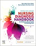
Ackley and Ladwig’s Nursing Diagnosis Handbook: An Evidence-Based Guide to Planning Care
We love this book because of its evidence-based approach to nursing interventions. This care plan handbook uses an easy, three-step system to guide you through client assessment, nursing diagnosis, and care planning. Includes step-by-step instructions showing how to implement care and evaluate outcomes, and help you build skills in diagnostic reasoning and critical thinking.
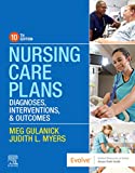
Nursing Care Plans – Nursing Diagnosis & Intervention (10th Edition)
Includes over two hundred care plans that reflect the most recent evidence-based guidelines. New to this edition are ICNP diagnoses, care plans on LGBTQ health issues, and on electrolytes and acid-base balance.
Nurse’s Pocket Guide: Diagnoses, Prioritized Interventions, and Rationales
Quick-reference tool includes all you need to identify the correct diagnoses for efficient patient care planning. The sixteenth edition includes the most recent nursing diagnoses and interventions and an alphabetized listing of nursing diagnoses covering more than 400 disorders.

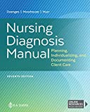
Nursing Diagnosis Manual: Planning, Individualizing, and Documenting Client Care
Identify interventions to plan, individualize, and document care for more than 800 diseases and disorders. Only in the Nursing Diagnosis Manual will you find for each diagnosis subjectively and objectively – sample clinical applications, prioritized action/interventions with rationales – a documentation section, and much more!
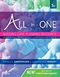
All-in-One Nursing Care Planning Resource – E-Book: Medical-Surgical, Pediatric, Maternity, and Psychiatric-Mental Health
Includes over 100 care plans for medical-surgical, maternity/OB, pediatrics, and psychiatric and mental health. Interprofessional “patient problems” focus familiarizes you with how to speak to patients.
See also
Other recommended site resources for this nursing care plan:
- Nursing Care Plans (NCP): Ultimate Guide and Database MUST READ!
Over 150+ nursing care plans for different diseases and conditions. Includes our easy-to-follow guide on how to create nursing care plans from scratch. - Nursing Diagnosis Guide and List: All You Need to Know to Master Diagnosing
Our comprehensive guide on how to create and write diagnostic labels. Includes detailed nursing care plan guides for common nursing diagnostic labels.
Other nursing care plans related to respiratory system disorders:
- Asthma
- Aspiration Risk & Aspiration Pneumonia
- Airway Clearance Therapy & Coughing
- Bronchiolitis
- Bronchopulmonary Dysplasia (BPD)
- Chronic Obstructive Pulmonary Disease (COPD)
- Cystic Fibrosis
- Hemothorax and Pneumothorax
- Influenza (Flu)
- Ineffective Breathing Pattern (Dyspnea)
- Impairment of Gas Exchange
- Lung Cancer
- Mechanical Ventilation
- Near-Drowning
- Pleural Effusion
- Pneumonia
- Pulmonary Embolism
- Pulmonary Tuberculosis
- Tracheostomy
References and Sources
Recommended sources, interesting articles, and references about ineffective or altered breathing pattern (dyspnea) to further your reading.
- Amorim Beltrão, B., da Silva, V. M., de Araujo, T. L., & de Oliveira Lopes, M. V. (2011). Clinical indicators of ineffective breathing pattern in children with congenital heart diseases. International Journal of Nursing Terminologies and Classifications, 22(1), 4-12.
- Cavalcante, J. C. B., Mendes, L. C., de Oliveira Lopes, M. V., & de Oliveira Lima, L. H. (2010). Clinical indicators of ineffective breathing pattern in children with asthma. Rev Rene, 11(1).
- Gouna, G., Rakza, T., Kuissi, E., Pennaforte, T., Mur, S., & Storme, L. (2013). Positioning effects on lung function and breathing pattern in premature newborns. The Journal of pediatrics, 162(6), 1133-1137.
- Lopes, M. V. O., Silva, V. M. D., & VEC, S. F. (2020). A Content Analysis of Clinical Indicators of the Nursing Diagnosis Ineffective Breathing Pattern. International Journal of Nursing Knowledge.
- Pascoal, L. M., Lopes, M. V. D. O., da Silva, V. M., Beltrão, B. A., Chaves, D. B. R., de Santiago, J. M. V., & Herdman, T. H. (2014). Ineffective breathing pattern: defining characteristics in children with acute respiratory infection. International journal of nursing knowledge, 25(1), 54-61.
- Silveira, U. A., de Oliveira Lima, L. H., & de Oliveira Lopes, M. V. (2008). Defined characteristics of the nursing diagnoses ineffective airway clearanceand ineffective breathing pattern in asthmatic children. Rev Rene, 9(4).
- Campbell, M. L. (2017). Dyspnea. Critical Care Nursing Clinics, 29(4).
- Hashmi, M. F., Modi, P., Basit, H., & Sharma, S. (2023, February 19). Dyspnea – StatPearls. NCBI.
- Hinkle, J. L., & Cheever, K. H. (2018). Brunner & Suddarth’s Textbook of Medical-surgical Nursing. Wolters Kluwer.
- Hoffman, M., Augusto, V. M., Eduardo, D. S., Silveira, B. M. F., Lemos, M. D., & Parreira, V. F. (2021). Inspiratory muscle training reduces dyspnea during activities of daily living and improves inspiratory muscle function and quality of life in patients with advanced lung disease. Physiotherapy Theory and Practice, 37(8).
- Hulya, S., Ilknur, N., & Gulru, P. (2020). Effect of exercise capacity on perception of dyspnea, psychological symptoms and quality of life in patients with chronic obstructive pulmonary disease. Heart & Lung.
- Lenferink, A., van der Palen, J., & Effing, T. (2018). The role of social support in improving chronic obstructive pulmonary disease self-management. Expert Review of Respiratory Medicine, 12(8).
- Oh, H., Park, K., Yoon, S., Kim, Y., Lee, S.-H., Choi, Y. Y., & Choi, K.-H. (2018). Clinical Utility of Beck Anxiety Inventory in Clinical and Nonclinical Korean Samples. Frontiers in Psychiatry, 4(9).
- Reuter, S., Mosan, C., & Baack, M. (2014). Respiratory Distress in the Newborn – PMC. NCBI.
- Tramontano, A., & Palange, P. (2023). Nutritional State and COPD: Effects on Dyspnoea and Exercise Tolerance. Nutrients, 15(7).
- Whited, L., & Graham, D. D. (2023, March 9). Abnormal Respirations – StatPearls. NCBI. Retrieved June 9, 2023, from
- Yohannes, A. M., Junkes-Cunha, M., Smith, J., & Vestbo, J. (2017). Management of Dyspnea and Anxiety in Chronic Obstructive Pulmonary Disease: A Critical Review. Journal of the American Medical Directors Association, 18(12).
- Zhou, Y. H., & Mak, Y. W. (2017). Psycho-Physiological Associates of Dyspnea in Hospitalized Patients with Interstitial Lung Diseases: A Cross-Sectional Study. International Journal of Environmental Research and Public Health, 17.
- Zimmerman, B., & Williams, D. (2022). Lung Sounds – StatPearls. NCBI.
Amazingly helpful.Thanks very much
Super grateful for this tool…