Learn about the nursing care management of patients with fractures in this nursing study guide.
Table of Contents
- What is Fracture?
- Classification
- Causes
- Clinical Manifestations
- Complications
- Assessment and Diagnostic Findings
- Medical Management
- Nursing Management
- Practice Quiz: Fracture
- See Also
What is Fracture?
Injury to one part of the musculoskeletal system results in the malfunction of adjacent muscles, joints, and tendons.
- A fracture is a complete or incomplete disruption in the continuity of the bone structure and is defined according to its type and extent.
- Fractures occur when the bone is subjected to stress greater than it can absorb.
- When the bone is broken, adjacent structures are affected, resulting in soft tissue edema, hemorrhage into muscles and joints, joint dislocations, ruptured tendons, severed nerves, and damaged blood vessels.
Classification
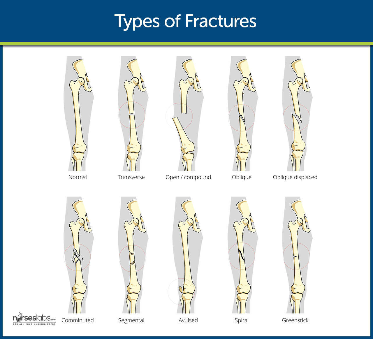
There are several kinds of fracture that may occur in a bone:
- Complete fracture. A complete fracture involves a break across the entire cross-section of the bone and is frequently displaced.
- Incomplete fracture. An incomplete fracture involves a breakthrough only part of the cross-section of the bone.
- Comminuted fracture. A comminuted fracture is one that produces several bone fragments.
- Closed fracture. A closed fracture is one that does not cause a break in the skin.
- Open fracture. An open fracture is one in which the skin or mucous membrane wound extends to the fractured bone.
Causes
Fractures may be caused by the following:
- Direct blows. Being hit directly by a great force could cause fractures in the bones.
- Crushing forces. Forces that come into contact with the bones and crush them could also result in fractures.
- Sudden twisting motions. Twisting the joints in a sudden motion leads to fractures.
- Extreme muscle contractions. When the muscles have reached their limit in contraction, it could lead to serious fractures.
Clinical Manifestations
The clinical signs and symptoms of a fracture may include the following but not all are present in every fracture:
- The pain is continuous and increases in severity until the bone fragments are immobilized.
- Loss of function. After a fracture, the extremity cannot function properly because the normal function of the muscles depends on the integrity of the bones to which they are attached.
- Displacement, angulation, or rotation of the fragments in a fracture of the arm or leg causes a deformity that is detectable when the limb is compared with the uninjured extremity.
- There is an actual shortening of the extremity because of the compression of the fractured bone.
- When the extremity is gently palpated, a crumbling sensation, called crepitus, can be felt.
- Localized edema and ecchymosis. Localized edema and ecchymosis occur after a fracture as a result of trauma and bleeding into the tissues.
Complications
Complications of fractures may either be acute or chronic.
- Hypovolemic shock resulting from hemorrhage is more frequently noted in trauma patients with pelvic fractures and in patients with displaced or open femoral fractures.
- Fat embolism syndrome. After fracture of long bones and or pelvic bones, or crush injuries, fat emboli may develop.
- Compartment syndrome. Compartment syndrome in an extremity is a limb-threatening condition that occurs when perfusion pressure falls below tissue pressure within a closed anatomic compartment.
Assessment and Diagnostic Findings
To determine the presence of fracture, the following diagnostic tools are used.
- X-ray examinations: Determines location and extent of fractures/trauma, may reveal preexisting and yet undiagnosed fracture(s).
- Bone scans, tomograms, computed tomography (CT)/magnetic resonance imaging (MRI) scans: Visualizes fractures, bleeding, and soft-tissue damage; differentiates between stress/trauma fractures and bone neoplasms.
- Arteriograms: May be done when occult vascular damage is suspected.
- Complete blood count (CBC): Hematocrit (Hct) may be increased (hemoconcentration) or decreased (signifying hemorrhage at the fracture site or at distant organs in multiple trauma). Increased white blood cell (WBC) count is a normal stress response after trauma.
- Urine creatinine (Cr) clearance: Muscle trauma increases the load of Cr for renal clearance.
- Coagulation profile: Alterations may occur because of blood loss, multiple transfusions, or liver injury.
Medical Management
Management of a patient with a fracture can belong to either emergent or post-emergent.
- Immediately after injury, if a fracture is suspected, it is important to immobilize the body part before the patient is moved.
- Adequate splinting is essential to prevent the movement of fracture fragments.
- In an open fracture, the wound should be covered with a sterile dressing to prevent contamination of the deeper tissues.
- Fracture reduction refers to the restoration of the fracture fragments to anatomic alignment and positioning and can be open or closed depending on the type of fracture.
Nursing Management
Nursing management for close and open fractures should be differentiated.
Nursing Assessment
Assessment of the fractured area includes the following:
- Close fracture. The patient with close fracture is assessed for absence of opening in the skin at the fracture site.
- Open fracture. The patient with open fracture is assessed for risk for osteomyelitis, tetanus, and gas gangrene.
- The fractured site is assessed for signs and symptoms of infection.
Diagnosis
Based on the assessment data gathered, the nursing diagnoses developed include:
- Acute pain related to fracture, soft tissue injury, and muscle spasm.
- Impaired physical mobility related to fracture.
- Risk for infection related to opening in the skin in an open fracture.
Planning & Goals
Main Article: 8 Fracture Nursing Care Plans
Planning and goals developed for a patient with fracture are:
- Relief of pain.
- Achieve a pain-free, functional, and stable body part.
- Maintain asepsis.
- Maintain vital signs within normal range.
- Exhibit no evidence of complications.
Nursing Interventions
Nursing care of a patient with fracture include:
- The nurse should instruct the patient regarding proper methods to control edema and pain.
- It is important to teach exercises to maintain the health of the unaffected muscles and to increase the strength of muscles needed for transferring and for using assistive devices.
- Plans are made to help the patients modify the home environment to promote safety such as removing any obstruction in the walking paths around the house.
- Wound management. Wound irrigation and debridement are initiated as soon as possible.
- Elevate extremity. The affected extremity is elevated to minimize edema.
- Signs of infection. The patient must be assessed for presence of signs and symptoms of infection.
Evaluation
The following should be evaluated for a successful implementation of the care plan.
- Pain was relieved.
- Achieved a pain-free, functional, and stable body part.
- Maintained asepsis.
- Maintained vital signs within normal range.
- Exhibited no evidence of complications.
Discharge and Home Care Guidelines
After completion of the home care instructions, the patient or caregiver will be able to:
- Control swelling and pain. Describe approaches to reduce swelling and pain such as elevating the extremity and taking analgesics as prescribed.
- Care of the affected area. Describe management of immobilization devices or care of the incision.
- Consume diet to promote bone healing.
- Mobility aids. Demonstrate use of mobility aids and assistive devices safely.
- Avoid excessive use of injured extremity and observe weight-bearing limits.
Documentation Guidelines
The focus of documentation should include:
- Client’s description of response to pain and acceptable level of pain.
- Prior medication use.
- Level of function.
- Ability to participate in specific or desired activities.
- Signs and symptoms of infectious process.
- Wound/ incision site.
- Plan of care.
- Teaching plan.
- Response to interventions, teaching, and actions performed.
- Attainment or progress toward desired outcomes.
- Modifications to plan of care.
- Long term needs.
Practice Quiz: Fracture
Here’s a 5-item quiz about the study guide. Please visit our nursing test bank for more NCLEX practice questions.
1. The following are the different types of fractures except for:
A. Open fracture.
B. Diagonal fracture.
C. Closed fracture.
D. Comminuted fracture.
2. The most definitive diagnostic tool used in a patient with a fracture is:
A. Blood studies.
B. SGPT and SGOT tests.
C. X-ray.
D. MRI.
3. Which of the following is a nursing diagnosis for a patient with a fracture?
A. Risk for electrolyte imbalance.
B. Situational low self-esteem.
C. Acute pain.
D. Impaired breathing pattern.
4. The fractured part should be elevated above the level of what organ?
A. Brain.
B. Heart.
C. Liver.
D. Kidney.
5. What kind of shock is most commonly found in a patient with a fracture?
A. Hypovolemic shock.
B. Cardiogenic shock.
C. Neurologic shock.
D. Septic shock.
Answers and Rationale
1. Answer: B. Diagonal fracture.
- B: Diagonal fracture is not a type of fracture.
- A: Open fracture is one of the types of fractures.
- C: Closed fracture is one of the types of fractures.
- D: Comminuted fracture is one of the types of fractures.
2. Answer: C. X-ray.
- C: X-ray is the most definitive diagnostic tool in assessing for fracture as it allows visualization of the affected part.
- A: Blood studies are not used in a patient with fracture.
- B: SGPT and SGOT tests are not used in patient with fracture.
- D: MRI may be used but it is not the most definitive tool in assessing fractures.
3. Answer: C. Acute pain.
- C: Acute pain is the most appropriate nursing diagnosis for a patient with fracture.
- A: Risk for electrolyte imbalance is not a nursing diagnosis for a patient with fracture.
- B: Situational low self-esteem is not a nursing diagnosis for a patient with fracture.
- D: Impaired breathing pattern is not a nursing diagnosis for a patient with fracture.
4. Answer: B. Heart.
- B: The fractured area should be elevated above the level of the heart to promote venous return.
- A: The fractured area should not be elevated above the level of the brain.
- C: The fractured area should not be elevated above the level of the liver.
- D: The fractured area should not be elevated above the level of the kidney.
5. Answer: A. Hypovolemic shock.
- A: Hypovolemic shock resulting from hemorrhage is more frequently noted in trauma patients with pelvic fractures and in patients with displaced or open femoral fractures.
- B: Cardiogenic shock is not frequently found in patients with fracture.
- C: Neurologic shock is not frequently found in patients with fracture.
- D: Septic shock is not frequently found in patients with fracture.
See Also
Posts related to this study guide:


Perfect and reliable
Your care plans are so helpful even to us students
so perfect notes for students but can you guy add the pathophysiology part.
Thank you! Could you please upload quizzes and practice questions on Musculoskeletal disorders?
This is noted. In the meantime, please check NCLEX Practice Questions test bank.
Hi Dractive, please check out Musculoskeletal Disorders NCLEX Practice Quiz (70 Questions)
That’s wonderful but i wish if i could get all musculoskeletal disorders in the form of pdf
You can save the webpage as a PDF by simply going to File > Print > Save as PDF.
Great Read!!! Thanks for sharing such a great blog, blog like these is really helpful.
It is good application for the students because it gives thinking capacity to the part you learn
What are the pertinent labs to monitor?