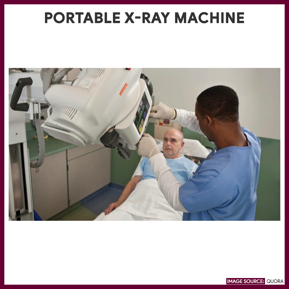
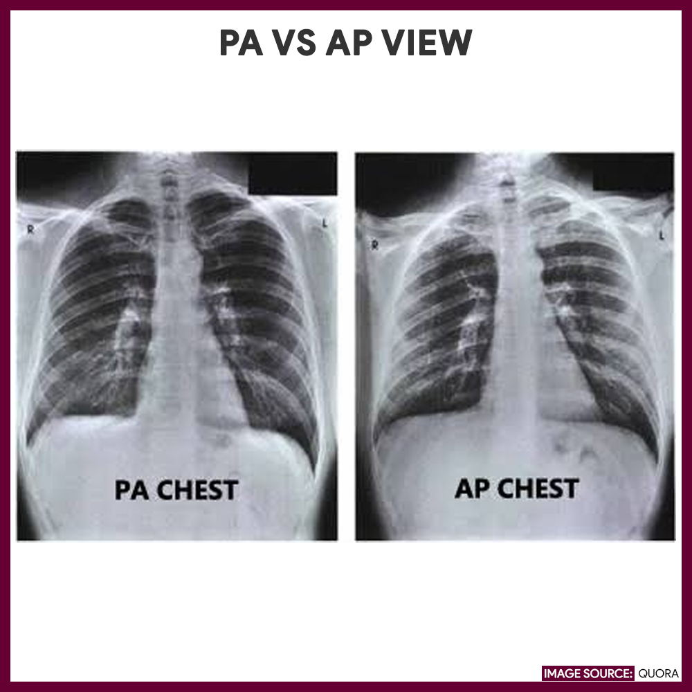
Chest X-ray (Chest radiography, CXR) is one of the most frequently performed radiological examinations. A chest x-ray is a painless, non-invasive test that uses electromagnetic waves to produce visual images of the heart, lungs, bones, and blood vessels of the chest. Air spaces normally seen in the lungs appear dark on the chest films. A basic chest x-ray includes a posteroanterior (PA) view, in which x-rays pass from the back to the front of the body and a left lateral view. Other projections such as lateral decubitus, lordotic views, or oblique views can also be requested. For critically ill patients who cannot leave the nursing unit, a portable x-ray machine is performed at the bedside using anteroposterior (AP) projections with an addition of a lateral decubitus view if a free flow fluid or air is suspected.
Chest images should be examined in full inspiration and erect if feasible to reduce cardiac magnification and demonstrate fluid levels. Expiration images may be needed to identify a pneumothorax or locate foreign materials. Rib detail images may be taken to delineate bone pathology, helpful when chest radiographs illustrate metastatic lesions or fractures. In the onset of the disease process of asthma, tuberculosis, and chronic obstructive pulmonary disease, chest x-ray results may not correlate with the patient’s clinical status and may even be normal.
Nurses are responsible for ensuring the patient’s comfort while at the x-ray room since some may experience pain from injury or symptoms from a disease condition, as well as the apprehension about what the result may show. In addition, producing a good quality image relies on the ability of the patient to cooperate, such as holding breath for a while. Providing a calm and relaxed environment for the patient is indeed vital.
This diagnostic and laboratory procedure study guide can help nurses understand their tasks and responsibilities during a chest x-ray.
Indications of Chest X-ray
Here are some of the reasons why a Chest x-ray is performed:
- Assist in the diagnosis of diaphragmatic hernia, lung tumors, and metastasis
- Detect known or suspected pulmonary, cardiovascular, and skeletal disorders
- Identify the presence of chest trauma
- Confirm correct placement and position of the endotracheal tube, tracheostomy tube, chest tubes, central venous catheters, nasogastric feeding tube, pacemaker wires, intraortic balloon pump, Swan-Ganz catheters, and automatic implantable cardioverter defibrillator
- Evaluate positive purified protein derivative (PPD) or Mantoux test for pulmonary tuberculosis.
- Monitor progressions, resolutions, or maintenance of disease
- Evaluate the patient’s response to a therapeutic regimen (antibiotic, chemotherapy)
Contraindication
Chest X-ray is not advisable for:
- Patients who are pregnant or suspected of being pregnant unless the potential benefits of a procedure using radiation outweigh the risk of maternal and fetal damage
Interfering Factors
These are factors or conditions that may alter the outcome of Chest X-ray:
- Presence of metallic objects within the area of examination
- Excessive or unnecessary movements made by the patient during the procedure
- Incorrect position of the patient, which may produce poor exposure of the area to be examined
- Inability of the patient to take full inspiration
- Improper adjustment of the radiographic equipment to serve thin or obese patients, which can result in underexposure or overexposure of the films
Procedure
The procedure for chest x-rays is as follows:
- Items are removed
Patients will be asked to remove any clothing, jewelry, or other articles that may interfere with the study. - Appropriate clothing is given
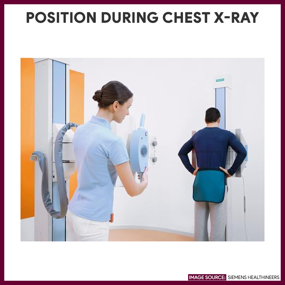
Patients will be provided by an X-ray gown to wear. - Positioning the patient
The patient in a standing or sitting position will face the cassette or image detector with hands on hips, inhale deeply, hold one’s breath until the X-ray image is made. For a lateral view, the chest is position on the left side against the image holder with hands raised above the head. - Images are taken
The x-ray technician will stand behind a protective shield while the films are being developed within a few minutes.
Nursing Responsibilities for Chest X-ray
The following are the nursing interventions and nursing care considerations for the patient
Before Chest X-ray
The following are the nursing interventions before chest x-ray:
- Remove all metallic objects. Items such as jewelry, pins, buttons etc can hinder the visualization of the chest.
- No preparation is required. Fasting or medication restriction is not needed unless directed by the health care provider.
- Ensure the patient is not pregnant or suspected to be pregnant. X-rays are usually not recommended for pregnant women unless the benefit outweighs the risk of damage to the mother and fetus.
- Assess the patient’s ability to hold his or her breath. Holding one’s breath after inhaling enables the lungs and heart to be seen more clearly in the x-ray.
- Provide appropriate clothing. Patients are instructed to remove clothing from the waist up and put on an X-ray gown to wear during the procedure.
- Instruct patient to cooperate during the procedure. The patient is asked to remain still because any movement will affect the clarity of the image.
After Chest X-ray
The nurse should note the following nursing interventions after chest x-ray:
- No special care. Note that no special care is required following the procedure
- Provide comfort. If the test is facilitated at the bedside, reposition the patient properly.
Normal Results
Normal findings in a chest x-ray will show a:
Abnormal Results
The following abnormalities can be seen on a chest x-ray test. These includes:
- Atelectasis (collapse or incomplete expansion of pulmonary parenchyma)
- Bronchitis (inflammation of the bronchial tube)
- Cardiomegaly (enlargement of the heart)
- Flattened diaphragm associated with hyperinflation of the lung (indicator for COPD)
- Foreign bodies lodged in the pulmonary system as seen by a radiopaque object
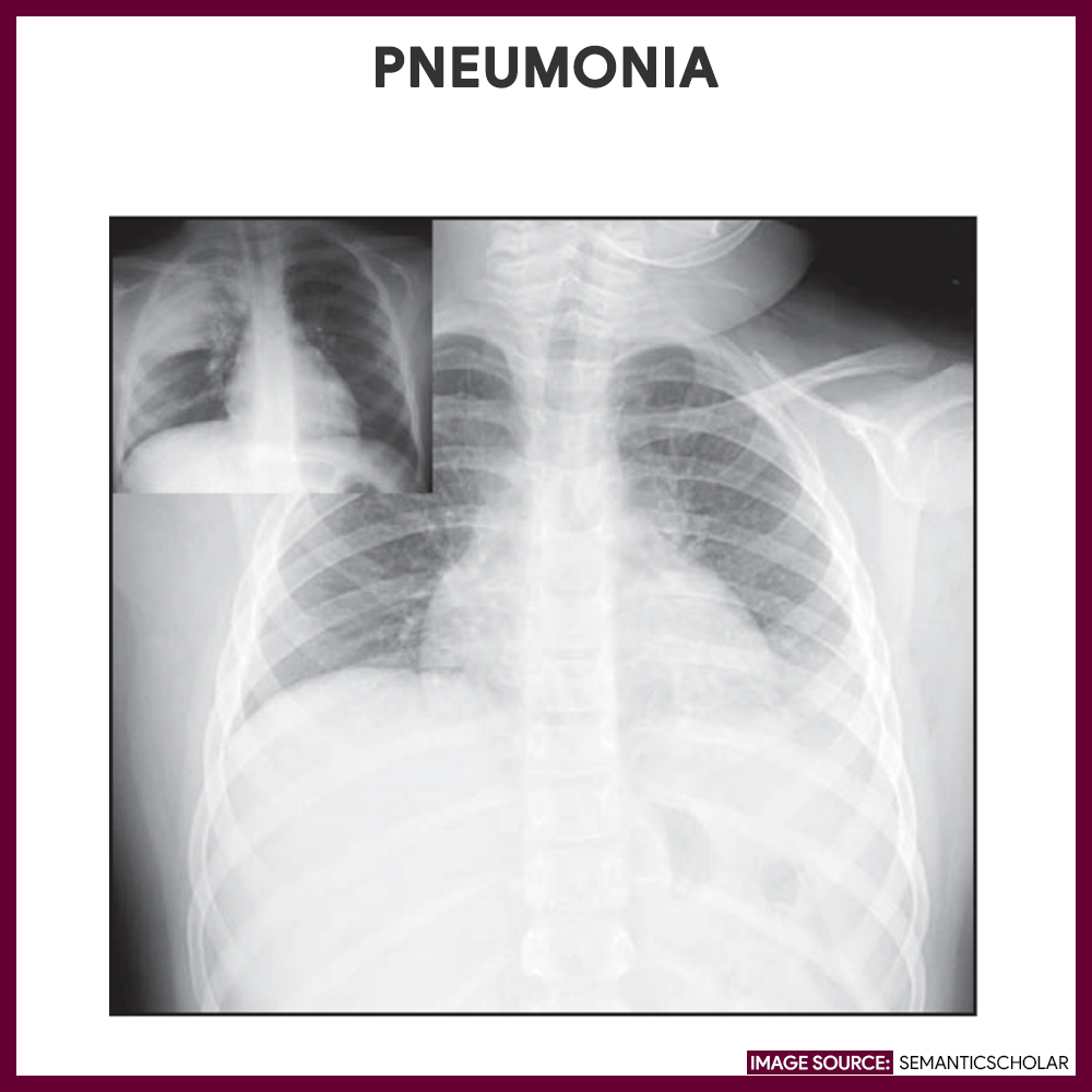
- Irregular patchy infiltrates in the lung fields (suggestive of pneumonia)
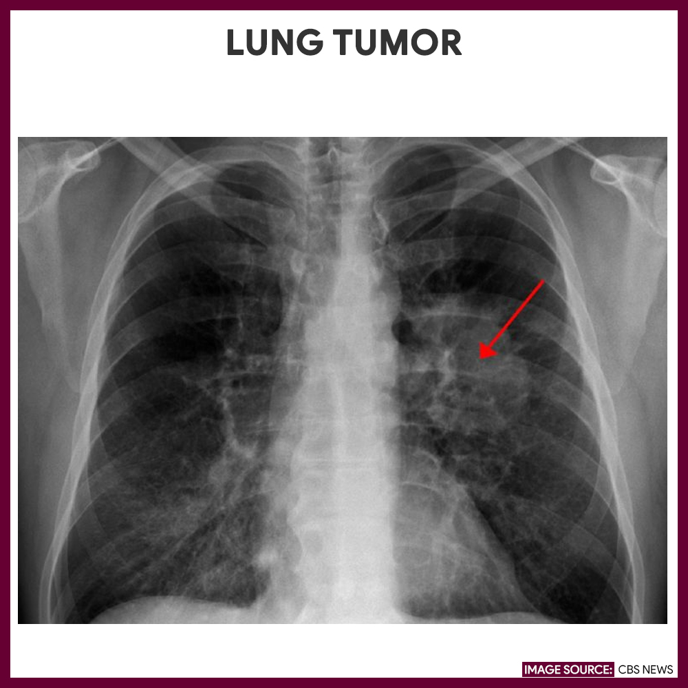
- Lung tumors (irregular and abnormal white shadow on the lung fields)
- Malposition of tubes or wires
- Misalignment or break of bones (indicating fracture)
- Pericardial effusion (fluid accumulation around the heart)
- Pericarditis (inflammation of the pericardium)
- Pleural effusion (fluid accumulation within the pleural space)
- Pneumothorax (presence of air within the pleural space)
- Pulmonary bases, infiltrates, fibrosis,
- Scoliosis (curvature of the spinal column)
- Swollen lymph nodes
- Tuberculosis (patchy, nodular infiltrates usually located on the upper lobe lung fields; cavities in the lung)
- Widened mediastinum (suggesting neoplasm or aortic aneurysm)
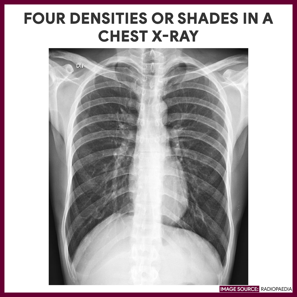
Gallery
Images and photographs related to chest x-ray:


References and Sources
Additional resources and references for this Chest X-ray nursing study guide:
- Suzanne C. Smeltzer. Brunner & Suddarth’s Handbook of Laboratory and Diagnostic Tests: Lippincott Williams & Wilkins
- Dan L. Hobbs, M.S.R.S., R.T.(R)(CT)(MR): Chest Radiography for Radiologic Technologists
- Anne M. Van Leeuwen, Mickey Lynn Bladh. Laboratory & Diagnostic Tests with Nursing Implications: Davis’s
- Swingler, G. H., & Zwarenstein, M. (2008). Chest radiograph in acute respiratory infections. Cochrane Database of Systematic Reviews, (1). [Link]


Leave a Comment