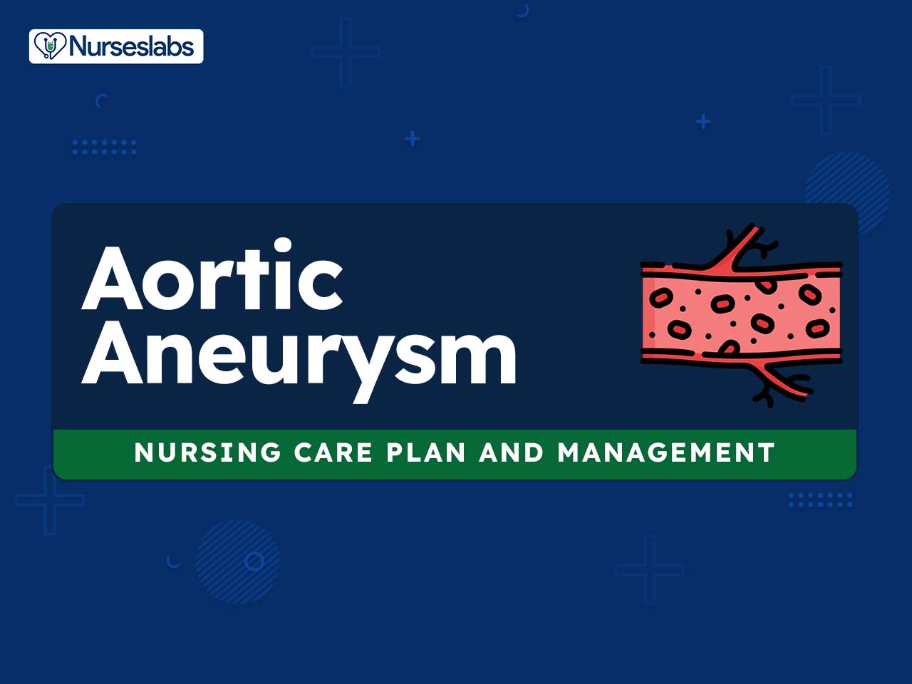
Use this nursing care plan and management guide to help care for patients with aortic aneurysm. Enhance your understanding of nursing assessment, interventions, goals, and nursing diagnosis, all specifically tailored to address the unique needs of individuals facing aortic aneurysm.
What is Aortic Aneurysm?
Aortic aneurysm (Abdominal Aneurysm; Dissecting Aneurysm; Thoracic Aneurysm;) is a localized, circumscribed, blood-filled abnormal dilation of an artery caused by disease or weakening of the vessel wall.
True aneurysms involve the dilation of all layers of the vessel wall. The two types of true aneurysms are (1) saccular, which is characterized by a bulbous out-pouching of one side of the artery resulting in localized stretching of the artery wall, and (2) fusiform, which is characterized by a uniformly shaped dilation of the entire circumference of the artery. True aneurysms are asymptomatic and are typically diagnosed by physical examination or a diagnostic ultrasound or computed tomography (CT) scan. The natural history of an aneurysm is enlargement; as a rule, the larger it is, the greater the chance of rupture. Aneurysms are most commonly seen in the abdominal aorta. Abdominal aortic aneurysms (AAAs) account for about 75% and thoracic aneurysms for about 25% of all cases. They occur more often in men than in women. Risk factors include smoking and familial history of aneurysms. When an aneurysm becomes large enough for risk for rupture, it can be repaired by open surgical repair or a less-invasive endograft-covered stent repair.
Dissecting aneurysms occur when the inner layer of the blood vessel wall tears and splits, creating a false channel and cavity of blood between the intimal and adventitial layers. They are typically classified according to location. According to the Stanford classification, type A involves the ascending aorta and its transverse arch and type B involves the descending aorta. A dissecting AAA is the most catastrophe involving the aorta, and it has a high mortality rate if not detected early and treated with surgery. More than 90% of clients present with sudden onset of severe pain which is usually described, as sharp, tearing, or stabbing in nature. Symptoms depend on the size and location of the dissection or rupture. Risk factors for dissection include congenital, inflammatory, hypertension, pregnancy, trauma, and Marfan syndrome.
Nursing Care Plans and Management
Nursing Assessment
Aortic aneurysms can often be silent, producing no symptoms until they rupture. However, if symptoms do occur, they can include:
Assess for the following subjective and objective data:
- Abdominal Aortic Aneurysm (AAA).
- Thoracic Aortic Aneurysm:
- Tenderness or pain in the chest.
- Back pain.
- Shortness of breath or trouble breathing or swallowing.
- Coughing, possibly with blood.
- Hoarseness.
- Signs of a Ruptured Aortic Aneurysm:
- Sudden, intense and persistent abdominal or back pain, which can be described as a tearing sensation.
- Pain radiating to back or legs.
- Sweatiness.
- Clamminess.
- Dizziness.
- Nausea.
- Vomiting.
- Low blood pressure.
- Rapid heart rate.
- Shock or loss of consciousness.
Nursing Diagnosis
Following a thorough assessment, a nursing diagnosis is formulated to specifically address the challenges associated with aortic aneurysm based on the nurse’s clinical judgement and understanding of the patient’s unique health condition. While nursing diagnoses serve as a framework for organizing care, their usefulness may vary in different clinical situations. In real-life clinical settings, it is important to note that the use of specific nursing diagnostic labels may not be as prominent or commonly utilized as other components of the care plan. It is ultimately the nurse’s clinical expertise and judgment that shape the care plan to meet the unique needs of each patient, prioritizing their health concerns and priorities.
Nursing Goals
Goals and expected outcomes may include:
- The client will verbalize strategies to reduce his anxiety level.
- The client will demonstrate a positive coping method.
- The client or significant others verbalize understanding of disease process, treatment options, and goals of therapy.
- The client will maintain adequate cardiac output, as evidenced by HR of 60 to 100 beats per minute, normotensive BP, palpable pulse, clear lung sounds, urine output of more than 30 ml/hr, and normal level of consciousness.
- The client will maintain adequate tissue perfusion as evidenced by strong palpable pulses; warm, dry extremities; BP within the normal range for the client; urinary output greater than or equal to 30 ml/hr; alert, normal level of consciousness; normal bowel sounds; absence of abdominal or chest pain.
- The client will have a reduced risk for complications from progressive dissection or rupture s a result of early detection of symptoms and appropriate intervention.
Nursing Interventions and Actions
Therapeutic interventions and nursing actions for patients with aortic aneurysm may include:
1. Reducing Anxiety and Fear
Clients with an aortic aneurysm may experience anxiety due to the potentially life-threatening nature of the condition and the uncertainty surrounding their prognosis. The fear of sudden rupture and the need for surgery or other invasive procedures can also contribute to anxiety. Nurses play a vital role in addressing these emotional concerns by providing clear and comprehensive information about the condition, its treatment options, and the expected outcomes. Empathetic communication, active listening, and reassurance help create a supportive environment where patients feel comfortable expressing their fears and concerns.
1. Assess the client’s anxiety level (mild, severe). Note signs and symptoms, especially nonverbal communication.
Aortic dissection and/or rupture can result in an acute life-threatening situation that will produce high levels of anxiety in the client as well as in significant others.
2. Acknowledge awareness of the client’s anxiety.
Acknowledgment of the client’s feelings validates the feelings and communicates acceptance of those feelings.
3. Provide a quiet, private place for significant others to wait.
A quiet environment can reduce anxiety.
4. Reduce unnecessary external stimuli.
Anxiety may escalate with an excessive conversation, noise, and equipment around the client.
5. Explain all procedures as appropriate, using simple, concrete words.
The information helps allay anxiety. Clients who are anxious may not be able to comprehend anything more than simple, clear, brief instructions.
2. Initiating Health Teachings and Patient Education
Initiating health teachings and patient education in individuals diagnosed with aortic aneurysm is of utmost importance in promoting their understanding of the condition and empowering them to actively participate in their own care. By providing comprehensive and tailored education, healthcare professionals can equip patients with the knowledge and skills necessary to make informed decisions about their treatment options, manage their symptoms, and adopt a healthy lifestyle to minimize the risk of complications. Effective patient education involves clear communication, using accessible language, and utilizing visual aids to enhance comprehension. Furthermore, it is essential to create a supportive environment that encourages patients to ask questions, express concerns, and actively engage in their learning process.
1. Assess the client’s knowledge of the disease and treatment options.
This information provides an important starting point in education.
2. Instruct medically treated clients about the following: goals of therapy (avoidance of excess BP and strain on the disease arterial wall), the importance of follow-up computed tomography scanning, signs and symptoms to report, side effects of the drug, and use of antihypertensive medications as prescribed; the importance of compliance.
Clients treated medically need to maintain goal BP levels and comply with scheduled CT scans to monitor the size of the aneurysm. Knowledge of early warning signs facilitates rapid treatment. These may include pain in the chest, back, groin, and abdomen; decreased urine output; cool, pale extremities.
3. Instruct surgical clients about the following: activity restrictions, signs and symptoms to report, wound care, and avoidance activities that are isometric or abruptly can raise BP (e.g., lifting and carrying heavy objects, straining for bowel movements).
Discharge instructions guide clients regarding self-care measures; Heavy lifting of more than 5 to 10 pounds is restricted for 4 to 6 weeks after surgical repair of an aortic aneurysm. These restrictions reduce strain on suture lines until they are completely healed; Clients need to be aware of the warning signs that warrant medical attention.
4. Instruct endograft clients about the need for follow-up CT scans at 1 and 6 months and yearly for the rest of their lives.
The endograft may incur a leak; ongoing evaluation is needed so appropriate treatment can be initiated.
3. Managing Decrease in Cardiac Output
Clients with aortic aneurysms are at risk for decrease in cardiac output due to progressive dissection, which can impair blood flow to vital organs, as well as aortic rupture, which can cause life-threatening bleeding. In addition, certain medications used to treat the condition, such as beta-blockers, can also contribute to decreased cardiac output by lowering heart rate and blood pressure. Nurses play a critical role in monitoring the patient’s cardiac status and intervening promptly to address any decrease in cardiac output. Treatment strategies may include optimizing blood pressure and heart rate through medication, ensuring adequate fluid balance, and implementing measures to reduce cardiac workload. In some cases, surgical intervention, such as repairing or replacing the aneurysm, may be necessary to restore normal blood flow and improve cardiac output.
1. Assess for signs of myocardial ischemia: chest pain, tachycardia, or ST-segment and T-wave changes on electrocardiogram (ECG).
ECG changes help guide the timing of interventions.
2. Assess the client’s hemodynamic status. Monitor for signs of decreasing cardiac output, such as tachycardia, decreased urine output, and restlessness.
A dissecting abdominal aortic aneurysm (AAA) is the most common catastrophe involving the aorta, and it has a high mortality rate if not detected early and treated with surgery. Clients with decreasing or rupturing aneurysms are hemodynamically compromised. Early evaluation of warning signs facilitates prompt intervention.
3. Send a blood specimen for type and crossmatch and other routine preoperative blood work.
Blood replacement therapy may be required to maintain effective blood volume.
1. If decreased cardiac output is related to further dissection (severe aortic insufficiency) or ruptured aorta, anticipate emergency angiography and surgery.
Rapid, efficient intervention is critical to preserve circulation and life
2. Administer medications, intravenous fluids, and blood as ordered.
These maintain adequate cardiac output before surgical intervention.
3. Stay with the client.
The presence of competent, calm staff may provide emotional support and reduce fear.
4. Prepare the client for surgical repair.
The information helps to allay anxiety. Clients who are anxious may not be able to comprehend anything more than simple, brief instructions and explanations.
5. If decreased cardiac output is drug induced, anticipate the following:
- 5.1. For beta-blocker: May stop the drug or reduce the dose.
Beta-blockers have a negative inotropic effect, which can potentiate heart failure. The presence of crackles and S3 indicates heart failure.
- 5.2. For vasodilators: Stop the drug and administer isotonic solution (0.9% normal saline solution) or plasma expanders.
Fluids are usually required to maintain increased intravascular volume.
4. Promoting Effective Tissue Perfusion
Clients with aortic aneurysms are at risk for ineffective tissue perfusion due to a variety of factors, including iatrogenic causes such as surgical complications, trauma to the vessel wall during medical procedures, defects in the vessel wall that impair blood flow, and conditions that increase stress on the arterial wall, such as hypertension and atherosclerosis. Impaired tissue perfusion can lead to a range of complications, including organ damage and ischemia. Nurses focus on strategies to enhance tissue perfusion in these patients. This may involve monitoring blood pressure and heart rate to maintain optimal cardiac function. Additionally, medications may be administered to promote vasodilation and improve blood flow.
1. Assess and monitor the location and characteristics of pain such as in the thoracic area (pain in the neck, low back, shoulders, or abdomen) and in the abdominal area (pain in the abdomen or back, flank, or groin caused by the pressure on adjacent structures).
A description of pain can help differentiate the location and determine treatment. More than 90% of clients with abdominal aortic aneurysms have a sudden onset of severe pain described as sharp, tearing, or stabbing in nature.
2. Obtain a thorough history regarding risk factors for dissection or rupture.
Most clients are asymptomatic unless their aneurysm is dissecting or ruptures. History aids in ruling out cerebrovascular, cardiac, renal, vascular, and occlusive disease. Poorly controlled hypertension increases stress on the aortic wall and its risk for dissection or rupture.
3. Monitor for signs and symptoms indicating progressive dissection.
A high index suspicion can determine the treatment to reduce mortality. Clinical signs and symptoms indicate the site and progression of dissection. Acute aortic dissection usually occurs along the thoracic aorta. Pain is severe and may mimic the pain associated with myocardial infarction. Pain may be located both above and below the diaphragm if the dissection is extensive. Changes in the level of consciousness and diminished carotid pulses are associated with the dissection of the aortic arch. Dissection of the abdominal aorta can cause decreased urine output, diminished motor and sensory function in the lower extremities, abdominal pain, and bloody diarrhea.
For abdominal aneurysms:
4. Assess the lower extremities for signs of peripheral ischemia and insufficiency (such as paralysis, pain, paresthesia, pallor, pulselessness, and poikilothermia [decreased temperature, coolness]).
Dissection can cause reduced sensory and motor function in the lower extremities.
5. Monitor for abdominal distention, diarrhea, or severe abdominal pain and/or fever.
These signs rule out embolization or decreased perfusion to the mesenteric artery and rupture into the abdominal cavity.
6. Gently palpate the abdomen for a midline mass or pulsation.
An enlarging abdominal aortic aneurysm may present as a midline pulsatile abdominal mass. The pulsations may equal the apical heart rate. The technique for pulsation should be as gentle as possible to avoid trauma to the aneurysm.
7. Monitor urine output.
Decrease urine output may result from the compression of the renal arteries from the infrarenal abdominal aneurysm, and cross-clamping of the aorta during surgery, or embolization. Urine output may not be affected if the aneurysm is above the renal artery. However, most aneurysms are located below the renal artery.
For thoracic aneurysms:
8. Assess the quality of peripheral pulses.
Peripheral pulses assure distal perfusion. A suggested grading system is as follows:
- 0 = absent
- 1+ = present
- 2+ = strong
9. Assess for respiratory compromise.
Respiratory compromise is a result of compression of the trachea or bronchus.
10. Assess for hemoptysis.
Hemoptysis results from compression of the trachea or lung.
11. Assess for dysphagia.
Dysphagia may be caused by esophageal compression.
12. Monitor BP for hypertension.
Hypertension is a risk factor for rupture. Differential arm BP may be present as a result of compression of the subclavian artery.
13. Assess for upper extremity and head swelling with cyanosis.
These signs can be caused by the obstruction of the superior vena cava.
14. Anticipate further diagnostic studies such as chest x-ray study and abdominal or lateral x-ray study of the abdominal spine, ultrasonography, aortography, computed tomography (CT) angiography scan, and magnetic resonance imaging (MRI) scan.
Tests are required to confirm the diagnosis and delineate anatomy (location, shape, and size of aneurysms).
15. Provide nonpharmacological measures to manage stress such as relaxation techniques, meditation, and deep breathing exercises.
Chronic stress can contribute to inflammation in the body, which can worsen AAA. These measures can help manage stress levels and decrease inflammation.
16. Administer antihypertensive medications as indicated: angiotensin-converting enzyme (ACE) inhibitor, and beta-blocker.
Bp control is imperative for maintaining tissue perfusion. The goal is to maintain a systolic BP of less than 120 mm Hg. These medications reduce the stress applied to the arterial walls and may reduce the risk of dissection in hypertensive clients.
17. Teach the patient about lifestyle modifications, such as smoking cessation.
Smoking is a strong and established major risk factor for aneurysm formation, as it can damage the blood vessels and increase inflammation in the body. Quitting smoking can slow down the growth of the aneurysm and reduce the risk of rupture.
18. Instruct the client regarding dietary restrictions.
High levels of cholesterol can contribute to the development of AAA. Eating a healthy diet, low in saturated and trans fats, and getting regular exercise can help manage cholesterol levels.
19. Stress the importance of maintaining a healthy weight and exercising regularly.
Being overweight can increase the risk of AAA and other cardiovascular conditions. Maintaining a healthy weight through proper diet and exercise can manage blood pressure, cholesterol levels, and overall cardiovascular health, which can help slow the growth of AAA.
20. For type A dissections (involving ascending aorta or transverse arch), prepare the client for surgical intervention.
The surgical procedure involves the replacement of the ascending aorta to prevent aortic rupture or retrograde progression of the dissection.
21. For type B dissection (involving descending thoracic aorta), anticipate chronic medical treatment, which consists of the following long-term measures: reduce factors that will increase BP and HR, pace activities (eating, personal hygiene, visitors) appropriately, provide a quiet environment as much as possible, and administer sedatives as indicated.
The major treatment approach for type B involves a pharmacological regimen to control BP. It may require surgical treatment if hypertension is uncontrollable, persistent pain occurs, compromise to major organ occurs, or the aorta ruptures.
5. Assessing and Monitoring for Potential Complications
Due to the inherent risks associated with this condition, nurses must remain vigilant in their observation and evaluation. Regular assessments of vital signs, including blood pressure and heart rate, help identify any changes that may indicate complications such as aortic dissection or rupture. Imaging studies, such as ultrasounds or CT scans, may be performed to monitor the size and stability of the aneurysm. Additionally, monitoring for symptoms such as chest or abdominal pain, shortness of breath, or signs of internal bleeding is essential. Prompt recognition of potential complications allows for timely intervention, which may include medical management or surgical intervention to prevent further deterioration.
1. Monitor vital signs regularly.
Regular assessment of blood pressure, heart rate, respiratory rate, and oxygen saturation helps detect any changes that may indicate potential complications such as aortic dissection or rupture. Early recognition allows for timely intervention and prevents further deterioration.
2. Assess pain.
Frequent assessment of pain levels and characteristics is important, as severe or sudden-onset chest or abdominal pain may indicate aortic dissection or rupture. Prompt intervention can be initiated to manage pain and prevent further complications.
3. Monitor cardiac rhythm.
Continuous monitoring of the patient’s cardiac rhythm and ECG helps identify any arrhythmias or changes in cardiac function. This allows for early detection and intervention in case of cardiac complications.
4. Monitor serial imaging studies.
Regular imaging studies, such as ultrasounds or CT scans, are performed to monitor the size and stability of the aneurysm. Serial imaging helps identify any significant changes or progression of the aneurysm, enabling timely intervention to prevent complications.
5. Assess neurological status.
Regular assessment of neurological status, including level of consciousness, motor strength, and sensory function, is crucial. Neurological changes may indicate compromised blood flow to the brain and prompt further evaluation and intervention.
6. Observe for signs of internal bleeding.
Close observation for signs of internal bleeding, such as hypotension, tachycardia, pallor, or abdominal distension, is essential. Early recognition allows for prompt medical or surgical intervention to control bleeding and stabilize the patient.
7. Monitor fluid balance.
Regular monitoring of fluid intake and output, along with assessment of hydration status, helps ensure adequate tissue perfusion and prevent hypovolemia or fluid overload, which can contribute to complications.
8. Monitor laboratory studies.
Periodic laboratory tests, including complete blood count, coagulation profile, and renal and hepatic function tests, help assess organ function and detect any abnormalities or complications that may arise.
9. Provide information about condition.
Providing information to patients about potential complications, their signs and symptoms, and the importance of reporting any changes promptly is crucial. Educating patients on self-monitoring and encouraging compliance with follow-up appointments can help facilitate early detection and intervention.
10. Collaborate with interdisciplinary team.
Collaborating with the healthcare team, including surgeons, cardiologists, radiologists, and other specialists, ensures a comprehensive approach to assessing and monitoring potential complications. Regular communication and sharing of information contribute to effective management and prompt intervention when needed.
6. Administering Medications and Pharmacologic Support
Administering medications and providing pharmacologic support is an important aspect of caring for patients with aortic aneurysm. Medications are often prescribed to manage symptoms, prevent complications, and improve overall cardiovascular health. The choice of medication and dosage is individualized based on the patient’s specific needs and underlying conditions. Nurses administer medications, monitor their effects, and adjust dosages as prescribed. It is important to educate patients about their medications, including potential side effects and the importance of adherence.
1. Beta-blockers (metoprolol, propranolol)
Used to lower blood pressure and reduce the stress on the weakened aortic wall, helping to decrease the risk of aneurysm enlargement or rupture.
2. Calcium channel blockers (amlodipine, diltiazem)
These medications can relax and dilate blood vessels, improving blood flow and reducing the strain on the aorta.
3. Angiotensin-converting enzyme (ACE) inhibitors (lisinopril, enalapril) or angiotensin receptor blockers (ARBs) (losartan, valsartan)
These drugs are used to control blood pressure and reduce the workload on the heart, helping to prevent complications and slow the progression of the aneurysm.
4. Statins (atorvastatin, simvastatin)
Prescribed to manage cholesterol levels and reduce the risk of atherosclerosis, which can contribute to the development or progression of aortic aneurysm.
5. Antiplatelet agents (aspirin, clopidogrel)
These medications are used to prevent blood clot formation and reduce the risk of thrombosis within the aneurysm.
6. Antihypertensive medications
Various classes of antihypertensive drugs may be used to achieve and maintain optimal blood pressure levels, reducing the strain on the aortic wall. Examples include diuretics (e.g., hydrochlorothiazide), alpha-blockers (e.g., doxazosin), and vasodilators (e.g., hydralazine).
7. NSAIDs (Nonsteroidal anti-inflammatory drugs)
These medications are generally avoided in patients with aortic aneurysm due to their potential to increase the risk of aneurysm rupture. Safer alternatives for pain relief may be recommended, such as acetaminophen.
7. Monitoring Laboratory and Diagnostic Procedures
Various laboratory tests and diagnostic procedures may be conducted to gather important information and monitor the patient’s condition. Monitoring laboratory and diagnostic procedures in patients with aortic aneurysm allows healthcare providers to track important indicators of cardiovascular health, detect potential complications, and make informed decisions regarding treatment and management strategies.
1. Complete blood count (CBC)
This test provides information about the patient’s red blood cell count, white blood cell count, and platelet levels. Changes in these parameters can indicate anemia or potential bleeding risks.
2. Coagulation profile
Monitoring clotting factors, such as prothrombin time (PT), activated partial thromboplastin time (aPTT), and international normalized ratio (INR), helps assess the patient’s blood clotting ability and guides the management of anticoagulation therapy.
3. Lipid profile
Measuring cholesterol levels, including total cholesterol, LDL cholesterol, HDL cholesterol, and triglycerides, helps evaluate the patient’s cardiovascular risk and guides the use of lipid-lowering medications.
4. Renal function tests
Assessing kidney function through tests like serum creatinine and blood urea nitrogen (BUN) helps monitor the impact of medications on renal function and identify any potential complications related to impaired kidney function.
5. Liver function tests
Monitoring liver enzymes, such as alanine aminotransferase (ALT) and aspartate aminotransferase (AST), along with bilirubin levels, helps evaluate liver function and detect any medication-related hepatotoxicity or liver dysfunction.
6. Imaging studies
Diagnostic imaging, such as ultrasounds, computed tomography (CT) scans, or magnetic resonance imaging (MRI), may be performed to assess the size, location, and stability of the aneurysm. Serial imaging studies help monitor changes in the aneurysm’s characteristics and guide treatment decisions.
7. Echocardiogram
This test uses sound waves to visualize the heart’s structure and function, providing valuable information about the heart’s size, function, and the presence of any valve abnormalities or complications related to the aneurysm.
Recommended Resources
Recommended nursing diagnosis and nursing care plan books and resources.
Disclosure: Included below are affiliate links from Amazon at no additional cost from you. We may earn a small commission from your purchase. For more information, check out our privacy policy.
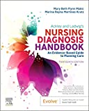
Ackley and Ladwig’s Nursing Diagnosis Handbook: An Evidence-Based Guide to Planning Care
We love this book because of its evidence-based approach to nursing interventions. This care plan handbook uses an easy, three-step system to guide you through client assessment, nursing diagnosis, and care planning. Includes step-by-step instructions showing how to implement care and evaluate outcomes, and help you build skills in diagnostic reasoning and critical thinking.
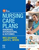
Nursing Care Plans – Nursing Diagnosis & Intervention (10th Edition)
Includes over two hundred care plans that reflect the most recent evidence-based guidelines. New to this edition are ICNP diagnoses, care plans on LGBTQ health issues, and on electrolytes and acid-base balance.
Nurse’s Pocket Guide: Diagnoses, Prioritized Interventions, and Rationales
Quick-reference tool includes all you need to identify the correct diagnoses for efficient patient care planning. The sixteenth edition includes the most recent nursing diagnoses and interventions and an alphabetized listing of nursing diagnoses covering more than 400 disorders.

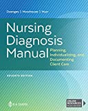
Nursing Diagnosis Manual: Planning, Individualizing, and Documenting Client Care
Identify interventions to plan, individualize, and document care for more than 800 diseases and disorders. Only in the Nursing Diagnosis Manual will you find for each diagnosis subjectively and objectively – sample clinical applications, prioritized action/interventions with rationales – a documentation section, and much more!
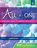
All-in-One Nursing Care Planning Resource – E-Book: Medical-Surgical, Pediatric, Maternity, and Psychiatric-Mental Health
Includes over 100 care plans for medical-surgical, maternity/OB, pediatrics, and psychiatric and mental health. Interprofessional “patient problems” focus familiarizes you with how to speak to patients.
See also
Other recommended site resources for this nursing care plan:
- Nursing Care Plans (NCP): Ultimate Guide and Database MUST READ!
Over 150+ nursing care plans for different diseases and conditions. Includes our easy-to-follow guide on how to create nursing care plans from scratch. - Nursing Diagnosis Guide and List: All You Need to Know to Master Diagnosing
Our comprehensive guide on how to create and write diagnostic labels. Includes detailed nursing care plan guides for common nursing diagnostic labels.
Other care plans for hematologic and lymphatic system disorders:
























Leave a Comment