Utilize this comprehensive nursing care plan and management guide to provide effective care for patients undergoing pacemaker therapy. Gain valuable insights on nursing assessment, interventions, goals, and nursing diagnosis specifically tailored for pacemaker therapy in this guide.
What are pacemakers?
Pacemakers are electronic devices that deliver controlled electrical stimuli to the heart muscle to regulate its rhythm. They are used in patients with slower-than-normal impulse formation, conduction disturbances, or certain tachydysrhythmias. Biventricular pacing, known as cardiac resynchronization therapy (CRT), is used for advanced heart failure. Pacemakers can be permanent or temporary, with temporary ones being used in hospital settings until a permanent pacemaker can be implanted. The pacemaker consists of a pulse generator and leads that are implanted in the heart, delivering electrical stimuli to maintain a stable heart rate.
Pacemakers consist of an electronic pulse generator and pacemaker electrodes, which deliver controlled electrical stimuli to the heart muscle. The pulse generator contains circuitry and batteries that determine the rate and strength of the electrical stimulus. Sensitivity settings allow the device to detect intracardiac electrical activity. The pacemaker leads can be endocardial (placed inside the heart) or epicardial (placed outside the heart during open-heart surgery). Temporary pacemakers use leads connected to a temporary generator, while permanent pacemakers have leads connected to a subcutaneously implanted generator. Permanent pacemakers have insulation to protect against moisture and filters to prevent electrical interference. Battery life varies but typically lasts 6 to 12 years. If the battery is depleted, the entire generator is replaced. In emergency situations, transcutaneous pacing can be performed using large ECG electrodes connected to a defibrillator. Transcutaneous pacing is temporary and uncomfortable, and its use requires hospitalization.
Nursing Care Plans & Management
The nursing care planning goals for patients on pacemaker therapy include ensuring proper pacemaker function through regular assessment of heart rate, rhythm, and pacemaker settings, monitoring for any signs of pacemaker malfunction or complications, promoting wound healing, and preventing infections at the insertion site, educating the patient and the family about pacemaker function, activity restrictions, and signs of complications, providing psychosocial support to address emotional concerns and facilitate adjustment to lifestyle changes, and collaborating with the healthcare team to optimize patient outcomes and promote a high quality of life.
Nursing Problem Priorities
The following are the nursing priorities for patients with a pacemaker:
1. Monitoring the patient’s cardiac rhythm and pacemaker function. Regular evaluation of the patient’s cardiac rhythm and pacemaker function is important to ensure optimal heart function and the effectiveness of pacemaker therapy.
2. Proper wound care. Maintaining a clean and sterile wound environment promotes healing, reduces the risk of wound-related complications, and ensures the pacemaker continues to function optimally without interruption or the need for further interventions.
3. Observing for pacemaker-related complications. Pacemaker-related complications can include infection at the insertion site, lead dislodgement or fracture, hematoma formation, pneumothorax, thrombosis or embolism, malfunction or electrical issues, and allergic reactions or interference with other devices.
4. Providing emotional support. This involves actively listening to the patient’s concerns and fears, validating emotions, and offering reassurance and empathy.
5. Patient education and teaching. Providing education and support to the patient and the family about the pacemaker function, activity restrictions, and signs of complications.
Nursing Assessment
Nursing assessments for patients on pacemaker therapy include monitoring cardiac rhythm and pacemaker function through continuous telemetry or periodic ECG monitoring, assessing the pacemaker insertion site for signs of infection, hematoma, or wound healing complications, and evaluating the patient’s tolerance to activity and any symptoms suggestive of pacemaker malfunction or complications.
Assess for the following subjective and objective data:
- Decreased heart rate
- Decreased cardiac output, stroke volume Increased peripheral vascular resistance
- Changes in the level of consciousness
- Inappropriate pacing or sensing
- Disruption of skin tissue
- Insertion site
- Skin layer destruction
Assess for factors related to the problems of patients on pacemaker therapy:
- Cardiac dysrhythmias
- Heart blocks
- Tachydysrhythmias
- Pacemaker battery failure
- Lead malfunction or inadequate pacing settings
- Insertion of pacemaker
- Pacemaker failure
- Puncture or perforation of heart tissues
- Lead migration
- Skin erosion
- Invasive procedure
- Pacemaker insertion
- Redness and heat at the site of insertion
- Pain and swelling
- Loss of control of heart function
Nursing Diagnosis
Following a thorough assessment, a nursing diagnosis is formulated to specifically address the challenges associated with pacemaker therapy based on the nurse’s clinical judgement and understanding of the patient’s unique health condition. While nursing diagnoses serve as a framework for organizing care, their usefulness may vary in different clinical situations. In real-life clinical settings, it is important to note that the use of specific nursing diagnostic labels may not be as prominent or commonly utilized as other components of the care plan. It is ultimately the nurse’s clinical expertise and judgment that shape the care plan to meet the unique needs of each patient, prioritizing their health concerns and priorities.
Nursing Goals
Goals and expected outcomes may include:
- The client will be free of dysrhythmias with an adequate cardiac output to perfuse all body organs.
- The client will be able to adhere to all activity restrictions.
- The client’s permanent pacemaker will function without complications, with no lead dislodgement or competitive rhythms noted.
- The client will have a well-healed incision with no signs or symptoms of infection.
- The client will be able to demonstrate appropriate wound care prior to discharge.
- The client will be free of life-threatening complications that may be associated with pacemaker insertion.
- The client will regain optimal mobility within the limitations of the disease process and will have increased strength and function of limbs.
- The client will be able to deal effectively with body image disturbances in the current situation.
- The client will be able to talk with family, therapist, or others about emotional or psychological problems.
Nursing Interventions and Actions
Therapeutic interventions and nursing actions for patients on pacemaker therapy may include:
- 1. Improving Cardiac Tissue Perfusion and Cardiovascular Monitoring
- 2. Maintaining Skin Integrity
- 3. Preventing Infection and Injury
- 4. Improving Physical Mobility
- 5. Managing Body Image Disturbance and Self-Esteem
1. Improving Cardiac Tissue Perfusion and Cardiovascular Monitoring
Ineffective cardiac tissue perfusion in patients on pacemaker therapy can occur due to underlying cardiovascular conditions, such as coronary artery disease or heart failure, which can impair blood flow to the heart muscle. The following are assessment and nursing interventions to improve cardiac tissue perfusion:
Monitor ECG for changes in rhythm, rate, and presence of dysrhythmias. Treat as indicated.
Observation for pacemaker malfunction promotes prompt treatment. Pacer electrodes may irritate the ventricle and promote ventricular ectopy.
Obtain and observe rhythm strip every 4 hours and PRN. Notify the physician of abnormalities.
Identifies proper functioning of pacemakers, with appropriate capture and sensing.
Monitor vital signs every 15 minutes until stable; repeat every 2 hours or PRN.
Assures adequate perfusion and cardiac output.
Monitor for sudden complaints of chest pain, and auscultate for pericardial friction rub or muffled heart tones. Observe for JVD and pulsus paradoxes.
This may indicate perforation of the pericardial sac and impending or present cardiac tamponade.
Monitor the patient for complaints of dizziness, weakness, fatigue, syncope, edema, chest pain, palpitations, pulsations in neck veins, or dyspnea.
During ventricular pacing, AV synchrony may cease and cause a sudden decrease in cardiac output. This may indicate “pacemaker syndrome” or failure of the pacer to function which results in decreased perfusion.
Limit movement of extremity involved near the insertion site as ordered.
Prevents accidental disconnection and dislodgement of lead wires immediately after placement.
If the patient arrests and requires defibrillation, attempt to avoid the pacemaker battery location as a site for defibrillation. If the patient is successfully resuscitated, prepare for potential reprogramming of the pacemaker.
Defibrillator shock may result in pacemaker damage and potential diversion of electrical current. Pacemakers may be damaged, or settings may be altered by the application of electrical current required to resuscitate the patient.
2. Maintaining Skin Integrity
The skin around the pacemaker insertion site is vulnerable to complications such as infection, hematoma, or skin breakdown. Nurses ensure skin integrity by regularly assessing the site, providing proper wound care, promoting hygiene, and educating patients on the signs of skin-related issues to avoid infections and promote healing, ultimately ensuring the effective functioning of the pacemaker and minimizing the risk of complications. The following are assessment and nursing interventions to maintain skin integrity:
Inspect the pacemaker insertion site for redness, edema, warmth, drainage, or tenderness.
Prompt detection of problems helps promote prompt treatment or nursing intervention.
Change dressing daily, or per hospital protocol, using a sterile technique.
Allows for the observation of the site and detection of inflammation or infection. The sterile technique is recommended because of the close proximity of the portal or opening to the heart, increasing the potential for systemic infection.
Instruct on wound care to pacer site and to avoid taking showers for 2 weeks post insertion.
Promotes compliance with care to decrease the potential for infection. Moisture can promote bacterial growth.
Instruct the patient to avoid wearing constrictive clothing until the site has healed completely.
May cause discomfort at the incision site from pressure and rubbing against the skin.
3. Preventing Infection and Injury
Injury and complications related to pacemaker therapy can include infection at the insertion site, hematoma formation, lead dislodgement or fracture, pneumothorax, thrombosis or embolism, malfunction or electrical issues, allergic reactions to device components, and interference with other medical devices. The following are assessment and nursing interventions to prevent infection and injury:
Assess the presence of hematoma, redness, and swelling at the site, temperature elevation, and/or skin erosion.
Indicates the presence of infection or potential for skin breakdown.
Monitor the patient for bleeding at the pacemaker site.
Bleeding at the incision site may occur based on the patient’s coagulation status. Pressure dressings or manual pressure may be required to control bleeding.
Monitor for the presence of pulses at the site distal to pacer insertion.
Hemorrhage may promote tissue edema and compression to arterial blood flow resulting in diminished or absent pulses.
Instruct the patient on signs and symptoms, such as restlessness, syncope, chest pain, or dyspnea of which to notify the nurse.
Provides prompt identification of potential complications and allows for timely treatment May indicate malposition of lead irritating heart muscle tissue, which can then be promptly treated.
Instruct patient and/or family to notify physician for redness, swelling, or drainage at the site of pacemaker battery insertion.
Provides for the potential identification of infection and allows for prompt treatment with antimicrobials to reduce the possibility of sepsis.
Empty drainage device, if present.
Promotes wound drainage and healing.
Apply sterile dressing until the wound heals. Avoid dislodging the catheter during site care.
The nurse performs regular dressing changes, carefully assessing the insertion site for signs of infection, such as redness, swelling, soreness, or abnormal drainage. Any changes in the wound’s appearance, elevated temperature, or increased white blood cell count should be promptly reported to the primary provider.
Instruct the patient on the method to take the temperature.
Monitors for potential elevation associated with infection.
Instruct the patient of signs to observe indicating infection and to notify the physician.
Provides measures to take and information to report for prompt treatment.
Administer antibiotics as ordered.
Prevents or treats wound infection.
4. Improving Physical Mobility
Physical limitations for patients on pacemaker therapy may include avoiding high-intensity activities or contact sports that could potentially damage the pacemaker or lead wires. Patients are often advised to avoid placing excessive strain on the area where the pacemaker is implanted, to prevent dislodgement or damage to the device. To improve physical mobility, it is important to encourage regular physical activity within the recommended guidelines provided by the healthcare provider. The following are assessment and nursing interventions to improve physical mobility:
Evaluate the patient’s perception of his degree of immobility.
Psychological and physical immobility are interrelated. Psychological immobility is used as a defense mechanism when the patient has no control over his body, and this can lead to disproportionate fear and concern. If pacemaker insertion was an emergent procedure, the patient may have misperceptions regarding movement and may be afraid that any movement may result in his demise. Changes in body image promote psychological immobility and may result in emotional handicaps, rather than actual physical ones.
Maintain bed rest following pacemaker insertion for 24-48 hours or depending on the protocol.
Provides time for stabilization of leads and decreases the potential for dislodgement.
Immobilize the extremity proximal to the pacemaker insertion site with an arm board, sling, and so forth.
Prevents potential for dislodgement of lead caused by movement.
Resume range of motion exercises one week after permanent pacemaker insertion to the affected extremity. Provide ROM to unaffected extremity immediately after pacer insertion, as warranted.
Promotes a gradual increase in activity. Stretching should be avoided until the lead wire has been secured in the heart. ROM prevents stiffness of shoulders and joint immobility.
Instruct on extension-dorsiflexion exercises of feet every 1-2 hours.
Promotes venous return, prevents venous stasis, and decreased the potential for thrombophlebitis.
Monitor the patient for progression and improvement in stiffness or pain.
Physical therapy may be required if immobility results are severe.
Apply the trapeze bar to the bed.
Allows for easier movement; allows the patient to assist with movement in bed with the unaffected extremity.
Reposition every 2 hours and PRN.
Prevents potential for immobility hazards, such as pressure areas and atelectasis.
Instruct patient regarding deep breathing exercises to be done every 1-2 hours, and to avoid forceful coughing.
Facilitates lung expansion and decreases the potential for atelectasis. Coughing may dislodge the pacemaker lead.
Instruct patient and/or family regarding the need for immobilization of arm immediately post-pacemaker insertion, and how long immobility is to be expected.
Provides knowledge, and decreases fear that the patient may be immobilized for long periods of time.
5. Managing Body Image Disturbance and Self-Esteem
Patients with pacemaker therapy may experience body image disturbances and self-esteem issues due to the presence of a visible device and associated scars or limitations on physical activities. Patients may feel self-conscious, have concerns about body appearance, or fear societal judgment. The following are assessment and nursing interventions to manage body image disturbance and self-esteem:
Determine the level of the patient’s knowledge about the disease process, treatment, and anxiety level.
May identify the extent of the problem and interventions that will be required.
Evaluate the extent of loss to the patient or family, and what it means to them.
Depending on the time frame for patient teaching prior to the insertion of the pacemaker, the patient may not have received adequate information and may have difficulty dealing with changes in his body appearance, as well as generalized health condition and loss of control.
Assess the patient’s stage of grieving.
Provides recognition of appropriate versus inappropriate behavior. Prolonged grief may require further care.
Observe for withdrawal, manipulation, noninvolvement with care, or increased dependency. Set limits on dysfunctional behaviors and help the patient to seek positive behaviors that will assist with recovery.
May suggest problems with adjustment to the health condition, grief response to the loss of function, or worry about others accepting the patient’s new body status. Patients may deal with crises in the same manner as previously and may need redirection in behaviors to facilitate recovery and acceptance.
Provide positive reinforcement during care and with instruction and setting goals. Do not give false reassurance.
Promotes trust and establishes rapport with patients as well as an opportunity to plan for the future based on the reality of the situation.
Provide opportunities for patients to take an active role in wound care.
Promotes self-esteem and facilitates feelings of control of body and health.
Provide reassurance that pacemakers will not alter sexual activity.
Promotes knowledge and decreases fear.
Discuss the potential for mood changes, anger, grief, and so forth, after discharge, and to seek help if persisting for a lengthy time.
Facilitates identification that feelings are not unusual and must be recognized in order to deal with them effectively.
Identify support groups for patients and/or families to contact.
Provides ongoing support for patient and family and allows for ventilation of feelings.
Consult counselor or therapist, if needed.“
May be required for further interventions to resolve emotional or psychological issues.
6. Managing Potential Complications
Specific management strategies vary depending on the type of complication but may include the administration of appropriate antibiotics for infection, hematoma evacuation or compression for hematoma, lead repositioning or replacement for lead-related issues, and close collaboration with the healthcare team for further evaluation or surgical intervention as needed. The following are assessment and nursing interventions to manage potential complications:
Monitor for signs of failure to sense the patient’s own rhythm, and correct the problem.
Potential causes are lead dislodgement, battery failure, low, sensitivity, wire fracture, or improper placement of the catheter.
Check the patient for low blood sugar levels, and use glucocorticoids or sympathomimetics, mineralocorticoids, or anesthetics.
May impair the pacemaker stimulation threshold.
Monitor for dyspnea, chest pain, pallor, cyanosis, absent or diminished breath sounds, tracheal deviation, and the patient’s feeling of impending doom.
This may indicate a puncture of the lung and the presence of pneumothorax, requiring immediate treatment.
Monitor the patient for the presence of muscle twitching and hiccups.
This may indicate perforation of the heart with pacing to the chest wall or diaphragm.
Observe for signs and symptoms of cardiac tamponade.
This may indicate perforation of the pericardial sac and impending cardiac tamponade which requires immediate medical attention.
Monitor vital signs; observe for diaphoresis, dyspnea, and restlessness.
Hypotension and these other signs may indicate puncture of subclavian vasculature and potential hemothorax.
Protect patients from microwave ovens, radar, diathermies, etc.
Environmental electromagnetic interference may impair demand pacemaker function by disrupting the electrical stimulus.
Ensure that all electrical equipment is grounded. Avoid touching equipment and the patient at the same time.
Prevents the potential for microshock and accidental electrocution. Electric current seeks the path of least resistance, and the potential for stray current to travel through the electrode into the patient’s heart may precipitate ventricular fibrillation.
7. Initiating Health Teaching and Patient Education
The focus on patient education among patients with pacemakers includes educating patients regarding the purpose and function of the pacemaker, activity restrictions and limitations to avoid damage or dislodgement, signs and symptoms of pacemaker malfunction or complications, the importance of regular follow-up appointments, and self-care measures such as proper wound care and hygiene. The following are assessment and nursing interventions to provide health teaching and patient education:
Instruct the patient on the technique for changing the dressing.
Sterility can be maintained if the proper technique is used.
Instruct the patient in checking pulse rate every day for 1 month, then every week, and notify the physician if the rate varies more than 5 beats/minute.
Provides the patient with some control over the situation. Assists in promoting a sense of security; allows prompt recognition of deviations from preset rate and potential pacemaker failure.
Instruct on limitations on activity: avoid excessive bending, stretching, lifting heavy, strenuous exercise, or contact sports.
A full range of motion can be recovered in approximately 2 months after fibrosis stabilizes the pacemaker lead. Excessive activity may cause lead to dislodgement.
Instruct to avoid shoulder-strap purses, suspenders, or firing rifles resting over the generator site.
May promote irritation over implanted generator site.
Instruct to wear a MedicAlert bracelet with information about the type of pacemaker and rate.
Provides, necessary information about the patient, condition, and ethical considerations on the pacemaker should the patient be incapacitated and unable to speak.
Instruct the patient to notify the physician if radiation therapy is needed, and to wear a lead shield if it’s required.
Radiation therapy can cause the failure of the silicone chip in the pacer with repeated radiation treatment.
Instruct patient/family regarding avoiding electromagnetic fields, magnetic resonance imaging, radio transmitters, and the like. If the patient notices dizziness or palpitations, the patient should try to move away from the area, and if symptoms persist, seek medical attention.
May affect the function of the pacemaker and alter the programmed settings.
Instruct the patient on the need for a pacemaker, the procedures involved, expected outcomes, etc.
Provides knowledge decreases fear and anxiety and provides a baseline for further instruction.
Instruct the patient and/or family to observe for signs of redness, drainage, fever, pain, tenderness, and swelling at the site. Have them report to their primary care provider immediately.
Prompt recognition of complications and facilitates timely treatment.
Instruct patient/family regarding pacemaker use, need for removal or replacement, and signs and symptoms to report to the physician.
Pulse generators may require removal for battery replacement, fracture of lead wires, pacemaker failure, and so forth. Knowledge of potential problems can help facilitate timely identification, notification of physicians, and appropriate care.
Evaluation
The expected patient outcomes and evaluation for patients on pacemaker therapy may include the following:
- Stable cardiac rhythm and improved heart rate.
- Improved symptoms and enhanced functional capacity.
- Improved ability to participate in physical activities and maintain functional independence.
- Improved self-perception and acceptance of the body with the presence of the pacemaker.
- Optimal pacemaker function and absence of complications.
- Patient understanding and adherence to self-care measures.
Discharge and Home Care Guidelines
The goals of education for patients with pacemakers are to ensure the patient’s understanding of the purpose and function of the pacemaker, promote self-care and safety measures, and recognize and respond appropriately to potential complications or symptoms requiring immediate medical attention. Here are some of the guidelines:
- Incision care. This includes maintaining a clean and dry incision site, limiting excessive water exposure, and changing the dressing. Teach the patient to observe for signs of infection, such as redness, swelling, or drainage.
- Activity restrictions. Advise the patient to avoid heavy lifting, contact sports, and activities that may involve repetitive arm or chest movements that could potentially dislodge or damage the pacemaker.
- Electromagnetic precautions. Inform the patient about potential sources of electromagnetic interference, such as certain devices, strong magnetic fields, or electrical equipment, and provide guidelines on maintaining a safe distance from these sources to avoid interference with the pacemaker function.
- Follow-up appointments. Provide the patient with information about scheduling appointments to monitor pacemaker function, adjust programming if needed, and assess the patient’s overall cardiac health.
- Emergency preparedness. Instruct the patient and the family on recognizing signs of pacemaker malfunction or complications, provide emergency contact information, and teach measures to take in case of an emergency.
- Emotional support and resources. Offer emotional support and provide information about support groups or counseling services that can help the patient and the family cope with the emotional and psychosocial aspects of living with a pacemaker.
Recommended Resources
Recommended nursing diagnosis and nursing care plan books and resources.
Disclosure: Included below are affiliate links from Amazon at no additional cost from you. We may earn a small commission from your purchase. For more information, check out our privacy policy.
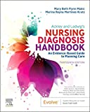
Ackley and Ladwig’s Nursing Diagnosis Handbook: An Evidence-Based Guide to Planning Care
We love this book because of its evidence-based approach to nursing interventions. This care plan handbook uses an easy, three-step system to guide you through client assessment, nursing diagnosis, and care planning. Includes step-by-step instructions showing how to implement care and evaluate outcomes, and help you build skills in diagnostic reasoning and critical thinking.
Nursing Care Plans – Nursing Diagnosis & Intervention (10th Edition)
Includes over two hundred care plans that reflect the most recent evidence-based guidelines. New to this edition are ICNP diagnoses, care plans on LGBTQ health issues, and on electrolytes and acid-base balance.

Nurse’s Pocket Guide: Diagnoses, Prioritized Interventions, and Rationales
Quick-reference tool includes all you need to identify the correct diagnoses for efficient patient care planning. The sixteenth edition includes the most recent nursing diagnoses and interventions and an alphabetized listing of nursing diagnoses covering more than 400 disorders.

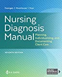
Nursing Diagnosis Manual: Planning, Individualizing, and Documenting Client Care
Identify interventions to plan, individualize, and document care for more than 800 diseases and disorders. Only in the Nursing Diagnosis Manual will you find for each diagnosis subjectively and objectively – sample clinical applications, prioritized action/interventions with rationales – a documentation section, and much more!
All-in-One Nursing Care Planning Resource – E-Book: Medical-Surgical, Pediatric, Maternity, and Psychiatric-Mental Health
Includes over 100 care plans for medical-surgical, maternity/OB, pediatrics, and psychiatric and mental health. Interprofessional “patient problems” focus familiarizes you with how to speak to patients.

See also
Other recommended site resources for this nursing care plan:
- Nursing Care Plans (NCP): Ultimate Guide and Database MUST READ!
Over 150+ nursing care plans for different diseases and conditions. Includes our easy-to-follow guide on how to create nursing care plans from scratch. - Nursing Diagnosis Guide and List: All You Need to Know to Master Diagnosing
Our comprehensive guide on how to create and write diagnostic labels. Includes detailed nursing care plan guides for common nursing diagnostic labels.
Other nursing care plans for cardiovascular system disorders:
- Angina Pectoris (Coronary Artery Disease)
- Cardiac Arrhythmia (Digitalis Toxicity)
- Cardiac Catheterization
- Cardiogenic Shock
- Congenital Heart Disease
- Decreased Cardiac Output & Cardiac Support
- Heart Failure
- Hypertension
- Hypovolemic Shock
- Impaired Tissue Perfusion & Ischemia
- Myocardial Infarction
- Pacemaker Therapy


























Leave a Comment