Cranial nerves play a vital role in various sensory and motor functions. A thorough assessment of cranial nerves helps identify neurological deficits and aids in diagnosing neurological conditions. This guide offers a comprehensive overview and a handy cheat sheet for quick reference during assessments.
What are Cranial Nerves?
Cranial nerves are 12 pairs of nerves identified by Roman numerals (CN I through XII) or by name. These nerves can be sensory, motor, or both in function. These nerves are essential for transmitting sensory information from vision, hearing, taste, and smell to the brain, enabling perception and response to the environment. They also control motor functions by innervating muscles responsible for facial expressions, eye movements, chewing, swallowing, and speaking, promoting coordinated and effective physical actions.
Equipment
Equipment needed for cranial nerve assessment:
- Snellen chart or visual acuity chart
- Penlight
- Ophthalmoscope
- Cotton swab or cotton ball
- Tuning fork (for Weber and Rinne tests)
- Tongue depressor
- Odorants (for olfactory assessment)
- Taste strips or solutions (for taste/gustatory assessment)
- Reflex hammer
- Ophthalmic ruler (for measuring pupil size)
- Gauze or tissue (for wiping excess eye drops)
- Gloves
Cranial Nerves Chart
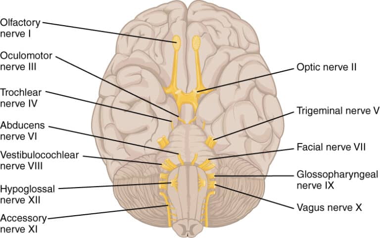
There are 12 cranial nerves, each with specific functions. They are numbered I through XII based on their position from the front to the back of the brain. Below is a chart of the 12 cranial nerves, the assessment technique used, if the response elicited is normal, and how to document it.
I: Olfactory Nerve
Originates from the olfactory epithelium in the nasal cavity and passes through the cribriform plate of the ethmoid bone. It is responsible for the sense of smell by transmitting olfactory stimuli from the nasal mucosa to the olfactory bulbs in the brain.
| Cranial Nerve Assessment | Normal Response | Documentation |
|---|---|---|
| Ask the client to smell and identify the smell of cologne with each nostril separately and with the eyes closed. | Client is able to identify different smell with each nostril separately and with eyes closed unless such condition like colds is present. | Client was able to describe the odor of the materials used. |
II: Optic Nerve
Arises from the retina of the eye and extends through the optic canal to the optic chiasm. It transmits visual information from the retina to the brain, allowing for the perception of light, color, and form.
| Assessment Technique | Normal Response | Documentation |
|---|---|---|
| Provide adequate lighting and ask client to read from a reading material held at a distance of 36 cm. (14 in.). | The client should be able to read with each eye and both eyes. | Client was able to read with each eye and both eyes. |
III: Oculomotor Nerve
Emerges from the midbrain and passes through the superior orbital fissure to innervate multiple muscles that control eye movements. Controls most of the eye movements, including raising the eyelid, constricting the pupil, and controlling the shape of the lens for near vision.
| Cranial Nerve Assessment | Normal Response | Documentation |
|---|---|---|
| Reaction to light: Using a penlight and approaching from the side, shine a light on the pupil. Observe the response of the illuminated pupil. Shine the light on the pupil again, and observe the response of the other pupil. | Illuminated and non-illuminated pupil should constrict. | PERRLA (pupils equally round and reactive to light and accommodation) |
| Reaction to accommodation: Ask client to look at a near object and then at a distant object. Alternate the gaze from the near to the far object. Next, move an object towards the client’s nose. | Pupils constrict when looking at a near object, dilate when looking at a distant object, converge when near object is moved towards the nose. | PERRLA (pupils equally round and reactive to light and accommodation) |
IV: Trochlear Nerve
Arises from the posterior aspect of the midbrain and passes through the superior orbital fissure. Innervates the superior oblique muscle of the eye, primarily responsible for downward and inward movement of the eye (depression and intorsion). Evaluate extraocular movements and assess for diplopia (double vision).
| Cranial Nerve Assessment | Normal Response | Documentation |
|---|---|---|
| Hold a penlight 1 ft. in front of the client’s eyes. Ask the client to follow the movements of the penlight with the eyes only. Move the penlight upward, downward, sideward and diagonally. | Client’s eyes should be able to follow the penlight as it moves. | Both eyes are able to move as necessary. |
V: Trigeminal Nerve
Emerges from the pons and has three divisions: ophthalmic (V1), maxillary (V2), and mandibular (V3), which exit through separate foramina. Sensory function involves facial sensation (touch, pain, and temperature) for the forehead, eyes, nose, cheeks, and lower jaw. It also innervates the muscles of mastication for chewing.
| Cranial Nerve Assessment | Normal Response | Documentation |
|---|---|---|
| While the client looks upward, lightly touch the lateral sclera of eye to elicit blink reflex. | Client should have a (+) corneal reflex, able to respond to light and deep sensation and able to differentiate hot from cold. | Client was able to elicit corneal reflex, sensitive to pain stimuli and distinguish hot from cold. |
| To test light sensation, have client close eyes, wipe a wisp of cotton over client’s forehead. | (same as above) | (same as above) |
| To test deep sensation, use alternating blunt and sharp ends of an object. Determine sensation to warm and cold object by asking client to identify warmth and coldness. | (same as above) | (same as above) |
VI: Abducens Nerve
Arises from the pons and passes through the superior orbital fissure. Innervates the lateral rectus muscle of the eye, responsible for abduction (outward movement) of the eye. Evaluate extraocular movements and assess for nystagmus (involuntary eye movements).
| Cranial Nerve Assessment | Normal Response | Documentation |
|---|---|---|
| Hold a penlight 1 ft. in front of the client’s eyes. Ask the client to follow the movements of the penlight with the eyes only. Move the penlight through the six cardinal fields of gaze. | Both eyes coordinated, move in unison with parallel alignment. | Both eyes move in coordination. |
VII: Facial Nerve
Emerges from the pons and exits the skull through the stylomastoid foramen. Controls facial expression and taste sensation. Assess facial symmetry, taste perception on the anterior two-thirds of the tongue, and ability to close the eyes tightly.
| Assessment Technique | Normal Response | Documentation |
|---|---|---|
| Ask client to smile, raise the eyebrows, frown, and puff out cheeks, close eyes tightly. Ask client to identify various tastes placed on the tip and sides of tongue. | Client should be able to smile, raise eyebrows, and puff out cheeks and close eyes without any difficulty. The client should also be able to distinguish different tastes. | Client performed various facial expressions without any difficulty and able to distinguish varied tastes. |
VIII: Vestibulocochlear Nerve
Arises from the brainstem and enters the inner ear. Controls hearing and balance. Evaluate auditory acuity, Weber and Rinne tests, and assess balance and coordination.
- Rinne test. Compares air conduction (tuning fork near the ear) to bone conduction (tuning fork on the mastoid bone). Normally, air conduction is heard longer. If bone conduction is heard longer, it indicates conductive hearing loss; if air conduction is heard longer but sound doesn’t reach the inner ear properly, it indicates sensorineural hearing loss.
- Weber test. Involves placing a vibrating tuning fork on the forehead or midline of the skull to assess sound localization. If the sound is heard equally in both ears, hearing is normal or symmetric. If the sound lateralizes to one ear, it may indicate conductive hearing loss in that ear or sensorineural hearing loss in the opposite ear.
| Cranial Nerve Assessment | Normal Response | Documentation |
|---|---|---|
| Have the client occlude one ear. Out of the client’s sight, place a tickling watch 2 to 3 cm. ask what the client can hear and repeat with the other ear. | Client should be able to hear the tickling of the watch in both ears. | Client was able to hear tickling in both ears. |
| Ask the client to walk across the room and back and assess the client’s gait. | The client should have upright posture and steady gait and able to maintain balance. | The client was able to stand and walk in an upright position and able to maintain balance. |
IX: Glossopharyngeal Nerve
Emerges from the medulla oblongata and exits through the jugular foramen. Controls taste sensation for the posterior one-third of the tongue, assists in swallowing and innervates the carotid body and sinus for blood pressure regulation.
| Cranial Nerve Assessment | Normal Response | Documentation |
|---|---|---|
| Ask the client to say “ah” and have the patient yawn to observe upward movement of the soft palate. | Client should be able to elicit gag reflex and swallow without any difficulty. | Client was able to elicit gag reflex and able to swallow without difficulty. |
| Elicit gag response. | (same as above) | (same as above) |
| Note ability to swallow. | (same as above) | (same as above) |
X: Vagus Nerve
Emerges from the medulla oblongata and exits through the jugular foramen. Involved in autonomic functions such as heart rate, digestion, and respiratory rate. Also, innervates muscles of the pharynx and larynx for speech and swallowing.
| Cranial Nerve Assessment | Normal Response | Documentation |
|---|---|---|
| Ask the patient to swallow and speak (note hoarseness) | The client should be able to swallow without difficulty and speak audibly. | Client was able to swallow without difficulty and speak audibly. |
XI: Accessory Nerve
Originates from the medulla oblongata and the upper spinal cord, ascending through the foramen magnum and exiting through the jugular foramen. Controls the sternocleidomastoid and trapezius muscles, enabling head movement and shoulder shrugging.
| Cranial Nerve Assessment | Normal Response | Documentation |
|---|---|---|
| Ask client to shrug shoulders against resistance from your hands and turn head to side against resistance from your hand (repeat for other side). | Client should be able to shrug shoulders and turn head from side to side. | Client was able to shrug his shoulders and turn his head from one side to the other. |
XII: Hypoglossal Nerve
Emerges from the medulla oblongata and exits through the hypoglossal canal. Controls the movements of the tongue, including speech, swallowing, and manipulation of food during chewing.
| Cranial Nerve Assessment | Normal Response | Documentation |
|---|---|---|
| Ask client to protrude tongue at midline and then move it side to side. | The client should be able to move tongue without any difficulty. | The client was able to move tongue in different directions. |
Cranial Nerves Cheat Sheet Download
In this section, you can download the cranial nerves cheat sheet.

References and Sources
- Chhabra, A., Bajaj, G., Wadhwa, V., Quadri, R. S., White, J., Myers, L. L., … Zuniga, J. R. (2018). MR Neurographic Evaluation of Facial and Neck Pain: Normal and Abnormal Craniospinal Nerves below the Skull Base. RadioGraphics, 38(5), 1498–1513.
- Fisch, A. (2015). Clinical Examination of the Cranial Nerves. Nerves and Nerve Injuries, 195–225.
- Kozier B., et al., (2018). Fundamentals of Canadian Nursing
Originally published on January 3, 2012.


Leave a Comment