Intravenous therapy, commonly administered through peripheral venous catheters, often leads to catheter removal due to complications, completion of treatment, or lack of use. Local complications such as hematoma, thrombosis, phlebitis, thrombophlebitis, infiltration, and extravasation are frequently associated with peripheral IV catheters. Nurses need to possess strong technical and medical knowledge of evidence-based practices. This ensures treatment efficacy, high-quality care, and informed decision-making regarding the appropriate use of peripheral venous catheters in patients undergoing intravenous therapy.
This article provides a guide for managing local and systemic complications and provides general nursing considerations for preventing these complications.
Table of Contents
- Risk Factors for Peripheral IV Site Complications
- Local Complications of Peripheral IV Therapy
- Phlebitis
- Thrombophlebitis
- Infiltration
- Extravasation
- Local Infection
Risk Factors for Peripheral IV Site Complications
Each risk factor requires careful consideration and appropriate nursing interventions, such as using the correct catheter size, securing the IV properly, rotating sites regularly, and closely monitoring patients for early signs of complications.
- Patient factors (age, vein quality, chronic illness). Older patients or those with poor vein quality (e.g., fragile or sclerotic veins) are more susceptible to complications like infiltration, extravasation, and thrombophlebitis. Chronic illnesses such as diabetes or vascular diseases can also compromise vein integrity.
- Dehydration. Dehydration can lead to poor vein filling, making veins more fragile and prone to rupture during catheter insertion, increasing the risk of hematoma formation or phlebitis.
- Improper catheter size. Using a catheter that is too large for the vein can cause irritation, increasing the risk of phlebitis and thrombosis. Larger catheters cause more mechanical irritation and trauma to the vein walls, leading to inflammation.
- Inadequate site monitoring. Failure to regularly assess and monitor the IV site for early signs of complications like redness, swelling, or discomfort increases the risk of more severe issues like infection, infiltration, or extravasation.
- Poor aseptic technique. Failure to use proper hand hygiene and aseptic technique during insertion, dressing changes, or IV site maintenance can introduce bacteria, leading to infection at the insertion site or systemic complications like catheter-related bloodstream infections (CR-BSI).
- Prolonged use of the same IV site. Keeping an IV catheter in place for an extended period increases the risk of phlebitis, thrombophlebitis, and infection. Guidelines recommend rotating IV sites every 72-96 hours to reduce these risks.
- Frequent movement of the catheter. Movement of the catheter within the vein, caused by patient activity or improper securement, can lead to irritation of the vein lining, increasing the risk of phlebitis and infiltration.
- Use of irritant or vesicant medications. Administering irritating or vesicant medications (e.g., chemotherapy drugs) through a peripheral IV increases the risk of phlebitis, infiltration, and extravasation. Vesicant drugs can damage surrounding tissues if leakage occurs.
Local Complications of Peripheral IV Therapy
Peripheral IV therapy can lead to localized issues at the insertion site, such as irritation, swelling, or inflammation. These complications require careful monitoring to prevent escalation and provide patient comfort.
Hematoma
A hematoma is the accumulation of blood outside the blood vessel, usually caused by the vein being punctured during catheter insertion or removal. This can occur due to improper insertion technique, using a catheter that is too large for the vein, or failing to apply sufficient pressure after IV removal.
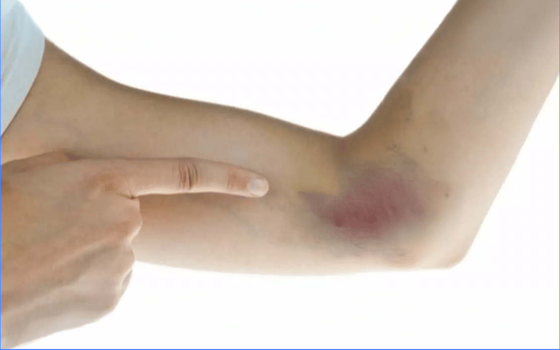
Types of Hematoma
Hematomas can be classified into two types:
- Superficial Hematoma. Results from minor trauma to a small vein, typically visible as a bruise at the IV site. This is due to fragile veins, improper insertion technique, or removing the IV without adequate pressure application.
- Deep Hematoma. It occurs when deeper veins are punctured or damaged during catheter insertion, causing a more significant accumulation of blood in the tissue. This is typically linked to with the use of larger-gauge needles or incorrect vein selection.
Clinical Criteria for Hematoma
- Grade 1 (Mild). Minor swelling, bruising, and discomfort at the IV site.
- Grade 2 (Moderate). More noticeable swelling and pain with moderate bruising.
- Grade 3 (Severe). Large, painful swelling, significant bruising, and loss of function in the affected limb.
Nursing Interventions and Prevention for Hematoma
- Gentle Insertion Techniques. Use care when inserting the catheter to reduce the risk of puncturing the vein.
- Appropriate Catheter Size. Select the right size catheter for the patient’s vein to avoid unnecessary trauma.
- Cold Compresses. Immediately apply cold compresses to the affected area to reduce blood flow, minimize swelling, and prevent the hematoma from worsening.
- Firm Pressure After Removal. Apply firm pressure after removing the catheter to prevent blood leakage into the surrounding tissue.
- Warm Compresses. After initial cold treatment, use warm compresses to encourage reabsorption of the blood and improve circulation for faster healing.
Phlebitis
Phlebitis is the inflammation of a vein, often caused by irritation from the catheter on the vessel wall. It can result from mechanical trauma due to the catheter, chemical irritation from IV solutions or medications, or a bacterial infection. Common signs and symptoms include redness, heat, purulent drainage, tenderness along the vein, and a palpable cord-like structure under the skin.
Types of Phlebitis
Phlebitis can occur due to irritation from the catheter, IV medications, or bacterial contamination, each involving different risk factors. Here are the following types and causes:
- Mechanical Phlebitis. Mechanical Phlebitis occurs when the catheter irritates the vein wall due to factors like its size, position, or movement within the vein. Risk factors include using large-gauge catheters, improper vein selection, or inadequate stabilization of the IV catheter, all of which can increase mechanical irritation.
- Chemical Phlebitis. Chemical Phlebitis is caused by irritation from IV medications or solutions, particularly those that are hypertonic, acidic, or administered too rapidly. Common risk factors include medications like potassium chloride, antibiotics, or chemotherapy agents, and improper dilution of these drugs, which can damage the vein lining.
- Bacterial (Infectious) Phlebitis. Bacterial Phlebitis results from bacterial contamination at the IV insertion site, often due to poor aseptic technique. Risk factors include inadequate site hygiene, improper handwashing, and failure to rotate the IV site, which can introduce bacteria and lead to infection and vein inflammation.
Phlebitis Grading Scale
This grading of phlebitis provides a structured method for assessing and managing phlebitis, ensuring appropriate interventions based on severity.
- Grade 0—The insertion site appears healthy with no signs of phlebitis; only continued observation is needed.
- Grade 1—Slight pain or redness near the IV site may indicate early signs of phlebitis. Observing the cannula should continue.
- Grade 2—Pain, redness, or swelling at the IV site suggests early-stage phlebitis, requiring the catheter to be repositioned.
- Grade 3—Pain along the cannula, redness, and swelling indicate medium-stage phlebitis, necessitating catheter repositioning and possible treatment.
- Grade 4— Pain, redness, swelling, and a palpable venous cord more than 1 inch in length suggest advanced phlebitis or early thrombophlebitis, requiring catheter repositioning and treatment consideration.
- Grade 5—Pain, redness, swelling, palpable venous cord, purulent drainage at insertion siteand fever indicate advanced thrombophlebitis, requiring immediate treatment and catheter repositioning.
Nursing Interventions and Prevention for Phlebitis
- Discontinue the IV. Remove the IV to eliminate the irritant causing inflammation.
- Apply Warm Compresses. Use warm compresses to reduce inflammation and promote blood flow in the affected area.
- Elevate the Affected Limb. Elevating the limb helps to reduce swelling and discomfort.
- Monitor Symptoms. Watch for signs of worsening symptoms to ensure timely intervention if the condition progresses.
- Smallest Gauge Catheter. Use the smallest gauge catheter suitable for the therapy to minimize irritation to the vein.
- Rotate IV Sites. Regularly change IV sites to avoid repeated trauma to the same vein.
- Follow Hospital Protocols. Adhere to established protocols for catheter maintenance to reduce the risk of phlebitis.
Thrombophlebitis
Thrombophlebitis is the formation of a blood clot in an inflamed vein, often resulting from prolonged IV use. It can be caused by extended use of a catheter at the same site, irritation from the catheter itself, or reduced blood flow due to patient immobility. Signs and symptoms typically include pain, redness, swelling, and the presence of a hard, cord-like vein in the area surrounding the IV site.
Nursing Intervention and Prevention for Thrombophlebitis
1. Discontinue the IV. Removing the IV catheter halts the source of vein irritation or injury, preventing further damage and lessening the risk of clot progression.
2. Elevate the affected limb. Elevating the limb promotes better circulation, reduces swelling, and helps alleviate discomfort, which aids in reducing inflammation around the site.
3. Apply warm compresses. Warm compresses help dilate blood vessels, increase blood flow, reduce pain, and promote healing by easing inflammation.
4. Proper management reduces the risk of clot migration or further vascular complications. Timely and appropriate interventions prevent the clot from migrating and reduce the risk of more severe complications like embolism or deep vein thrombosis.
5. Rotate IV sites as needed. Regularly rotating IV sites minimizes prolonged vein irritation and prevents complications like phlebitis and thrombophlebitis.
6. Secure the catheter properly to minimize movement. Stabilizing the catheter reduces vein trauma from movement, preventing irritation and the potential formation of clots.
7. Assess for early signs of clot formation. Monitoring for early symptoms allows for prompt intervention, reducing the risk of severe complications like clot migration or embolism.
Infiltration
Infiltration occurs when non-vesicant solutions leak into the surrounding tissues rather than being administered into the vein. It is commonly caused by dislodgment of the catheter from the vein, improper IV insertion, or weakened vein integrity. Signs and symptoms of infiltration include swelling, coolness, discomfort, and blanching of the skin around the IV site, indicating that the fluid is not being properly infused into the bloodstream.

Infiltration Assessment Tool
Infiltration is graded based on the severity of symptoms and the extent of fluid leakage into surrounding tissues. The commonly used grading scale is as follows:
- Grade 0— No symptoms; IV site appears healthy with no signs of infiltration.
- Grade 1— Mild swelling, slight pain, or tightness at the IV site, but no significant fluid leakage or visible signs of injury.
- Grade 2— Moderate swelling with mild pain and coolness to the touch, fluid leakage is present but with no lasting tissue damage.
- Grade 3— Significant swelling, moderate pain, coolness, and possible blanching of the skin. Infiltration of non-vesicant fluids causes visible tissue effects.
- Grade 4— Severe swelling, pain, skin tightness, and blanching, with infiltration of a large amount of fluid, including vesicant fluids, potentially causing tissue damage or necrosis.
Nursing Intervention and Prevention for Infiltration
1. Stop the IV infusion and remove the cannula. Terminating the infusion immediately prevents further leakage of fluids into the surrounding tissues, minimizing potential damage.
2. Elevate the affected limb. Elevating the limb reduces swelling and discomfort by promoting fluid reabsorption and improving circulation.
3. Apply cold compresses. Cold compresses help control swelling and soothe discomfort by constricting blood vessels, reducing fluid accumulation in the tissue.
4. Properly secure the IV catheter with tape or a stabilization device. Securing the catheter helps prevent displacement, ensures that it remains in the vein, and reduces the risk of fluid leakage into surrounding tissues.
5. Monitor the site regularly for signs of displacement. Regular monitoring allows for early detection of catheter movement, preventing infiltration and minimizing patient discomfort and tissue damage.
Extravasation
Extravasation occurs when vesicant (tissue-damaging) fluids, such as chemotherapy drugs, leak into the surrounding tissue, potentially causing severe tissue damage. This complication is similar to infiltration but involves more harmful medications. Signs and symptoms of extravasation include pain, burning, stinging, swelling, blistering, and tissue necrosis at or around the IV site.
Extravasation Assessment Tool
This assessment tool describes the progressive severity of extravasation through skin characteristics, edema, mobility, and the presence of fever.
- Grade 0 (No Extravasation)— Normal color and integrity, temperature normal, no edema, mobility unaffected, no fever.
- Grade 1 (Mild)— Slight redness or discoloration, intact integrity, slight warmth, minimal swelling, mobility normal or slightly reduced, no fever.
- Grade 2 (Moderate)— Pronounced redness or pinkish discoloration, intact but firm, warm to touch, moderate swelling, reduced mobility due to discomfort, no fever.
- Grade 3 (Severe)— Dark red, purple, or bruised, compromised integrity with possible blistering, hot to touch, significant swelling and hardening, limited mobility, possible fever.
- Grade 4 (Life-threatening or Permanent Damage)— Deep purple, black, or necrotic, severely compromised with ulceration or necrosis, hot or cold, severe swelling and hardening, mobility severely reduced or lost, possible fever.
Nursing Intervention and Prevention for Extravasation
1. Stop the infusion immediately. Halting the infusion prevents further leakage of vesicant drugs into surrounding tissues, minimizing tissue damage.
2. Apply an antidote (if available). Administering an antidote can neutralize the vesicant medication, reducing the extent of tissue injury.
3. Elevate the limb. Elevating the affected limb promotes better circulation, reducing swelling and facilitating healing.
4. Notify the physician for further evaluation. Immediate consultation with a physician ensures appropriate follow-up care, such as wound management or surgical intervention, if needed.
5. Ensure proper vein selection for vesicant drugs. Selecting an appropriate vein reduces the risk of catheter dislodgement, lowering the chances of extravasation.
6. Closely monitor IV sites during administration. Regular monitoring allows for early detection of any leakage, minimizing the likelihood of severe tissue damage and enhancing patient safety.
Local Infection
Local infection refers to an infection that occurs at the IV insertion site, which can worsen if not treated promptly. It is typically caused by bacterial contamination due to poor aseptic technique, prolonged catheter dwell time, or inadequate care of the IV site. Signs and symptoms of a local infection include redness, swelling, pain, warmth at the site, and potentially purulent drainage.
Nursing Intervention and Prevention
1. Discontinue the IV. Removing the IV catheter stops the source of infection, preventing further site contamination.
2. Clean the site and apply a sterile dressing. Cleaning the site and applying a sterile dressing help control the infection, prevent its spread, and promote healing.
3. Notify the physician for possible antibiotic treatment. Consulting a physician ensures appropriate treatment, such as antibiotics, to address the infection and prevent systemic complications.
4. Use proper aseptic technique during insertion and care. Maintaining an aseptic technique minimizes bacterial contamination, reducing the risk of infection.
5. Change dressings regularly and rotate IV sites as per hospital policy. Regular dressing changes and site rotation ensure proper site hygiene, reducing the risk of infection and promoting faster recovery.
Systemic Complications of Peripheral IV Therapy
Along with the local complications that can arise at the IV site, nurses should be aware of systemic complications that may develop during peripheral IV therapy. Here is a comprehensive list of systemic complications:
Catheter-related bloodstream infection (CR-BSI)
Catheter-related bloodstream infection is a serious infection that occurs when bacteria or other pathogens enter the bloodstream through an intravenous (IV) catheter. It is typically caused by poor aseptic technique during catheter insertion or care, prolonged catheter use, or contamination of the catheter or IV fluids. Signs and symptoms of CR-BSI include fever, chills, redness or tenderness around the catheter site, and in more severe cases, hypotension, tachycardia, and signs of systemic infection.
Nursing Intervention and Prevention for Catheter-related bloodstream infection
1. Stop the infusion. Terminating the infusion immediately prevents further bloodstream contamination and reduces the infection’s risk of worsening.
2. Notify the physician. Prompt communication with the physician ensures timely intervention and appropriate treatment.
3. Administer antibiotics as ordered. Antibiotics quickly target the infection, reducing the risk of severe sepsis or shock.
4. Use strict aseptic technique. Proper aseptic technique during IV insertion and care minimizes the risk of bacterial contamination.
5. Change IV tubing as per hospital policy. Regular tubing changes minimize potential contamination, reducing the likelihood of infection.
6. Monitor the site for signs of infection. Regular monitoring allows for early detection of any infection, ensuring timely intervention and preventing more serious complications like sepsis.
Fluid overload
Fluid overload occurs when excessive fluid is administered, overwhelming the body’s capacity to manage it. Common causes include rapid infusion rates, improper fluid calculations, and failure to monitor fluid balance, especially in patients with kidney or heart issues. Signs include shortness of breath, elevated blood pressure, edema, jugular vein distention, and fine or coarse crackles. If untreated, fluid overload can potentially lead to pulmonary edema or heart failure.
Nursing Intervention and Prevention
1. Slow or stop the infusion. Slowing or stopping the infusion immediately prevents further fluid accumulation, reducing the risk of worsening fluid overload.
2. Elevate the head of the bed. Elevating the head of the bed helps ease breathing by reducing fluid pressure on the lungs, improving oxygenation.
3. Administer diuretics as ordered. Diuretics help remove excess fluid from the body, relieve symptoms, and prevent complications such as pulmonary edema or heart failure.
4. Monitor the rate of infusion and assess fluid balance regularly. Regularly monitoring infusion rates and fluid balance ensures that the patient receives the correct fluid amount, reducing the risk of fluid overload.
5. Ensures fluid administration based on individual needs. Tailoring fluid therapy to the patient’s specific needs prevents overwhelming the cardiovascular system, helping to avoid complications like pulmonary edema or heart failure.
Air embolism
Air embolism occurs when air enters the bloodstream, obstructing the blood vessels. This can happen due to improper priming of IV lines, accidental disconnection of the IV tubing, or failure to remove air bubbles from syringes or IV bags. Causes often include negligence in handling IV equipment. Signs and symptoms of an air embolism include sudden chest pain, shortness of breath, cyanosis, hypotension, anxiety, and confusion. In severe cases, it can lead to cardiovascular collapse or respiratory failure, requiring immediate medical intervention.
Nursing Intervention and Prevention for Air Embolism
1. Immediately clamp the line. Clamping the IV line stops further air from entering the bloodstream, preventing the situation from worsening.
2. Position the patient in Trendelenburg’s position (left side). This positioning helps trap the air in the right atrium, preventing it from traveling to the lungs or heart and reducing the risk of life-threatening complications.
3. Administer oxygen. Providing oxygen helps improve oxygenation and counteracts the effects of the air embolism on the respiratory system.
4. Immediately notify the physician. Prompt communication with the physician ensures rapid response and further treatment to mitigate the embolism.
5. Ensure proper priming of IV lines. Proper priming eliminates air bubbles, preventing them from entering the bloodstream.
6. Avoid air bubbles in syringes and IV tubing. Removing air bubbles from IV lines and syringes helps prevent air from entering the bloodstream, lowering the risk of developing an air embolism.
Catheter embolism
Catheter embolism is a systemic complication of IV therapy that occurs when a fragment of the catheter breaks off and enters the bloodstream, potentially traveling to critical organs such as the lungs or heart. This can be caused by improper catheter insertion, excessive force during catheter manipulation, or damage to the catheter during insertion or removal. Signs and symptoms of catheter embolism include sudden chest pain, shortness of breath, anxiety, hypotension, and signs of shock.
Nursing Intervention and Prevention for Catheter Embolism
1. Notify the physician immediately. Promptly informing the physician ensures timely intervention and preparation for diagnostic testing or treatment, reducing the risk of severe complications.
2. Prepare the patient for further diagnostic testing or treatment. Early preparation for testing or treatment allows for quick action to prevent serious outcomes like pulmonary embolism.
3. Inspect the catheter for damage before and after insertion. Checking the catheter for any signs of damage ensures that fragments do not break off and enter the bloodstream, preventing embolism.
4. Avoid excessive force during removal. Gentle removal of the catheter prevents breakage, reducing the risk of catheter fragments entering the bloodstream and causing serious complications such as pulmonary embolism or cardiac arrest.
General Nursing Considerations
Nurses should be knowledgeable about the best practices for IV site care to ensure safe and effective IV therapy. Here are some nursing considerations:
1. Always maintain a strict aseptic technique during IV insertion, care, and medication administration. Prevents the introduction of pathogens, reducing the risk of local and systemic infections.
2. Regularly assess the IV site for signs of complications, particularly in high-risk patients (e.g., children, elderly, those on vesicant drugs). Early detection allows for prompt intervention, minimizing patient harm.
3. Use the smallest gauge catheter appropriate for the patient’s therapy to minimize vein trauma. Reducing mechanical irritation lowers the risk of phlebitis, thrombophlebitis, and infiltration.
4. Secure the catheter and tubing properly to prevent dislodgement and complications like infiltration and extravasation. Proper stabilization ensures the catheter remains in place, reducing the risk of mechanical complications.
5. Follow hospital protocols for IV site rotation, tubing changes, and medication administration. Adhering to evidence-based guidelines prevents complications associated with prolonged catheter dwell times or improper medication administration.
6. Use proper venipuncture technique. Correct venipuncture technique minimizes trauma to the vein, reducing the risk of mechanical irritation and subsequent phlebitis.
7. Identify medications and solutions prone to tissue damage if they leak from the vein. Recognizing vesicant medications, such as chemotherapy drugs, potassium chloride, and certain antibiotics, helps prevent severe tissue damage by ensuring careful administration and early detection of infiltration or extravasation.
8. Teach patients about signs of complications, such as pain, swelling, redness, or fluid leakage. Involving patients in care empowers early reporting of potential problems, helping to prevent more serious complications.
Sources
- Alexandrou, E., Ray-Barruel, G., Carr, P. J., Frost, S. A., Inwood, S., Higgins, N., & Rickard, C. M. (2018). Use of short peripheral intravenous catheters: Characteristics, management and outcomes worldwide. Journal of Hospital Medicine,
- Braga, L. M., Parreira, P. M., Oliveira, A. D. S. S., Mónico, L. D. S. M., Arreguy-Sena, C., & Henriques, M. A. (2018). Phlebitis and infiltration: vascular trauma associated with the peripheral venous catheter. Revista Latino-Americana de Enfermagem, 26, e3002.
- Gorski LA, Hadaway L, Hagle ME, Broadhurst D, Clare S, Kleidon T, Meyer BM, Nickel B, Rowley S, Sharpe E, Alexander M. Infusion Therapy Standards of Practice, 8th Edition. J Infus Nurs.
- Kim, J. T., Park, J. Y., Lee, H. J., & Cheon, Y. J. (2020). Guidelines for the management of extravasation. Journal of educational evaluation for health professions, 17.
- Salma, U., Sarker, M. A. S., Zafrin, N., & Ahamed, K. S. (2019). Frequency of Peripheral Intravenous Catheter Related Phlebitis and Related Risk Factors: A Prospective Study. Journal of Medicine, 20(1), 29–33.
- Simin, D., Milutinović, D., Turkulov, V., & Brkić, S. (2018). Incidence, severity and risk factors of peripheral intravenous cannula-induced complications: an observational-prospective study. Journal of Clinical Nursing.
- Urbanetto, J. D. S., Peixoto, C. G., & May, T. A. (2016). Incidence of phlebitis associated with the use of peripheral IV catheter and following catheter removal. Revista Latino-Americana de Enfermagem, 24, e2746.
- Zhang, L., Cao, S., Marsh, N., Ray-Barruel, G., Flynn, J., Larsen, E., & Rickard, C. M. (2016). Infection risks associated with peripheral vascular catheters. Journal of infection prevention, 17(5), 207-213.













Leave a Comment