Review this study guide and learn more about meningococcemia including its types, signs and symptoms, diagnostic tests, treatment, and nursing management.
Meningococcemia is a life-threatening but highly preventable disease that usually affects children and young adults. Within hours of onset of symptoms, it may lead to shock and even death if prompt and appropriate medical care is not given.
What is Meningococcemia?
Meningococcemia is defined as dissemination of meningococci.
- Neisseria meningitidis is an encapsulated gram-negative diplococcus.
- Patients with acute meningococcemia may present with 1 of 3 syndromes: meningitis, meningitis with meningococcemia, or meningococcemia without obvious meningitis.
Pathophysiology
The fundamental pathologic change in meningococcemia is widespread vascular injury characterized by endothelial necrosis, intraluminal thrombosis, and perivascular hemorrhage.
- The clinical picture of meningococcemia is the product of compartmental intravascular infection and intracranial bacterial growth and inflammation.
- The pathogen binds tightly to the endothelial cells by type IV pili.
- From this arises microcolonies on the apical portion of the endothelial cell.
- These bacteria invade the subarachnoid space with resultant meningitis in 50%-70% of cases.
- Multiple organ failure, shock, and death may ensue as a result of anoxia in vital organs and massive disseminated intravascular coagulation (DIC).
Statistics and Incidences
From 2006-2015, 7,924 cases of meningococcal disease were reported (average annual incidence of 0.26 cases per 100,000 population).
- Of cases of invasive meningococcal disease, 30%-50% present with meningitis alone, 40% have meningitis with bacteremia, and 7%-10% have invasion of the bloodstream alone.
- The mortality rate remains around 10%, although, in some specialist centers, it has decreased to less than 5% overall.
- Approximately 2% of children younger than 2 years, 5% of children up to 17 years, and 20-40% of young adults are carriers of Neisseria meningitidis.
- Meningococcal infections in the United States and Northern Europe are most common in the winter, while cases of meningococcal disease in the African Meningitis Belt increase at the end of the dry season.
- Meningococcal disease is somewhat more prevalent in males (1.2 cases per 100,000) than in females (1 case per 100,000).
- Endemic meningococcal disease is most common in children aged 6-36 months.
Clinical Manifestations
Patients with acute meningococcemia may present with meningitis alone, meningitis and meningococcemia, meningococcemia without clinically apparent meningitis. The following are the signs and symptoms of the disease:
- A nonspecific prodrome of cough, headache, and sore throat.
- The above followed by a few days of upper respiratory symptoms, increasing temperature, and chills.
- Subsequent malaise, weakness, myalgias, headache, nausea, vomiting, and arthralgias.
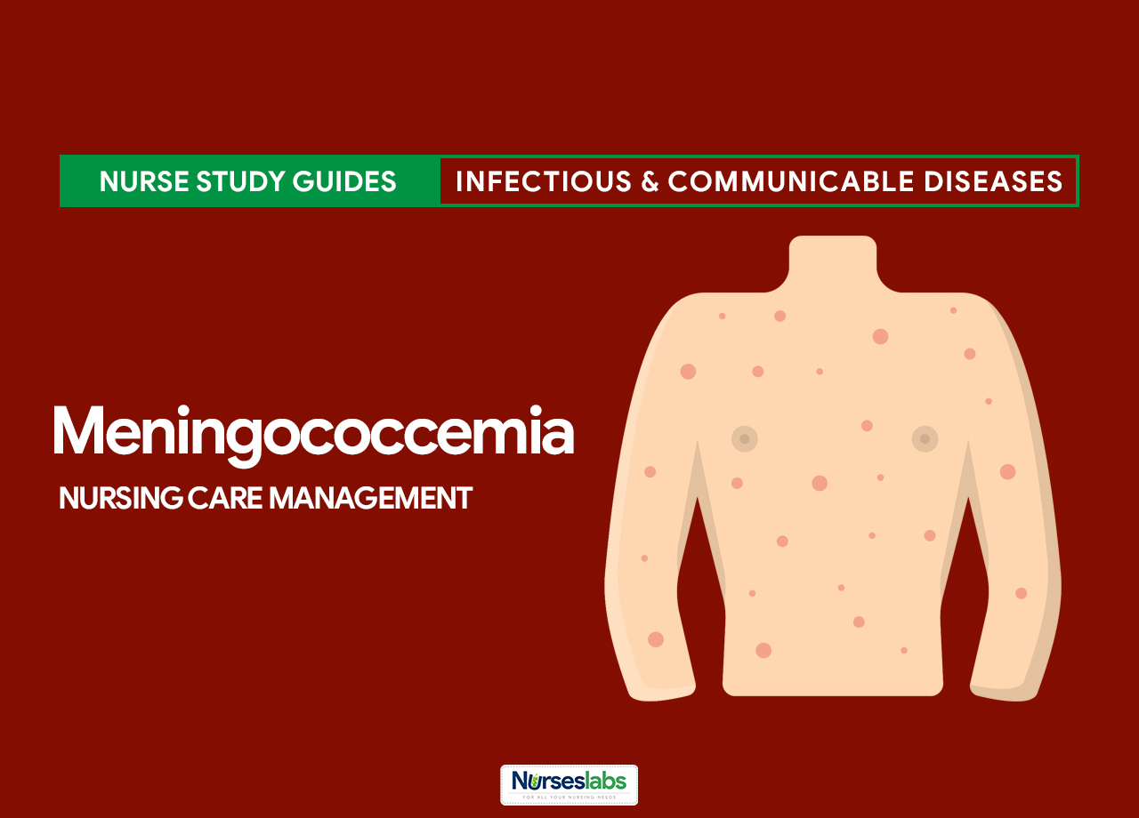
- The characteristic petechial skin rash is usually located on the trunk and legs and may rapidly evolve into purpura.
Assessment and Diagnostic Findings
Laboratory findings in the early stages of meningococcal disease are often nonspecific. Definitive diagnosis requires retrieval of meningococci from blood, cerebrospinal fluid, joint fluid, or skin lesions. Studies may include the following:
- Hematologic studies. A complete blood count (CBC), platelet count, blood urea nitrogen (BUN) study, and creatinine clearance evaluation, as well as a series of coagulation studies, can be used to evaluate a consumptive coagulopathy.
- Needle aspirates and skin biopsy. Gram-negative diplococci may be observed in punch biopsy and needle aspiration specimens of skin lesions or buffy coat preparations; Gram-negative diplococci may also be recovered from joint fluid.
- Histology. Leukocytoclastic vasculitis, thrombosis, and organisms are often demonstrated in biopsy specimens collected from patients with acute meningococcemia.
- Meningococcal polymerase chain reaction assay. Meningococcal PCR is a rapid method for diagnosing CSF infection.
- Slide agglutination test. With serogrouping, polysaccharide antigens on the capsule are identified by a slide agglutination test using polyclonal antibodies.
- Enzyme-linked immunosorbent assay. With serotyping and serosubtyping, outer membrane proteins (PorB and PorA) can be identified by an enzyme-linked immunosorbent assay (ELISA) using monoclonal antibodies.
- Lumbar puncture. Perform LP for CSF evaluation; organisms can be observed in the CSF in approximately half of patients who present with meningococcal meningitis.
Medical Management
Because mortality may be reduced with early antibiotic therapy, patients with a meningococcal rash should receive parenteral antibiotics by means of an intravenous (IV) or intramuscular (IM) route as soon as the diagnosis is suspected.
- Chemoprophylaxis. Chemoprophylaxis for meningococcal infection should be administered to intimate household, daycare center, and nursery school contacts of sporadic cases. Ciprofloxacin, ceftriaxone, and rifampicin are the most common antibiotic given as chemoprophylaxis.
- Diet. Patients with meningitis or fulminant meningococcemia are at risk of vomiting and should be prevented from taking anything by mouth prior to substantial clinical improvement with antimicrobial therapy.
- Activity. The level of patient activity is determined by the severity of the presentation; bed rest is recommended for patients suspected of having meningococcal disease; in most severe cases, patients are bed bound.
- Vaccination. The conjugate vaccine for group C meningococci in which the serogroup C meningococcal polysaccharide is conjugated to the protein CRM197 appears to provide immunogenic protection to young children. It was administered to all children during 1999-2000 in the United Kingdom.
Pharmacologic Management
Antimicrobial therapy is directed toward treatment of active infection or is used prophylactically to protect persons exposed to Neisseria meningitidis through close contact.
- Antimicrobial therapy. Mortality in meningococcal infections may be reduced with early antibiotic therapy; regarding community management, because mortality may be reduced with early antibiotic therapy, patients with a meningococcal rash should receive parenteral benzyl penicillin by means of an IV or IM route as soon as the diagnosis is suspected.
- Inotropic agents. These agents are used to support circulation in patients with shock.
- Diuretics, osmotic agents. These agents are used to control ICP during elective intubation; osmotic diuretics raise the osmolality of plasma and renal tubular fluid, which creates an osmotic inhibition of water transport in the proximal tubule.
- Diuretics, loop. Mannitol may reduce subarachnoid-space pressure by creating an osmotic gradient between CSF in the arachnoid space and plasma; it is not for long-term use.
- Corticosteroids. These agents elicit anti-inflammatory and immunosuppressive properties and cause profound and varied metabolic effects; they modify the body’s immune response to diverse stimuli.
- Vaccines, inactivated, bacterial. These agents may be used to prevent and control outbreaks of serogroup C meningococcal disease.
Nursing Management
Nursing management of a client with meningococcemia include the following:
Nursing Assessment
In 2015, CDC implemented enhanced meningococcal disease surveillance. The most recent Council of State and Territorial Epidemiologists (CSTE) case classification (2015) for meningococcal disease is:
- Suspected. Clinical purpura fulminans in the absence of a positive blood culture; or Gram-negative diplococci, not yet identified, isolated from a normally sterile body site (e.g., blood or cerebrospinal fluid [CSF]).
- Probable. Detection of Neisseria meningitidis antigen in formulin-fixed tissue by immunohistochemistry (IHC); or in CSF by latex agglutination.
- Confirmed. Detection of N. meningitidis-specific nucleic acid in a specimen obtained from a normally sterile body site (e.g., blood or CSF), using a validated polymerase chain reaction (PCR) assay; or isolation of N. meningitidis from a normally sterile body site (e.g., blood or CSF, or less commonly, synovial, pleural, or pericardial fluid); or from purpuric lesions.
Nursing Diagnosis
Based on the assessment data, the major nursing diagnosis for a patient with meningococcemia are:
- Ineffective Tissue Perfusion (Cerebral) related to cerebral edema.
- Hyperthermia related to abnormal temperature regulation.
- Acute Pain related to increased intracranial pressure.
- Disturbed Sensory Perception related to decreased LOC.
- Anxiety related to threat to or change in health status of child.
- Deficient Knowledge related to lack of exposure to information.
- Risk for Injury related to internal factor of altered neurologic regulatory function.
Nursing Care Planning and Goals
The major nursing goals for a child with meningococcemia:
- Child will have vital signs return to normal; child is alerted and oriented: motor, cognitive, and sensory function are within acceptable parameters for the child’s age; normal specific urine gravity.
- Child will regain and maintain body temperature within a normal range.
- Child will express feelings of comfort and relief of pain.
- Child will maintain normal LOC.
- Parents will experience decreased anxiety.
- Parents verbalize understanding of cause and treatment plan.
- Child will not experience injury.
Nursing Interventions
Nursing interventions for the patient with meningococcemia include:
- Increase cerebral perfusion. Monitor vital signs and neurological status; observe for any signs of increased intracranial pressure; observe for increasing restlessness, moaning, and guarding behaviors; maintain head or neck in midline position, provide small pillow for support; during reposition, avoid bending of the knee and pushing heels against the mattress; elevate the head of the bed 30°, and avoid neck flexion and hip flexion.
- Improve body temperature. Assess for signs of dehydration such as dry mouth, sunken eyes, sunken fontanelle, low concentrated urine output; gradually decrease temperature by performing tepid sponge bath; maintain adequate fluid intake as tolerated; and administer antipyretics as indicated.
- Decrease pain. Maintain a quiet environment and keep child’s room darkened; turn the client often and position the client carefully; prevent stimulation and restrict visitors; control environment to encourage rest; and administer antibiotic and corticosteroid as prescribed.
- Maintain normal LOC. Assess level of consciousness using pediatric Glasgow coma scale; assess for signs of cerebral edema such as dizziness, headache, irregular breathing, neck pain, nausea or vomiting; elevate head of bed up to 30° to 45° with the client’s head in neutral position; reorient the client to the environment, as needed; and administer and monitor anticonvulsants drug levels.
- Decrease anxiety. Encourage to express concerns and ask to express concerns and ask questions regarding the condition of ill child; encourage the parent to stay with the child or visit when able and call when concerned if hospitalized; assist in care (hold, feed, bathe, clothe and diaper), and provide information about child’s daily routines; encourage to be involved in care and decision-making regarding needs; and clarify any misinformation and answer questions in lay terms when parents able to listen, give same explanation as other staff and/or physician gave regarding disease process and transmission.
- Educate the caregivers and family. Assess knowledge of disease and method
to control and resolve disease; willingness and interest of parents to implement care; provide information and explanations in clear language that is understandable; use pictures, pamphlets, video tapes, model in teaching about disease; teach about administration of medications including (specify: action of drugs, dosages times frequency, side effects, expected results, methods to give medications); provide written instructions and schedule to follow and inform to administer full course of antibiotic to child; and teach to report elevated temperature, poor feeding or anorexia, irritability or other changes in behavior or level of consciousness, decrease in hearing acuity.
Evaluation
Nursing goals are met as evidenced by:
- Child’s vital signs returned to normal; child is alerted and oriented: motor, cognitive, and sensory function are within acceptable parameters for the child’s age; normal specific urine gravity.
- Child regained and maintained body temperature within a normal range.
- Child expressed feelings of comfort and relief of pain.
- Child maintained normal LOC.
- Parents experienced decreased anxiety.
- Parents verbalized understanding of cause and treatment plan.
- Child did not experience injury.
Documentation Guidelines
Documentation in a client with meningococcemia include the following:
- Individual findings, including factors affecting, interactions, nature of social exchanges, specifics of individual behavior.
- Cultural and religious beliefs, and expectations.
- Plan of care.
- Teaching plan.
- Responses to interventions, teaching, and actions performed.
- Attainment or progress toward the desired outcome.















Leave a Comment