Explore this comprehensive nursing care plan and management guide to effectively provide care for clients diagnosed with impaired tissue perfusion or ischemia. Gain knowledge about essential nursing assessments, evidence-based nursing interventions, goal-setting strategies, and nursing diagnoses related to tissue perfusion. This guide equips you with the necessary tools and information to deliver optimal care and promote improved tissue perfusion in clients.
What is Impaired Tissue Perfusion?
Blood, as a connective tissue composed of plasma and cells, plays a crucial role in delivering oxygen and nutrients to body cells. When there is insufficient arterial blood flow, cellular nutrition, and oxygenation are compromised, posing potential harm to the body. Atherosclerosis, the primary cause of impaired blood flow, narrows and obstructs blood vessels, leading to ischemia or inadequate blood supply due to blockage. This can give rise to various conditions such as angina, heart attack, stroke, and peripheral vascular disease.
The coronary circulation consists of the heart muscle moving blood to the lungs and the peripheral tissues. However, they do not receive oxygen or nourishment from the blood within the heart chambers. The coronary arteries fill with oxygen-rich blood during ventricular relaxation. If these arteries become clogged, the myocardium is deprived of oxygen, leading to chest pain or myocardial infarction.
The arterial circulation, on the other hand, moves blood from the heart to the tissues. It maintains the constant flow of blood to the capillary beds. Blood always moves from an area of higher pressure to an area of lower pressure. Blood pressure is the force exerted on arterial walls by the blood flowing within the vessel. The mean arterial pressure (MAP) maintains blood flow to the tissues throughout the cardiac cycle.
Signs of impaired circulation include diminished pulses, pain, pale skin, cool extremities, and hair loss. Risk factors for impaired circulation encompass smoking, high fat intake, obesity, sedentary lifestyle, hypertension, and diabetes. Additionally, inflammation, blood clots, and venous insufficiency contribute to impaired blood flow. Nursing care focuses on improving blood flow, educating clients, and preventing complications.
Tissue perfusion, on the other hand, refers to the continuous delivery of an adequate blood supply containing oxygen, nutrients, and hormones to the body’s tissues and organs. This process ensures that each cell receives the essential resources required for optimal function and metabolism. However, various conditions such as vascular diseases, blood clots, heart failure, or blockages can impede tissue perfusion, leading to insufficient oxygenation and nutrition of tissues, potentially causing damage or dysfunction. Nursing interventions and management strategies aim to enhance tissue perfusion, optimize blood flow, and prevent complications associated with impaired circulation.
Causes
The following are the common causes or related factors for patients with impaired tissue perfusion:
- Narrowing and hardening of arteries due to plaque buildup (e.g., atherosclerosis)
- Formation of blood clots within blood vessels obstructing blood flow (e.g., thrombosis)
- Blockage of an artery by a clot or foreign material traveling through the bloodstream (e.g. embolism).
- Abnormally low blood pressure reducing blood flow to tissues.
- Inadequate pumping of the heart leading to reduced blood circulation.
- Critical condition resulting from insufficient blood flow and oxygen delivery to tissues (e.g., shock).
- Reduced hemoglobin levels impairing oxygen transport to tissues (e.g., anemia).
- Reduced blood flow to the limbs due to narrowed arteries.
- Severe infection causing widespread inflammation and impaired blood flow (e.g., sepsis).
- Irregular heartbeats disrupting effective blood circulation.
- Narrowing of blood vessels decreasing blood flow to tissues.
- Physical damage to blood vessels hindering normal blood flow.
- Low blood volume limiting adequate tissue perfusion.
- Conditions affecting the autonomic regulation of blood flow.
- Excess body weight increasing the strain on the cardiovascular system.
Signs and Symptoms
The following are the common signs and symptoms or defining characteristics for impaired tissue perfusion:
- Pallor. Unusual paleness of the skin indicating reduced blood flow.
- Cool, Clammy Skin. Skin feels cold and moist due to inadequate perfusion.
- Delayed Capillary Refill. Slow return of color to the nail bed after pressure is applied.
- Weak or Rapid Pulse. Thready or tachycardic pulse reflecting compromised circulation.
- Hypertension or Hypotension. Elevated or abnormally low blood pressure affecting tissue perfusion.
- Chest Pain or Angina. Discomfort due to insufficient blood flow to the heart muscles.
- Shortness of Breath. Difficulty breathing resulting from impaired oxygen delivery to tissues.
- Fatigue and Weakness. Generalized tiredness caused by inadequate oxygen and nutrient supply.
- Edema. Swelling in extremities due to fluid accumulation from poor perfusion.
- Confusion or Altered Mental Status. Cognitive disturbances from insufficient blood flow to the brain.
- Numbness or Tingling. Sensory changes in limbs indicating reduced nerve perfusion.
- Delayed Wound Healing. Slower recovery of injuries due to inadequate blood supply.
- Muscle Cramps or Pain. Discomfort from oxygen-deprived muscles during activity.
- Bluish Discoloration (Cyanosis). Blue tint to lips, nails, or skin from poor oxygenation.
- Dizziness or Lightheadednes. Feeling faint due to decreased cerebral perfusion.
- Weak Pulse in Peripheral Vessels. Reduced strength of pulse in areas like wrists or ankles.
- Altered Urine Output. Decreased or irregular urination patterns from impaired kidney perfusion.
- Elevated Lactate Levels. Increased blood lactate as a marker of anaerobic metabolism due to hypoxia.
Nursing Care Plans and Management
Nurses play an essential role in enhancing tissue perfusion. Through continuous assessment, timely interventions, and close monitoring, nurses can contribute significantly to successful client outcomes. The nursing care plan for clients with impaired tissue perfusion encompasses a thorough assessment of the client’s condition, the formulation of realistic and measurable goals, evidence-based interventions to improve tissue perfusion, and regular evaluation of progress. Considerations for client education, collaboration with the healthcare team, and emotional support can also help ensure a holistic and client-centered approach to care.
Nursing Problem Priorities
The following are nursing problem priorities for clients with impaired tissue perfusion:
- Inadequate tissue oxygenation. Addressing inadequate oxygen delivery to tissues is of utmost importance. The nurse may initiate oxygen therapy, monitor oxygen saturation levels, and address any signs of circulatory insufficiency promptly.
- Pain management. Impaired tissue perfusion can lead to ischemic pain or discomfort. Assessing and managing the client’s pain is crucial to promote comfort and improve the client’s quality of life.
- Risk of tissue necrosis and shock. Impaired tissue perfusion can lead to tissue damage, necrosis, and shock. Preventive measures, including repositioning immobile clients and performing regular skin assessments may help mitigate the risk of necrosis and shock.
- Client and caregiver education. Clients and their families may have a limited understanding of impaired tissue perfusion and its management. Providing education about the condition, treatment plan, and lifestyle modifications is essential to promote client adherence to the care plan.
Nursing Assessment
It is important for nurses to recognize and assess these signs and symptoms promptly as they may indicate compromised tissue perfusion and the need for further interventions to restore adequate blood flow and oxygenation. The following are common signs and symptoms for clients with poor tissue perfusion:
- Pallor. Pallor refers to the paleness of the skin, which can occur due to decreased blood flow and oxygenation to the tissues. It is often seen in areas affected by ischemia.
- Pain or discomfort. Ischemia can cause pain or discomfort in the affected area due to inadequate blood supply and oxygenation. The type and intensity of pain may vary depending on the location and severity of the ischemia.
- Diminished or absent pulses. Diminished or absent pulses in the affected area may indicate reduced arterial blood flow, which is characteristic of ischemia. Assessing peripheral pulses is an important nursing intervention to evaluate tissue perfusion.
- Delayed capillary refill. Capillary refill time refers to the time it takes for color to return to blanched skin when pressure is applied and released. Prolonged capillary refill time (>3 seconds) suggests reduced blood flow and perfusion to the area.
- Cyanosis. Cyanosis is the bluish discoloration of the skin and mucous membranes resulting from insufficient oxygenation. It may occur in areas affected by severe ischemia.
- Impaired sensation or numbness. Ischemia can lead to altered sensation or numbness in the affected area due to compromised nerve function resulting from inadequate blood supply.
- Weakness or loss of motor function. Ischemia affecting the central nervous system or peripheral nerves can lead to weakness or loss of motor function in the corresponding area.
Nursing Diagnosis
After thorough assessment, nursing diagnoses are formulated to address the challenges of impaired tissue perfusion, guided by the nurse’s clinical judgment and understanding of the patient’s unique condition. While nursing diagnoses help organize care, their use may vary across clinical settings. Ultimately, the nurse’s expertise and judgment shape the care plan to prioritize each patient’s needs. Here are examples of nursing diagnoses that may be useful for common concerns associated with impaired tissue perfusion:
- Impaired Tissue Perfusion related to arterial narrowing as evidenced by pallor, cool clammy skin, and delayed capillary refill secondary to coronary artery disease and atherosclerosis.
- Impaired Tissue Perfusion related to formation of blood clots obstructing blood vessels as evidenced by weak or rapid pulse, chest pain, and shortness of breath secondary to pulmonary embolism.
- Impaired Tissue Perfusion related to excessive fluid loss through vomiting and diarrhea as evidenced by cool, clammy skin, hypotension, and decreased urine output secondary to gastroenteritis.
- Impaired Tissue Perfusion related to vasoconstriction reducing blood flow as evidenced by numbness, tingling, and cold extremities secondary to Raynaud’s phenomenon.
- Impaired Tissue Perfusion related to third spacing of fluids as evidenced by abdominal distension, edema, and decreased skin turgor secondary to severe burns.
- Impaired (Cardiac) Tissue Perfusion related to impaired cardiac output as evidenced by fatigue, dyspnea, and elevated jugular venous pressure secondary to heart failure.
- Impaired Tissue Perfusion related to hypotension from hemorrhage as evidenced by rapid pulse, weak pulse, and confusion secondary to traumatic hemorrhage.
- Impaired Tissue Perfusion related to excessive intravenous fluid administration as evidenced by pulmonary edema, elevated blood pressure, and jugular vein distention secondary to post-operative care.
- Impaired Tissue Perfusion related to increased capillary permeability as evidenced by edema, weight gain, and elevated central venous pressure secondary to sepsis.
- Impaired (Renal) Tissue Perfusion related to impaired renal excretion as evidenced by weight gain, peripheral edema, and elevated serum creatinine secondary to acute kidney injury.
- Impaired (Cerebral) Tissue Perfusion related to neurological disorders affecting autonomic regulation as evidenced by altered blood pressure, irregular heart rate, and poor peripheral perfusion secondary to Parkinson’s disease.
- Impaired Tissue Perfusion related to hypoxia reducing oxygen delivery as evidenced by cyanosis, confusion, and increased respiratory rate secondary to chronic obstructive pulmonary disease (COPD).
Nursing Goals
The SMART goals aim to enhance the client’s knowledge about circulation improvement, increase their activity tolerance, ensure optimal tissue perfusion, promote active participation in interventions, and foster awareness of when to seek medical assistance. The following are the common goals and expected outcomes for this nursing problem:
- The client will demonstrate an understanding of factors that improve circulation, including lifestyle modifications, by verbalizing and implementing at least three specific strategies within 8 hours.
- The client will exhibit increasing tolerance to activity, gradually progressing in duration and intensity, as evidenced by weekly tracking logs and regular assessments.
- The client will maintain maximum tissue perfusion to vital organs, as evidenced by warm and dry skin, strong peripheral pulses, vital signs within the client’s normal range, balanced input and output, absence of edema, normal arterial blood gas values, alert level of consciousness, and absence of chest pain.
- The client will actively engage in behaviors and actions to improve tissue perfusion, following the prescribed treatment plan and participating in recommended therapies and exercises.
- The client will verbalize when to promptly seek medical attention from the healthcare professional for any signs or symptoms of impaired tissue perfusion.
Nursing Interventions and Actions
To promote optimal tissue perfusion and manage ischemia, nursing actions include monitoring vital signs regularly, maintaining proper positioning, encouraging physical activity and range of motion exercises, managing pain effectively, ensuring adequate hydration, administering prescribed medications, providing wound care, monitoring for complications, educating the client on lifestyle modifications, and collaborating with the healthcare team. These interventions aim to assess and improve perfusion, prevent complications, and support the client’s overall vascular health. The following are the therapeutic nursing interventions and actions for clients with poor tissue perfusion:
1. Monitoring Tissue Perfusion
The frequency and extent of monitoring are based on several factors, including the severity of the client’s symptoms, the presence of risk factors, and the purpose of the assessment. Regular monitoring allows early detection of impaired tissue perfusion, which enables the healthcare professionals to deliver interventions and prevent complications promptly.
See also: Decreased Cardiac Output & Cardiac Support Nursing Care Plan and Management
Assessing cardiovascular tissue perfusion
Assess for signs of decreased tissue perfusion.
Particular clusters of signs and symptoms occur with differing causes. The nurse begins cardiovascular assessment by evaluating blood pressure in both arms and palpating peripheral pulses for strength and equality. The apical pulse is auscultated for the rate, rhythm, and quality of heart sounds. Carotid arteries are auscultated for bruits indicate atherosclerosis. Lung sounds are assessed for adventitious sounds to indicate increased pulmonary vessel pressure. Skin assessment provides information on color, temperature, hair distribution, lesions, and edema. Cool feet with weak pulses and shiny, hairless shins and feet may indicate peripheral vascular disease, while pitting edema may be present in clients with heart failure.
Review laboratory data (ABGs, BUN, creatinine, electrolytes, international normalized ratio, and prothrombin time or partial thromboplastin time) if anticoagulants are utilized for treatment.
Blood clotting studies are being used to conclude or make sure that clotting factors stay within therapeutic levels. These may also serve as gauges of organ perfusion or function. Irregularities in coagulation may occur as an effect of therapeutic measures. Acidosis is the best indicator in early shock of ongoing oxygen imbalance at the tissue level. A blood gas with a pH of 7.30 to 7.35 is abnormal but tolerable in the acute setting (Udeani & Geibel, 2018).
Check respirations and absence of work of breathing.
Cardiac pump malfunction and/or ischemic pain may result in respiratory distress. Nevertheless, abrupt or continuous dyspnea may signify thromboembolic pulmonary complications. When a thrombus breaks and becomes an emboli, it can occlude the blood supply. Although alveolar ventilation to the affected area often remains adequate, no gas exchange occurs there because of impaired blood flow.
Record BP readings for orthostatic changes (a drop of 20 mm Hg systolic BP or 10 mm Hg diastolic BP with position changes).
Stable BP is needed to keep sufficient tissue perfusion. Medication effects such as altered autonomic control, decompensated heart failure, reduced fluid volume, and vasodilation are among many factors potentially jeopardizing optimal BP. Because of compensatory mechanisms, the effect of age, and the use of certain medications, some clients in shock will present with normal blood pressure and pulse.
Examine GI function, noting anorexia, decreased or absent bowel sounds, nausea or vomiting, abdominal distension, and constipation.
Decreased blood flow to mesentery can turn into GI dysfunction and loss of peristalsis, for example. Problems may be potentiated or provoked by the utilization of analgesics, diminished activity, and dietary changes. The GI system is most vulnerable to hypoperfusion among the splanchnic organs because disturbed perfusion in the mesenteric area leads to GI dysfunction that can end with vital injury to the client (Wang & Liu, 2021).
Use pulse oximetry to monitor oxygen saturation and pulse rate.
Pulse oximetry is a useful tool to detect changes in oxygenation. Due to the noninvasive nature and relative importance of pulse oximetry readings, there are very few situations that do not indicate its use. Pulse oximetry can provide a rapid tool to assess oxygenation accurately (Hafen & Sharma, 2022).
Check hemodynamic studies.
Hemodynamics is the study of pressures and forces involved in blood circulation. It includes parameters such as heart rate, blood pressure, central venous pressure, and pulmonary vascular pressure. Hemodynamic studies are performed to assess fluid status and cardiovascular function. Some parameters are measured directly using catheters, while others are calculated. In intensive and cardiac care units, continuous hemodynamic monitoring is conducted to evaluate the cardiovascular status and the impact of interventions. Nurses play a crucial role in obtaining accurate readings and ensuring the integrity of the monitoring system.
Check for pallor, cyanosis, and cool or clammy skin. Assess the quality of every pulse.
The nonexistence of peripheral pulses must be reported or managed immediately. Systemic vasoconstriction resulting from reduced cardiac output may be manifested by diminished skin perfusion and loss of pulses. Therefore, assessment is required for constant comparisons.
Note skin texture and the presence of hair, ulcers, or gangrenous areas on the legs or feet.
Thin, shiny, dry skin with hair loss; brittle nails; and gangrene or ulcerations on toes and anterior surfaces of feet are seen in clients with arterial insufficiency. If ulcerations are on the side of the leg, they are usually venous. The skin provides important information for clients with septic shock. As a visible and easily accessible organ, the skin allows simple observation of local microcirculatory perfusion through skin temperature alterations, perfusion, and color (Hariri et al., 2019).
Assess for the presence of mottling.
Mottling is a characteristic discoloration of the skin following reduced blood flow. Mottling score reliability reflects organ failure severity in clients with sepsis or septic shock and helps to identify critically ill clients with worse outcomes.
Measure ankle-brachial index (ABI)
The ankle-brachial index (ABI) is a noninvasive measure used to diagnose peripheral arterial disease (PAD). The ABI is determined by comparing arterial pressure in the ankle to that in the arm. The client is positioned supine, and systolic pressures are measured using a Doppler instrument and ultrasound gel. The higher ankle pressure and higher arm pressure are used to calculate the ABI.
Observe signs of deep vein thrombosis, including pain, tenderness, swelling in the calf and thigh, and redness in the involved extremity.
Thrombosis with clot formation is usually first detected as swelling of the involved leg and then as pain. Without the activity of the calf and leg muscles, blood pools in the veins of the lower extremities, which can allow blood clots to form. If these clots break loose and become emboli, they could lodge in small vessels and alter the circulation.
Note the results of the D-Dimer Test.
High levels of D-Dimer, a fibrin degradation fragment, are found in deep vein thrombosis, pulmonary embolism, and disseminated intravascular coagulation. Current evidence strongly supports the use of a D-dimer assay in the setting of suspected DVT. Studies indicate that the D-dimer test can be used as a rapid screening measure in cases where leg swelling exists (Patel & Chun, 2019).
Monitor for the development of gangrene, venous ulceration, and symptoms of cellulitis.
Cellulitis often indicates peripheral vascular disease and is related to poor tissue perfusion. Superficial, irregular ulcers at the medial malleolus or red to yellow granulation tissue may be associated with the rupture of small skin capillaries from chronic venous insufficiency.
Monitor peripheral pulses. Check for loss of pulses with bluish, purple, or black areas and extreme pain.
These are symptoms of arterial obstruction that can result in the loss of a limb if not immediately reversed. A reliable assessment of the pulses depends on accurate identification of the location of the artery and careful palpation of the area. Light palpation is essential; firm finger pressure can obliterate the temporal, dorsalis pedis, and posterior tibial pulses and confuse the nurse. The dorsalis pedis pulses are not palpable in 10% of clients.
Assess for capillary refill time.
The capillary refill time (CRT) measures the amount of time necessary for the skin to return to baseline color after applying pressure on soft tissue, generally the fingertips. It gives important information on skin perfusion and microcirculatory status. CRT is a reliable triage tool to identify critically ill clients at risk of negative outcomes.
Monitor the client for chest pain and assess the level and characteristics of pain.
Ischemia of the heart muscle may produce pain or other symptoms, such as mild indigestion or a choking, heavy sensation in the upper chest. Most clients who develop acute MI present with an abrupt onset of squeezing or heavy substernal chest pain, which may radiate to the left arm or the neck. The chest pain may be atypical, the location being epigastric or only in the neck or arm. The pain quality may be burning, sharp, or stabbing.
Determine the peripheral perfusion index as applicable.
The peripheral perfusion index (PPI) is the difference between the pulsatile and nonpulsatile portion of the pulse wave, measured by plethysmography. It gives information on peripheral vascular tonus by the pulsatility, decreasing in vasoconstriction and increasing in vasodilation. PPI is an early predictor of central hypovolemia and is significantly lower in clients with an alteration in peripheral perfusion.
Perform an electrocardiogram (ECG) and monitor its results.
A 12-lead ECG may show changes indicative of ischemia, such as T-wave inversion, ST-segment elevation, or the development of an abnormal Q wave. ECG must be performed as soon as possible to help diagnose MI or myocardial ischemia.
Assessing cerebral tissue perfusion
Check rapid changes or continued shifts in mental status.
Electrolyte/acid-base variations, hypoxia, and systemic emboli influence cerebral perfusion. In addition, it is directly related to cardiac output. Some clients may report fatigue or generalized lethargy. Others may arrive by ambulance or in the custody of the law for the evaluation of bizarre behaviors.
Evaluate motor reaction to simple commands, noting purposeful and non-purposeful movement. Document limb movement and note right and left sides individually.
This measures overall awareness and capacity to react to external stimuli and best signifies the condition of consciousness in the client whose eyes are closed due to trauma or who is aphasic. Consciousness and involuntary movement are incorporated if the client can both take hold of and let go of the tester’s hand or grasp two fingers on command. Purposeful movement can comprise grimacing or withdrawing from painful stimuli. Other movements (posturing and abnormal flexion of extremities) usually specify disperse cortical damage. The absence of spontaneous movement on one side of the body signifies damage to the motor tracts in the opposite cerebral hemisphere.
Evaluate verbal reaction. Observe if the client is oriented to person, place, and time; or is confused; uses inappropriate words or phrases that make little sense.
This measures the appropriateness of speech content and level of consciousness. If minimum damage has taken place in the cerebral cortex, the client may be stimulated by verbal stimuli but may show drowsy or uncooperative. More broad damage to the cerebral cortex may be manifested by a slow reaction to commands, lapsing into sleep when not aroused, disorientation, and stupor. Injury to the midbrain, pons, and medulla is evidenced by a lack of appropriate reactions to stimuli.
Monitor higher functions, as well as speech, if the client is alert.
Indicators of location or degree of cerebral circulation or perfusion are alterations in cognition and speech content. A decline in higher cortical function may indicate diminished perfusion of the brain, which leads to an altered mental status ranging from confusion and agitation to flaccid coma (Ren & Lenneman, 2019).
Monitor cerebral perfusion pressure (CPP).
CPP is the net pressure gradient that drives oxygen delivery to cerebral tissue. It is the difference between the mean arterial pressure (MAP) and the intracranial pressure (ICP). abnormalities in CPP are strongly associated with poor outcomes. Inadequate CPP leads to ischemic injury, while excessive CPP may worsen cerebral edema (Kramer et al., 2019).
Measure the client’s ICP.
ICP is usually measured invasively through an intracranial pressure transduction device. The most common and most accurate method is with an intraventricular monitor, which is the current gold standard. ICP can also be measured non-invasively through transcranial Doppler ultrasonography.
Assessing renal tissue perfusion
Monitor intake and observe changes in urine output. Record urine-specific gravity as necessary. Note urine output.
Reduced intake or unrelenting nausea may result in lowered circulating volume, which negatively affects perfusion and organ function. Hydration status and renal function are revealed by specific gravity measurements. Reduced renal perfusion may take place due to vascular occlusion. Renal compensation for reduced perfusion results in diminished glomerular filtration, causing oliguria and subsequent renal failure.
Monitor the client’s blood pressure.
Severe heart failure may also cause hypotension. Although clients with heart failure may have low blood pressure, volume expansion is present and effective renal perfusion is poor, which can result in acute kidney injury.
Submit the client to diagnostic testing as indicated.
A variety of tests are available depending on the cause of the impaired tissue perfusion. Angiograms, Doppler flow studies, segmental limb pressure measurements such as ankle-brachial index (ABI), and vascular stress testing are examples of these tests. Although increased levels of BUN and creatinine are the hallmarks of renal failure, the rate of rise depends on the degree of renal insult (Workeneh & Batuman, 2022).
Monitor the client’s kidney function test results.
Among the measures of kidney function, glomerular filtration rate (GFR) has been regarded as the gold standard and is clinically estimated from serum creatinine. Renal blood flow and perfusion provide alternative measures of kidney function that may be useful in assessing a wide range of renal diseases (Zhang & Lee, 2019).
Determining risk factors and potential causes
Assess for probable contributing factors related to temporarily impaired arterial blood flow. Some examples include compartment syndrome, constricting cast, embolism, indwelling arterial catheters, positioning, thrombus, and vasospasm.
Early detection of the source facilitates quick, effective management. Vascular disease may manifest acutely when thrombi, emboli, or acute trauma compromises perfusion. Multiple factors predispose clients to thrombosis. These include sepsis, hypotension, low cardiac output, aneurysms, aortic dissection, bypass grafts, and atherosclerotic narrowing of the arterial lumen (Stephens & Schraga, 2022).
Obtain the client’s medical history, especially their history of cardiovascular diseases.
Complications of acute MI, such as mitral regurgitation, large right ventricular infarction, rupture of the interventricular septum or LV-free wall, and tamponade can result in shock. Cardiogenic shock is also more likely to develop in older adults and clients with diabetes or who had a previous inferior MI. A complete clinical assessment is critical to understanding the cause of shock and to targeting the therapy for correcting the cause.
Assess for cardiac risk factors.
A client thought to have myocardial ischemia should be assessed for cardiac risk factors. The evaluation should reveal a history of hyperlipidemia, LV hypertrophy, hypertension, or cigarette smoking, or a family history of premature coronary artery disease. The presence of two or more risk factors increases the likelihood of acute MI.
2. Promoting Optimal Tissue Circulation
Atherosclerosis is the most common cause of impaired blood flow to organs and tissues. As vessels narrow and become obstructed, distal tissues receive less blood, oxygen, and nutrients. Promoting adequate circulation may involve position changes, ambulation, and regular exercise.
Interventions for cardiovascular tissue perfusion
Check for optimal fluid balance. Administer IV fluids as ordered.
Sufficient fluid intake maintains adequate filling pressures and optimizes cardiac output needed for tissue perfusion. The optimization phase of fluid management consists of the administration of a limited amount of IV fluid with careful evaluation of the client’s response. Signs of fluid responsiveness, before any additional fluid is given, can be very valuable to avoid giving fluids to clients who may not benefit from them (Vincent, 2019).
Maintain optimal cardiac output.
This ensures adequate perfusion of vital organs. The vessels that supply blood to the heart muscle may become occluded by atherosclerosis or a blood clot, shutting off the blood supply to a portion of the myocardium. When this happens, the tissue becomes necrotic and dies, leading to myocardial infarction or heart attack.
Advise the client to maintain bedrest as indicated.
Bed or chair rest during the initial phase of treatment helps reduce myocardial oxygen consumption. This limitation on mobility should remain until the client is pain-free and hemodynamically stable.
Administer nitroglycerin (NTG) sublingually for complaints of angina.
Administering nitroglycerin (NTG) sublingually is a vital nursing intervention for clients experiencing angina due to impaired tissue perfusion. NTG acts as a potent vasodilator, specifically targeting the coronary arteries and improving blood flow to the myocardium. By dilating the coronary arteries, NTG helps alleviate constriction and narrowing, enhancing myocardial perfusion.
Administer thrombolytics as prescribed.
Thrombolytic therapy is initiated when primary PCI is not available or the transport time to a PCI-capable hospital is too long. The purpose of these agents is to dissolve the thrombus in a coronary artery, allowing blood to flow through the coronary artery again, minimizing the size of the infarction, and preserving ventricular function. These should not be administered if the client is bleeding or has a bleeding disorder.
Administer vasopressor and inotropic agents as indicated.
Dopamine, norepinephrine, and epinephrine are vasoconstricting drugs that help maintain adequate blood pressure during life-threatening hypotension and help preserve perfusion pressure for optimizing flow in various organs.
Maintain oxygen therapy as ordered.
This is to enhance myocardial perfusion. Oxygen therapy is usually initiated at the onset of chest pain in an attempt to increase the amount delivered to the myocardium and decrease pain. The therapeutic effectiveness of oxygen is determined by observing the rate and rhythm of respirations and the color of the skin and mucous membranes.
Monitor results of echocardiograms.
The echocardiogram is used to evaluate ventricular function. It may be used to assist in diagnosing MI, especially when ECG is nondiagnostic. The echocardiogram can detect hypokinetic and akinetic wall motion and can determine the ejection fraction.
Assist in cardiovascular procedures, such as emergent percutaneous coronary intervention.
This procedure is used to open the occluded coronary artery and promote reperfusion to the area that has been deprived of oxygen. Superior outcomes have been reported with the use of PCI when compared to thrombolytic agents.
Assist in inserting a central line for reperfusion and invasive monitoring.
The placement of a central line may facilitate volume resuscitation, provide vascular access for multiple infusions and allow invasive monitoring of central venous pressure. Invasive monitoring with a pulmonary artery catheter may be helpful in guiding fluid resuscitation.
Interventions for cerebral tissue perfusion
If intracranial pressure is increased, elevate the head of the bed from 30 to 45 degrees.
This promotes venous outflow from the brain and helps reduce pressure. This is the least invasive method of lowering a client’s ICP. Some researchers have demonstrated improved ICP control with elevation to 45°, but evidence from the use of multimodality monitoring has suggested 30° head elevation for maximum benefit.
Monitor the client’s blood pressure strictly.
Greatly elevated BP is thought to lead to rebleeding and hematoma expansion in hemorrhagic stroke. This may result in loss of cerebral autoregulation of cerebral perfusion pressure. If systolic BP is over 200 mm Hg or MAP is over 150 mm Hg, consider aggressive reduction of BP with continuous IV infusion; check BP every five minutes.
See also: Bleeding Risk & Hemophilia Nursing Care Plan and Management
Avoid measures that may trigger increased ICP such as coughing, vomiting, straining at stool, neck in flexion, head flat, or bearing down.
These will further reduce cerebral blood flow. Activities that involve holding the breath and straining, such as weightlifting or intense coughing, can transiently cause increased ICP. certain body positions, especially those that lower the head below the level of the heart, can also increase ICP.
Administer anticonvulsants as needed.
These reduce the risk of seizure which may result from cerebral edema or ischemia. An anticonvulsant regimen should be started for clients with a moderate or severe head injury. The administration should cease if no seizure activity occurs within the first seven days after injury (Rangel & Kopell, 2022).
Control environmental temperature as necessary. Perform a tepid sponge bath when fever occurs.
Fever may be a sign of damage to the hypothalamus. Fever and shivering can further increase ICP. pyrexia commonly occurs in clients with head injuries, possibly because of posttraumatic inflammation, direct damage to the hypothalamus, or secondary infection. Fever should be avoided, as it increases cerebral metabolic demand and affects ICP.
Provide rest periods between care activities and prevent the duration of procedures.
Constant activity can further increase ICP by creating a cumulative stimulant effect. During rest, the brain’s metabolic activity decreases, resulting in lower oxygen and glucose consumption. This decrease in metabolic demand can lead to less production of metabolic waste products and a reduction in brain swelling, helping alleviate ICP.
Reorient to the environment as needed.
Decreased cerebral blood flow or cerebral edema may result in changes in the LOC. use clear and simple language when communicating with the client and repeat essential information as necessary. Create a quiet, low-stimulus environment to minimize stress and agitation; family members who are familiar to the client may be present, however, so that their presence can provide comfort and support.
Prepare the client for angiography.
Diagnosis of an aneurysm can be made by computed tomography angiography (CTA). CTAs are typically performed because they are widely available, can be completed rapidly, and can remove cardiac motion artifacts, enhancing their accuracy.
Administer oxygen therapy as indicated.
Supplemental oxygen is recommended when the client has a documented oxygen requirement of oxygen saturation below 95%. In the small proportion of clients with stroke who are relatively hypotensive, administration of IV fluid, vasopressor therapy or both may improve flow through critical stenoses.
Administer fibrinolytic therapy as prescribed.
Fibrinolytics restore cerebral blood flow in some clients with acute ischemic stroke and may lead to improvement or resolution of neurologic deficits. It should be administered 3 to 4.5 hours after symptom onset was found. The only fibrinolytic agent that has been shown to benefit selected clients with acute ischemic stroke is alteplase. While streptokinase may benefit clients with acute MI, in clients with acute ischemic stroke it has been shown to increase the risk of intracranial hemorrhage and death.
Administer antihypertensives as indicated.
The 2010 AHA/ASA guidelines strongly recommend the maintenance of BP below 140/90 mm Hg to prevent a first stroke. BP-lowering medications include thiazide diuretics, calcium channel blockers (CCB), angiotensin-converting enzyme inhibitors (ACEIs), and angiotensin receptor blockers (ARBs). For clients with diabetes, the use of ACEIs and ARBs to treat hypertension is a class I-A recommendation, according to the 2011 AHA/ASA primary prevention guidelines.
Institute measures to prevent the recurrence of stroke.
Secondary prevention refers to the treatment of clients who have already had a stroke. Measures may include the use of antiplatelet agents, anticoagulants, antihypertensives, statins, and lifestyle interventions. Smoking cessation, blood pressure control, diabetes control, a low-fat low-salt diet, weight loss, and regular exercise should be encouraged strongly as the medications described above.
Interventions for peripheral tissue perfusion
Assist with position changes.
Gently repositioning a client from a supine to a sitting/standing position can reduce the risk of orthostatic BP changes. Older adults are more susceptible to such drops of pressure with position changes. The nurse may position the client in a high-Fowler position to decrease preload and reduce pulmonary congestion. For the lower extremities, the nurse may elevate the client’s bed or have the client use a reclining chair, or sit with the feet resting on the floor to improve peripheral arterial circulation.
Encourage the client to ambulate as tolerated.
The nurse can assist the client with walking or other moderate or graded isometric exercises that may be prescribed to promote blood flow and encourage the development of collateral circulation. The nurse instructs the client to walk to the point of pain, rest until the pain subsides, and then resume walking so that endurance can be increased as collateral circulation develops.
Promote applications of warmth and avoid extremely cold temperatures.
Nursing interventions may involve applications of warmth to promote arterial flow and instructions to the client to avoid exposure to cold temperatures, which causes vasoconstriction. Adequate clothing and warm temperatures protect the client from chilling. If chilling occurs, a warm bath or drink is helpful, and a hot water bottle or heating pad may be applied to the abdomen to cause vasodilation throughout the lower extremities.
Promote active/passive ROM exercises.
Exercise prevents venous stasis and further circulatory compromise. Encourage leg exercises, such as flexion and extension of the feet, active contraction, and relaxation of the calf muscles for a client on bed rest, and promote ambulation as soon as possible.
Encourage the client to perform relaxation techniques to avoid stress.
Emotional upsets stimulate the sympathetic nervous system, resulting in peripheral vasoconstriction. Emotional stress can be minimized to some degree by avoiding stressful situations when possible or by consistently following a stress management program. Counseling services or alternative or complementary therapies, such as yoga, relaxation training, aromatherapy, or mindfulness-based stress reduction, may be indicated for clients who cannot cope with stress effectively.
Provide foot and leg care regularly.
Poorly perfused tissues are susceptible to damage and infection. When lesions develop, healing may be delayed or inhibited because of poor blood supply to the area. Inspect the client’s feet daily for redness, dryness, cuts, or blisters. If the client’s feet are dry, apply cream with lanolin, but never put cream in between toes to avoid maceration.
Minimize invasive procedures for the client.
The nurse should minimize the number of punctures for inserting IV lines and obtaining blood samples, avoid intramuscular injections, prevent any possible tissue trauma, and apply pressure at least twice as long as usual after any puncture is performed. This helps avoid prolonged bleeding in a client treated with thrombolytic therapy.
Administer medications as prescribed to treat the underlying problems. Note the response.
These medications facilitate perfusion for most causes of impairment.
- Antiplatelets/anticoagulants
These reduce blood viscosity and coagulation. Anticoagulants reduce thrombin generation and fibrin formation and minimize clot propagation. - Peripheral vasodilators
These enhance arterial dilation and improve peripheral blood flow. Vasodilators, such as nitroglycerin, decrease preload and/or afterload, and reduce systemic vascular resistance, allowing for improved forward flow and improving cardiac output. - Antihypertensives
These reduce systemic vascular resistance and optimize cardiac output and perfusion. Calcium channel blockers have been shown to decrease all cardiovascular events other than heart failure.
- Inotropes
Inotropic agents augment coronary and cerebral blood flow during the low-flow state associated with cardiogenic shock. They also improve cardiac output in refractory hypotension and shock. - Analgesics
Pain is frequently associated with peripheral arterial insufficiency, which can be chronic, continuous, and disabling. Analgesic agents such as hydrocodone plus acetaminophen, oxycodone, oxycodone plus acetylsalicylic acid, or oxycodone plus acetaminophen may be helpful in reducing pain so that the client can participate in therapies that can increase circulation and ultimately relieve pain more effectively.
Provide oxygen therapy as necessary.
This saturates circulating hemoglobin and augments the efficiency of blood that is reaching the ischemic tissues. Supplemental oxygen is recommended when the client has a documented oxygen requirement (oxygen saturation of 95%) (Jauch & Lutsep, 2022).
Position the client properly in a semi-Fowler’s to high-Fowler’s as tolerated.
Upright positioning promotes improved alveolar gas exchange. Blood flow is redistributed, with more blood flowing toward the base of the lungs due to the effect of gravity. This helps match the distribution of ventilation and perfusion, leading to better gas exchange.
Assist in the surgical management of arterial disorders.
Vascular surgical procedures are divided into two groups: inflow procedures, which improve blood supply from the aorta into the femoral artery, and outflow procedures, which provide blood supply to vessels below the femoral artery.
Consider the need for potential embolectomy, and heparinization, vasodilator therapy, thrombolytic therapy, and fluid rescue.
These facilitate perfusion when interference to blood flow transpires or when perfusion has gone down to such a serious level leading to ischemic damage. When conservative measures fail to improve quality of life and function, endovascular procedures are considered. The timing and need for revascularization are related to the general primary representations of critical and acute limb ischemia, in which urgent intervention is warranted.
Interventions for renal tissue perfusion
Monitor kidney function studies as appropriate.
The BUN level increases steadily at a rate that depends on the degree of catabolism, renal perfusion, and protein intake. Serum creatinine levels are useful in monitoring kidney function and disease progression and increase with glomerular damage. The glomerular filtration rate (GFR) declines, placing the client at risk of hyperkalemia.
Monitor for the use of medications that decrease renal blood flow.
Any agent that reduces renal blood flow, such as the long-term use of analgesics, may cause renal insufficiency. Chronic use of analgesic agents, particularly NSAIDs, may cause inflammation within the renal tissue and papillary necrosis.
Promote dietary modification, such as restriction of salt and fluid.
Restriction of salt and fluid is crucial in the management of oliguric kidney failure, wherein kidneys do not adequately excrete toxins or fluids. Because potassium and phosphorus are not excreted optimally in clients with AKI, blood levels of these electrolytes tend to be high. Restriction of these elements in the diet may be necessary, with guidance for frequent measurements.
Administer intravenous fluids or blood products as appropriate.
Adequate renal blood flow in clients with prerenal causes of AKI may be restored by IV fluids or transfusions of blood products. If AKI is caused by hypovolemia secondary to hypoproteinemia, an infusion of albumin may be prescribed.
Administer vasodilators as prescribed.
The rationale for vasodilator therapy in AKI is that improved renal perfusion may reduce kidney damage. A meta-analysis of 16 randomized studies concluded that the vasodilator fenoldopam reduces the need for renal replacement therapy and lowers the mortality rate in clients with AKI. dopamine in small doses causes selective dilatation of the renal vasculature, enhancing renal perfusion. It also reduces sodium absorption, which enhances urine flow.
Administer continuous renal replacement therapy as indicated.
Administer continuous renal replacement therapy in clients with stage 2 acute kidney injury and has a rise in serum creatinine level >2.0 times baseline and urine output <0.5 mL/kg/hour for >12 hours, or when “; life-threatening changes in fluid, electrolyte, and acid-base balance occurs.
3. Preventing Venous Stasis
When clients have limited mobility, venous return to the heart is impaired and the risk of venous stasis increases. Preventing venous stasis is an important nursing intervention to decrease the risk of complications following surgery, trauma, or medical problems.
Interventions for arterial insufficiency
Do not elevate the legs above the level of the heart.
With arterial insufficiency, leg elevation decreases arterial blood supply to the legs. Care should also be taken to avoid this position in clients with cardiac dysfunction because it will increase preload and may increase stress on the heart.
For early arterial insufficiency, encourage exercise such as walking or riding an exercise bicycle for 30 to 60 minutes per day.
Exercise enhances the development of collateral circulation, strengthens muscles, and provides a sense of well-being. The nurse can assist the client with walking or other moderate or graded isometric exercises that may be prescribed to promote blood flow and encourage the development of collateral circulation.
Keep the client warm, and have the client wear socks and shoes or sheepskin-lined slippers when mobile. Do not apply heat.
Clients with arterial insufficiency complain of being constantly cold; therefore keep extremities warm to maintain vasodilation and blood supply. Heat application can easily damage ischemic tissues. Adequate clothing and warm temperatures protect the client from chilling. Interventions that increase warmth to promote arterial flow and instructions to avoid cold exposure can be provided by the nurse.
Provide much attention to foot care. Refer to a podiatrist if the client has a foot or nail abnormality.
Ischemic feet are very vulnerable to injury; meticulous foot care can prevent further injury. Instruct the client to pat the feet dry instead of vigorous rubbing or scratching to avoid abrading the feet and creating sites for bacterial invasion. Fingernails and toenails should be carefully trimmed straight across and sharp corners filed to follow the contour of the nail. If the nails cannot be trimmed safely, it is necessary to consult a podiatrist, who can also remove corns and calluses.
Provide foods rich in protein and vitamins.
Good nutrition promotes healing and prevents tissue breakdown and is included in the overall therapeutic program for clients with peripheral vascular disease. Eating a diet that contains adequate protein and vitamins is necessary for clients with arterial insufficiency. Key nutrients play specific roles in wound healing.
Administer anticoagulant and fibrinolytic therapy as prescribed.
When the client has adequate collateral circulation, treatment may include IV anticoagulation with heparin, which can prevent the thrombus from spreading and reduce muscle necrosis. Intra-arterial thrombolytic medications are used to dissolve the thrombus. Fibrin-specific thrombolytic medications do not deplete circulating fibrinogen and plasminogen, which prevents the development of systemic fibrinolysis.
Interventions for venous insufficiency
If DVT is present, observe for symptoms of a pulmonary embolism, especially if there is a history of trauma.
Fatal pulmonary embolisms have been reported in one-third of trauma clients. Symptoms of PE depend on the size of the thrombus and the area of the pulmonary artery occluded by the rhombus; they may be nonspecific. Dyspnea is the most frequent symptom. Chest pain is common and is usually sudden and pleuritic in origin. The most frequent sign is tachypnea.
If the client is overweight, encourage weight loss to decrease venous disease.
Obesity is a risk factor for the development of chronic venous disease. Theories suggest that in clients who are obese, excessive adipose tissue may secrete mediators that lead to metabolic changes. Adipokines, free fatty acids, and other substances are known to modify insulin action and contribute to atherogenic changes in the cardiovascular system.
Discuss lifestyle with the client to see if the occupation requires prolonged standing or sitting.
These can result in chronic venous disease. The frequent causes of DVT are due to augmentation of venous stasis due to immobilization or central venous obstruction. Immobility can be as transient as that occurring during a transcontinental airplane flight or prolonged standing daily at work.
If the client is mostly immobile, consult with the healthcare provider regarding the use of a calf-high pneumatic compression device for the prevention of DVT.
Pneumatic compression devices can be effective in preventing deep vein thrombosis in an immobile client. Graduated compression stockings usually are prescribed for clients with venous disease. These are designed to apply 100% of the prescribed pressure gradient at the ankle and then decrease along the length of the stocking, increasing flow in the deep veins.
Elevate edematous legs as ordered and ensure that there is no pressure under the knee.
Elevation improves venous return and helps minimize edema. Pressure under the knee limits venous circulation. The nurse may position the client with legs elevated to promote venous return to the heart. This is particularly important with venous dysfunction. However, avoid pillows under the knees or more than 15 degrees of knee flexion to improve blood flow to the lower extremities and reduce venous stagnation.
Apply support hose as ordered.
Wearing a support hose helps decrease edema. A support hose should not be confused with graduated stockings because it provides less compression (12 to 20 mm Hg) than graduated stockings.
Encourage the client to walk with a support hose on and perform toe-up and point flex exercises.
Exercise helps increase venous return, build up collateral circulation, and strengthen the calf muscle pumps. Any type of stocking can inadvertently become a tourniquet or applied incorrectly. In such instances, the stockings produce rather than prevent stasis. For ambulatory clients, graduated compression stockings are removed at night and reapplied before the legs are lowered from the bed to the floor in the morning.
Administer anticoagulant and thrombolytic therapies.
Anticoagulant therapy delays the clotting of blood, prevents the formation of a thrombus, and forestall the extension of a thrombus after it has formed. Combining them with mechanical and ultrasonic-assisted thrombolytic therapy may eliminate venous obstruction, maintain venous patency, and prevent postthrombotic syndrome by early removal of the thrombus.
4. Promoting a Therapeutic Lifestyle
Therapeutic lifestyle changes include changes in dietary measures, weight loss, smoking cessation, and increased physical activity. These general recommendations may need to be adjusted for the individual client who has their own needs.
Provide foods rich in high-density lipoprotein (HDL), low in saturated fat, and high in soluble fiber.
HDL is known as good cholesterol because it transports other lipoproteins such as LDL to the liver, where they can be degraded and excreted. The Mediterranean diet, another diet that promotes vegetables and fish and restricts red meat, is also reported to reduce mortality from cardiovascular disease. These general recommendations may need to be adjusted for the individual client who has other nutritional needs, such as a client who has diabetes.
Encourage the client to engage in regular physical activity.
Regular, moderate physical activity increases HDL levels and reduces triglyceride levels, decreasing the incidence of coronary events and reducing overall mortality risk. The goal for most adults is to engage in moderate-intensity aerobic activity of at least 150 minutes per week or vigorous-intensity aerobic activity of at least 75 minutes per week.
Promote smoking cessation.
Cigarette smoking contributes to the development and severity of CAD. A client at risk for heart disease is encouraged to stop tobacco use through any means possible: educational programs, counseling, consistent motivation and reinforcement messages, support groups, and medications. Some clients find complementary therapies such as acupuncture and guided imagery, to be helpful.
5. Client and Caregiver Education
Education is a powerful tool in the management of impaired tissue perfusion. It empowers clients and caregivers, enhances treatment adherence, and promotes self-management, leading to positive client outcomes and improved quality of life.
Discuss with the client the difference between arterial and venous insufficiency.
Detailed diagnostic information clarifies clinical assessment and allows for more effective care. Arterial insufficiency occurs when there is inadequate blood flow through the arteries, usually due to atherosclerosis. Venous insufficiency occurs when the veins in the legs have difficulty returning blood to the heart, often due to damaged or weakened valves in the veins.
Educate clients about nutritional status and the importance of paying special attention to obesity, hyperlipidemia, and malnutrition.
Malnutrition contributes to anemia, which further compounds the lack of oxygenation to tissues. Obese clients encounter poor circulation in adipose tissue, which can create increased hypoxia in tissue. The association of a high blood cholesterol level with heart disease is well established, and the metabolism of fats is known to be an important contributor to the development of heart disease.
Encourage smoking cessation.
Smoking tobacco is also associated with catecholamine release resulting in vasoconstriction and poor tissue perfusion. A client at increased risk for heart disease is encouraged to stop tobacco use through any means possible: educational programs, counseling, consistent motivation and reinforcement messages, support groups, and medications.
Provide knowledge on normal tissue perfusion and possible causes of impairment.
Knowledge of causative factors provides a rationale for treatments. Atherosclerosis is by far the most common cause of impaired blood flow to organs and tissues. As vessels narrow and become constructed, distal tissues receive less blood, oxygen, and nutrients. Partial obstruction of the coronary arteries causes myocardial ischemia, often resulting in angina pectoris; if obstruction is complete a heart attack (MI) occurs.
Encourage changes in lifestyle that could improve tissue perfusion (avoiding crossed legs at the knee when sitting, changing positions at frequent intervals, rising slowly from a supine/sitting to a standing position, avoiding smoking, reducing risk factors for atherosclerosis [obesity, hypertension, dyslipidemia, inactivity]).
These measures reduce venous compression/venous stasis and arterial vasoconstriction. Venous thromboembolism can be prevented, especially if clients who are considered at high risk are identified and preventive measures are instituted without delay.
Explain all procedures and treatments.
Understanding expected events and sensations can help eliminate anxiety associated with the unknown. Knowledge deficit, frustration, fear, and depression can decrease the client’s and family’s compliance with the prescribed therapy; therefore, client and family education is necessary throughout the regimen.
Teach the client to recognize the signs and symptoms that need to be reported to the nurse.
The early assessment facilitates immediate treatment. Timely treatment can help prevent further deterioration and potentially reverse the underlying cause of impaired perfusion, improving the client’s outcomes.
When the client experiences dizziness due to orthostatic hypotension when getting up, educate methods to decrease dizziness, such as remaining seated for several minutes before standing, flexing the foot upward several times while seated, rising slowly, sitting down immediately if feeling dizzy, and trying to have someone present when standing.
Orthostatic hypotension results in temporary decreased cerebral perfusion. Orthostatic hypotension may develop when the client assumes a vertical position. Because of inadequate vasomotor reflexes, blood pools in the splanchnic area and in the legs, resulting in inadequate cerebral circulation. If indicators of orthostatic hypotension are present, advise the client to stop the activity and assist them to a supine position.
Provide the client with resources for dietary modifications.
Many resources are available to assist clients in controlling their cholesterol levels. The National Heart, Lung, and Blood Institute (NHLBI) and its National Cholesterol Education Program (NCEP), the AHA, and the American Diabetes Association (ADA), as well as CAD support groups and reliable internet resources, are a few examples of the available resources. Cookbooks and recipes that include the nutritional contents of foods can be included as resources for clients.
Instruct the client to read food and product labels.
Dietary control has been made easier because food manufacturers are required to provide nutritional data on product labels. The label information of interest to a person attempting to eat a heart-healthy diet is as follows: serving size (expressed in household measures), amount of total fat per serving, amount of saturated fats and trans fat per serving, amount of cholesterol per serving, and amount of fiber per serving.
Teach the client the appropriate exercises for daily physical activity.
Clients should be instructed to engage in an activity or a variety of activities that interest them to maintain motivation. They should be taught to exercise to an intensity that does not preclude their ability to talk; if they cannot have a conversation while exercising, they should slow down and switch to a less intensive activity.
Instruct the client to avoid temperature extremes during physical activity.
When the weather is hot and humid, the client should exercise during the early morning, or indoors, and wear loose-fitting clothing. When the weather is cold, they should layer clothing and wear a hat.
Caution the client with venous insufficiency in using warm or hot applications.
Clients are instructed to test the temperature of bath water and to avoid using hot water bottles and heating pads on the extremities. It is safer to apply a hot water bottle or a heating pad to the abdomen; this can cause reflex vasodilation in the extremities. Excess heat may increase the metabolic rate of the extremities and the need for oxygen beyond that provided by the reduced arterial flow through the diseased artery.
Instruct against the use of constrictive clothing and accessories, as well as activities that constrict blood flow to the extremities.
Constrictive clothing and accessories such as tight socks or shoelaces may impede circulation to the extremities and promote venous stasis and therefore should be avoided. Crossing the legs for more than 15 minutes at a time should be discouraged because it compresses vessels in the legs.
Provide information about medications that aid in smoking cessation.
The use of medications such as nicotine patches, varenicline, or bupropion may assist with stopping the use of tobacco. Products containing nicotine have some of the same effects as smoking: catecholamine release and increased platelet adhesion. These medications should be used for a short time and at the lowest effective doses.
Strongly advise avoidance of secondhand smoke exposure.
Exposure to other smokers’ smoke (passive or secondhand smoke) is believed to cause cardiovascular diseases in nonsmokers. Secondhand smoke also increases the risk of stroke by an estimated 20 to 30%. There is a concern that because these products expose clients to tobacco and its toxins, they may carry the risk of lung disease, cancer, and cardiovascular diseases.
Collaborate with the client to create a medication schedule.
The nurse collaborates with the client to develop a medication schedule and provide education about the name, dosage, actions, adverse effects, and any drug-drug or drug-food interactions of prescribed medications for heart failure, dysrhythmias, angina pectoris, or other symptoms. Specific precautions are emphasized, such as the risk to clients with aortic stenosis who experience angina pectoris and take nitroglycerin. Educate the client to monitor their weight.
The nurse should instruct the client to take a daily weight and report sudden weight gain, as defined by the healthcare provider. For example, in the past clients were instructed to call their providers if they gained 3 lbs in one day or 5 lbs in one week. Now weight gain parameters are more individualized for each client.
Recommended Resources
Recommended nursing diagnosis and nursing care plan books and resources.
Disclosure: Included below are affiliate links from Amazon at no additional cost from you. We may earn a small commission from your purchase. For more information, check out our privacy policy.
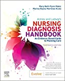
Ackley and Ladwig’s Nursing Diagnosis Handbook: An Evidence-Based Guide to Planning Care
We love this book because of its evidence-based approach to nursing interventions. This care plan handbook uses an easy, three-step system to guide you through client assessment, nursing diagnosis, and care planning. Includes step-by-step instructions showing how to implement care and evaluate outcomes, and help you build skills in diagnostic reasoning and critical thinking.
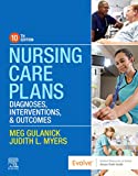
Nursing Care Plans – Nursing Diagnosis & Intervention (10th Edition)
Includes over two hundred care plans that reflect the most recent evidence-based guidelines. New to this edition are ICNP diagnoses, care plans on LGBTQ health issues, and on electrolytes and acid-base balance.
Nurse’s Pocket Guide: Diagnoses, Prioritized Interventions, and Rationales
Quick-reference tool includes all you need to identify the correct diagnoses for efficient patient care planning. The sixteenth edition includes the most recent nursing diagnoses and interventions and an alphabetized listing of nursing diagnoses covering more than 400 disorders.

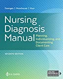
Nursing Diagnosis Manual: Planning, Individualizing, and Documenting Client Care
Identify interventions to plan, individualize, and document care for more than 800 diseases and disorders. Only in the Nursing Diagnosis Manual will you find for each diagnosis subjectively and objectively – sample clinical applications, prioritized action/interventions with rationales – a documentation section, and much more!
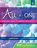
All-in-One Nursing Care Planning Resource – E-Book: Medical-Surgical, Pediatric, Maternity, and Psychiatric-Mental Health
Includes over 100 care plans for medical-surgical, maternity/OB, pediatrics, and psychiatric and mental health. Interprofessional “patient problems” focus familiarizes you with how to speak to patients.
See also
Other recommended site resources for this nursing care plan:
- Nursing Care Plans (NCP): Ultimate Guide and Database MUST READ!
Over 150+ nursing care plans for different diseases and conditions. Includes our easy-to-follow guide on how to create nursing care plans from scratch. - Nursing Diagnosis Guide and List: All You Need to Know to Master Diagnosing
Our comprehensive guide on how to create and write diagnostic labels. Includes detailed nursing care plan guides for common nursing diagnostic labels.
References
- Berman, A., Snyder, S., & Frandsen, G. (2016). Kozier and Erb’s Fundamentals of Nursing. Pearson.
- Hafen, B. B., & Sharma, S. (2022). Oxygen Saturation – Abstract. Europe PMC.
- Hariri, G., Joffre, J., Leblanc, G., Bonsey, M., Lavillegrand, J.-R., Urbina, T., Guidet, B., Maury, E., Bakker, J., & Ait-Oufella, H. (2019). Narrative review: clinical assessment of peripheral tissue perfusion in septic shock. Annals of Intensive Care, 9(37).
- Hinkle, J. L., & Cheever, K. H. (2018). Brunner & Suddarth’s Textbook of Medical-surgical Nursing. Wolters Kluwer.
- Jauch, E. C., & Lutsep, H. L. (2022, July 14). Ischemic Stroke Treatment & Management: Approach Considerations, Emergency Response and Transport, Acute Management of Stroke. Medscape Reference.
- Kramer, A. H., Couillard, P. L., Zygun, D. A., Aries, M. J., & Gallagher, C. N. (2019). Continuous Assessment of“Optimal” Cerebral Perfusion Pressure in Traumatic Brain Injury: A Cohort Study of Feasibility, Reliability, and Relation to Outcome. Neurocritical Care, 30.
- MA, S. M. (2023, April 3). Cerebral Perfusion Pressure – StatPearls. NCBI.
- Patel, K., & Chun, L. J. (2019, June 5). Deep Venous Thrombosis (DVT): Practice Essentials, Background, Anatomy. Medscape Reference.
- Rangel, L., & Kopell, B. H. (2022, May 4). Closed Head Injury: Practice Essentials, Pathophysiology, Epidemiology. Medscape Reference.
- Ren, X., & Lenneman, A. (2019, August 6). Cardiogenic Shock: Practice Essentials, Background, Pathophysiology. Medscape Reference.
- Stephens, E., & Schraga, E. D. (2022, May 24). Peripheral Vascular Disease: Background, Pathophysiology, Prognosis. Medscape Reference.
- Udeani, J., & Geibel, J. (2018, September 12). Hemorrhagic Shock: Background, Pathophysiology, Epidemiology. Medscape Reference.
- Vincent, J.-L. (2019). Fluid management in the critically ill. Kidney International, 96(1).
- Wang, X., & Liu, D. (2021). Hemodynamic Influences on Mesenteric Blood Flow in Shock Conditions. The American Journal of the Medical Sciences, 362(3).
- Workeneh, B. T., & Batuman, V. (2022, December 10). Acute Kidney Injury (AKI): Practice Essentials, Background, Pathophysiology. Medscape Reference.
- Zhang, J. L., & Lee, V. S. (2019). Renal Perfusion Imaging by MRI. Journal of Magnetic Resonance Imaging, 52(2).














Leave a Comment