Utilize this comprehensive nursing care plan and management guide to provide effective care for patients with pulmonary embolism. Gain valuable insights on nursing assessment, interventions, goals, and nursing diagnosis specifically tailored for pulmonary embolism in this guide.
What is pulmonary embolism?
Pulmonary embolism (PE) occurs when a blood clot (thrombus) becomes lodged in an artery in the lung and blocks blood flow to the lung (Ouellette & Mosenifar, 2020). In deep vein thrombosis (DVT), a thrombus develops within the deep veins, most commonly in the lower extremities. PE starts when a part of this thrombus breaks off and enters the pulmonary circulation. PE is a common and potentially lethal condition. Most clients who succumb to PE do so within the first few hours of the event (Ouellette & Mosenifar, 2020).
When a pulmonary artery is partially or completely obstructed, ventilation occurs without proper blood flow, leading to impaired gas exchange. The release of substances from the clot and surrounding area causes constriction of blood vessels, increasing pulmonary vascular resistance. This raises pulmonary arterial pressure and can lead to right ventricular failure, decreased cardiac output, and shock. Atrial fibrillation can also contribute to PE by causing clot formation in the right atrium. Massive PE is characterized by hemodynamic instability, while multiple small emboli can result in lung infarctions.
When a pulmonary embolism is identified, it is characterized as acute or chronic and central or peripheral.
- Acute pulmonary embolism. An embolus is acute if it is situated centrally within the vascular lumen or if it occludes a vessel. It causes distention of the affected vessel.
- Chronic pulmonary embolism. An embolus is chronic if it is eccentric and contiguous with the vessel wall, it reduces the arterial diameter by more than 50%, evidence of recanalization within the thrombus is present, and an arterial web is present.
- Central pulmonary embolism. Central vascular zones include the main pulmonary artery, the left upper lobe trunk, the right middle lobe artery, and the right and left lower lobe arteries. A PE is characterized as massive when it involves both pulmonary arteries or when it results in hemodynamic compromise.
- Peripheral pulmonary embolism. Peripheral vascular zones include the segmental and subsegmental arteries of the right upper lobe, the right middle lobe, the right lower lobe, the left upper lobe, the lingula, and the left lower lobe.
The clinical symptoms depend on the size and location of the embolus. The classic presentation of PE is the abrupt onset of pleuritic chest pain, shortness of breath, and hypoxia. However, most clients with PE have no obvious symptoms at presentation. Pulmonary embolism is not a disease in and of itself. Rather, it is a complication of underlying venous thrombosis (Ouellette & Mosenifar, 2020).
Careful analysis of risk factors aids in diagnosis; these include hypercoagulability, damage to the walls of the veins, prolonged immobility, recent surgery, deep vein thrombosis, postpartum state, and medical conditions such as polycythemia, heart failure, and trauma. Treatment approaches vary depending on the degree of cardiopulmonary compromise associated with PE. They can range from thrombolytic therapy in acute cases to anticoagulant therapy and general measures to optimize respiratory and vascular status (e.g., oxygen therapy and compression stockings).
Pulmonary embolism is a frequent hospital-acquired condition and one of hospitalized clients’ most common causes of death. Preventing thrombus formation is a critical nursing role.
Nursing Care Plans & Management
Nursing care planning and management for patients with pulmonary embolism (PE) include ensuring patient safety, optimizing gas exchange, preventing complications, and providing emotional support. This involves monitoring vital signs, administering supplemental oxygen, managing pain, implementing DVT prevention measures, providing education on medication compliance and follow-up care, collaborating with the healthcare team, and monitoring for complications.
Nursing Problem Priorities
The following are the nursing priorities for patients with pulmonary embolism:
- Minimizing risk for pulmonary embolism.
- Assessing potential for pulmonary embolism.
- Monitoring thrombolytic therapy.
- Preventing formation of thrombus.
Nursing Assessment
The symptoms of pulmonary embolism (PE) can vary depending on the size of the blood clot and the affected area of the pulmonary artery. Dyspnea (difficulty breathing) is the most common symptom, with its intensity and duration dependent on the extent of the embolism. Chest pain, often sudden and pleuritic, is also common and can mimic angina or a heart attack. Other symptoms include anxiety, fever, rapid heartbeat, cough, sweating, coughing up blood, and fainting. Tachypnea (rapid breathing) is the most frequent sign. PE can sometimes present with few symptoms or mimic other cardiopulmonary disorders, but severe cases can result in severe dyspnea, sudden chest pain, rapid weak pulse, shock, syncope, and even sudden death.
Assess for the following subjective and objective data:
- Confusion
- Decreased PaO2 and increased PaCO2
- Desaturation (Oxygen saturation below 90%)
- Dyspnea
- Headache
- Hypercapnia
- Hypoxia
- Pale skin
- Restlessness/Irritability
- Tachypnea
- Abnormal arterial blood gasses (ABGs)
- Impaired chest excursions
- Restlessness
- Tachycardia
- Use of accessory muscles
Assess for factors related to the cause of pulmonary embolism:
- Decreased lung perfusion caused by the obstruction of pulmonary arterial blood flow by the embolus
- Decreased bronchial airflow associated with bronchoconstriction
- Increased physiological shunting caused by the collapse of alveoli resulting from surfactant loss
- Increased alveolar dead space
- Anxiety and fear
Nursing Diagnosis
Following a thorough assessment, a nursing diagnosis is formulated to specifically address the challenges associated with pulmonary embolism based on the nurse’s clinical judgement and understanding of the patient’s unique health condition. While nursing diagnoses serve as a framework for organizing care, their usefulness may vary in different clinical situations. In real-life clinical settings, it is important to note that the use of specific nursing diagnostic labels may not be as prominent or commonly utilized as other components of the care plan. It is ultimately the nurse’s clinical expertise and judgment that shape the care plan to meet the unique needs of each patient, prioritizing their health concerns and priorities.
Nursing Goals
Goals and expected outcomes may include:
- The client will maintain adequate gas exchange, as evidenced by ABGs within the normal range, oxygen saturation of 90% or greater, alert response mentation or no further deterioration on the level of consciousness, and baseline HR for the client.
- The client will report or display resolution or absence of symptoms of respiratory distress.
- The client will maintain an effective breathing pattern, as evidenced by relaxed breathing at a normal rate and depth, and the absence of dyspnea.
- The client will verbalize understanding of desired content: the importance of medications, signs of excessive anticoagulation, and means to reduce the risk for bleeding and recurrence of emboli.
- The client will participate in the learning process.
Nursing Interventions and Actions
Therapeutic interventions and nursing actions for patients with Here are some common nursing diagnosis for pulmonary embolism: may include:
1. Minimizing Pulmonary Embolism Risk
Nurses play a vital role in preventing blood clot formation by promoting mobility, educating patients on leg exercises, and discouraging prolonged sitting or lying. They also emphasize the use of intermittent pneumatic compression (IPC) devices and proper fitting of inflatable sleeves to enhance leg blood flow. Nurses prioritize avoiding prolonged IV catheter placement to reduce the risk of clot formation. By implementing these interventions, nurses help prevent venous stasis and minimize the risk of blood clots in patients.
1. Monitor vital signs. Regularly assess and monitor the patient’s blood pressure, heart rate, respiratory rate, and oxygen saturation levels. This helps detect any signs of hemodynamic instability or respiratory distress.
Monitoring vital signs provides important information about the patient’s cardiovascular and respiratory status, allowing for early detection and prompt intervention in case of any deterioration.
2. Administer oxygen therapy. Provide supplemental oxygen as prescribed to maintain adequate oxygenation and alleviate respiratory distress.
Oxygen therapy helps improve oxygen levels in the blood, alleviates hypoxemia, and reduces the workload on the heart and lungs.
3. Assist with thrombolytic therapy. Collaborate with the healthcare team to administer thrombolytic medications as prescribed to dissolve blood clots and restore blood flow in the pulmonary arteries.
Thrombolytic therapy is often used in cases of severe pulmonary embolism to rapidly dissolve clots and improve pulmonary circulation, reducing the risk of further complications.
4. Support anticoagulant therapy. Monitor and facilitate the administration of anticoagulant medications, such as heparin or warfarin, as prescribed to prevent the formation of new blood clots and promote clot dissolution.
Anticoagulant therapy is crucial in preventing the progression of pulmonary embolism and reducing the risk of future clot formation.
5. Promote mobility and ambulation. Encourage early mobilization and ambulation as tolerated, and provide assistance as needed.
Promoting mobility helps prevent venous stasis, enhances blood flow, and reduces the risk of new clot formation. Early ambulation also aids in preventing complications associated with prolonged immobility.
6. Educate on medication adherence and side effects. Provide thorough education to the patient and family members about the prescribed medications, including the importance of adherence, potential side effects, and the need for regular monitoring of blood parameters.
Patient education ensures understanding of the treatment plan, promotes compliance with medication regimens, and empowers patients to actively participate in their care.
7. Implement deep breathing and coughing exercises. Instruct and assist the patient in performing deep breathing and coughing exercises to maintain effective gas exchange and prevent respiratory complications.
Deep breathing and coughing exercises help expand the lungs, mobilize secretions, and prevent atelectasis, thereby improving respiratory function and reducing the risk of pneumonia.
8. Provide emotional support and reassurance. Offer psychological support, address anxiety or fear, and provide emotional reassurance to the patient and family members during the recovery process.
Pulmonary embolism can be a traumatic experience for patients and their families. Emotional support helps reduce stress, promotes a positive mindset, and enhances overall well-being.
2. Managing Effective Gas Exchange & Oxygen Therapy
Minimizing the risk of pulmonary embolism (PE) and promoting effective gas exchange is of utmost importance in the management of patients diagnosed with this serious medical condition. One of the critical responsibilities of the nurse is to recognize patients who are at a high risk for pulmonary embolism (PE) and take steps to minimize the risk of PE in all patients. The nurse must maintain a heightened level of awareness for the possibility of PE in all patients, especially those with conditions that can lead to a reduced venous return.
1. Assess the skin color, nail beds, and mucous membranes for color changes.
Cool, pale skin occurs as a compensatory response to hypoxemia. When oxygen and perfusion become impaired, peripheral tissues become cyanotic. General “duskiness” and cyanosis in “warm tissues” such as earlobes, lips, tongue, and buccal membranes may indicate systemic hypoxemia.
2. Monitor for any changes in vital signs.
In initial hypoxia and hypercapnia, there is an increase in the respiratory rate, heart rate, and blood pressure. As the hypercapnia and hypoxia get worse, blood pressure may drop, heart rate tends to continue to be rapid and includes dysrhythmias, and respiratory failure ensues, with the client unable to maintain a rapid respiratory rate.
3. Assess for the signs and symptoms of hypoxia (such as confusion, headache, diaphoresis, restlessness, tachycardia, and pale skin).
Hypoxia results from increased dead space (ventilation without perfusion) that reduces effective gas exchange. Arterial hypoxemia is also a frequent, but not universal, finding in clients with acute embolism. The mechanisms of hypoxemia include ventilation-perfusion mismatch, intrapulmonary shunts, reduced cardiac output, and intracardiac shunt via a patent foramen ovale (Ouellette & Mosenifar, 2020).
4. Auscultate lung sounds, noting areas of decreased ventilation, and the presence of adventitious sounds.
Crackles are common clinical findings with pulmonary embolism. Crackles occur in fluid-filled tissues and airways or may reflect cardiac decompensation. Nonventilated areas may be identified by the absence of breath sounds.
5. Assess for the signs and symptoms of pulmonary infarction (such as fever, cough, bronchial breathing, hemoptysis, pleuritic pain, pleural friction rub, and consolidation).
A large pulmonary embolus or multiple small clots in a specific area of the lung can cause ischemic necrosis or infarction of the lung area. These clients present with acute onset of pleuritic chest pain, breathlessness, and hemoptysis. Although the chest pain may be clinically indistinguishable from ischemic myocardial pain, normal ECG findings and no response to nitroglycerin rule out myocardial pain (Ouellette & Mosenifar, 2020).
6. Assess for calf tenderness, redness, swelling, and hardened areas.
Pulmonary embolism often arises from a deep vein thrombosis and may have been previously overlooked. After traveling to the lung, large thrombi can lodge at the bifurcation of the main pulmonary artery or the lobar branches and cause hemodynamic compromise (Ouellette & Mosenifar, 2020).
7. Monitor for any changes in the ABGs.
ABG analysis characteristically reveals hypoxemia, hypocapnia, and respiratory alkalosis. The PaO2 and the calculation of the alveolar-arterial oxygen gradient contribute to the diagnosis in a general population thought to have a pulmonary embolism (Ouellette & Mosenifar, 2020).
8. Monitor oxygen saturation as indicated.
Pulse oximetry is a useful tool in the clinical setting to detect changes in oxygenation. Oxygen saturation should be at 90% or greater. Hypoxemia is present in varying degrees, depending on the amount of airway obstruction, usual cardiopulmonary function, and presence and degree of shock.
9. Assess the level of consciousness and evaluate mentation changes.
Systemic hypoxemia may be demonstrated initially by restlessness and irritability, then by progressively decreasing mentation. Syncope may occur and may be associated with a higher prevalence of hemodynamic instability and right ventricular dysfunction (Vyas & Goyal, 2022).
10. Assess activity intolerance, such as reports of weakness and fatigue, vital sign changes, or increased dyspnea during exertion.
These parameters assist in determining the client’s response to resumed activities and the ability to participate in self-care.
11. Maintain the client on bed rest. May resume activity gradually as tolerated.
This will decrease the oxygen demand during an episode of acute respiratory distress. Activity is recommended as tolerated, with early ambulation preferred over bed rest when feasible (Ouellette & Mosenifar, 2020).
12. Position the client properly to facilitate ventilation-perfusion matching.
Upright and sitting positions optimize diaphragmatic excursions and lung perfusion. When the client is positioned on one side, the affected area should not be dependent. Elevate the head of the bed as tolerated to promote maximal chest expansion and enhance the client’s comfort.
13. Maintain patent airways.
Institute interventions to maintain a patent airway such as deep breathing exercises, coughing, and suctioning. Plugged or collapsed airways reduce the number of functional alveoli, negatively affecting gas exchange.
14. Assist the client in dealing with fear and anxiety that may be present.
Feelings of fear and severe anxiety are associated with the inability to breathe and may actually increase oxygen consumption and demand. Encourage the client to express their feelings so that the client may regain a sense of control over their emotions.
15. Monitor frequently and arrange for someone to stay with the client as appropriate. Provide information as indicated.
This provides assurance that changes in condition will be noted and that assistance is readily available. Provide brief explanations of what is happening and the expected effects of the interventions to allay the client’s anxiety.
16. Ensure appropriate application of compression stockings.
For clients who have had a proximal DVT, the use of elastic compression stockings provides a safe and effective adjunctive treatment that can limit postphlebitic syndrome. True gradient compression stockings are highly elastic. Compression stockings of this type have been proven effective in the prophylaxis of thromboembolism and are also effective in preventing the progression of a blood clot in clients who already have DVT and PE (Ouellette & Mosenifar, 2020).
17. Prepare the client for a lung scan.
This may reveal a pattern of abnormal perfusion in areas of ventilation, reflecting ventilation and perfusion mismatch, confirming the diagnosis of PE and degree of obstruction.
18. Administer IV fluids or fluids by mouth as indicated.
Increased fluids may be given to reduce hyperviscosity of the blood, which can potentiate thrombus formation. Be aware that IV fluids may help or hurt the client who is hypotensive from PE, depending on which point on the Starling curve describes the client’s condition. A cautious trial of a small fluid bolus may be attempted, with careful surveillance of the systolic and diastolic blood pressures and immediate cessation if the situation worsens after the fluid bolus (Ouellette & Mosenifar, 2020).
19. Administer oxygen as indicated.
Supplemental oxygen maintains adequate oxygenation, decreases the work of breathing, relieves dyspnea, and promotes comfort. The appropriate amount of oxygen needs to be continuously delivered so the client does not become desaturated. Mechanical ventilation may be necessary to provide respiratory support and as adjunctive therapy for a failing circulatory system (Ouellette & Mosenifar, 2020).
20. Anticipate the need to start anticoagulant therapy and, if there is massive thromboembolism, the use of thrombolytic therapy.
Heparin or enoxaparin (Lovenox) is used to prevent the recurrence of emboli. These medications do not dissolve clots that already exist. If a massive thrombus is present or the client is hemodynamically unstable, thrombolytic therapy (e.g., alteplase or reteplase [Retavase]) is used to directly lyse or dissolve the clot. The current recommendation is anticoagulation for at least three months; the need for extending the duration of anticoagulation should be reevaluated at that time (Ouellette & Mosenifar, 2020).
21. Assist with chest physiotherapy techniques, such as postural drainage and percussion of nonaffected areas, blow bottles, and incentive spirometry.
This facilitates deeper respiratory effort and promotes drainage of secretions from lung segments into bronchi, where they may be more readily removed by coughing or suctioning.
22. Assist in surgical interventions as indicated.
Vena caval ligation or insertion of an intracaval umbrella may be useful for clients with recurrent emboli despite adequate anticoagulation. Either catheter embolectomy and fragmentation or surgical embolectomy is reasonable for clients with massive PE who have contraindications to fibrinolysis or who remain unstable after receiving fibrinolysis (Ouellette & Mosenifar, 2020).
3. Maintaining Patent Airway Clearance and Effective Breathing Pattern
Patent airway clearance is essential to prevent the accumulation of secretions, optimize oxygenation, and minimize the risk of respiratory complications. Nurses play a vital role in assessing and maintaining a patent airway, ensuring that the patient’s airway remains unobstructed and promoting the clearance of any accumulated secretions. In addition to airway clearance, promoting an effective breathing pattern is crucial for patients with pulmonary embolism. Prompt recognition of any abnormalities allows for timely intervention and support to optimize breathing and ventilation.
1. Assess the client’s anxiety level.
Pulmonary embolism is a sudden acute condition that can produce anxiety. Anxiety can result in rapid, shallow respirations and increased dyspnea. It can be a sign of decreasing hypoxemia. Impaired mental well-being in young adults with venous thromboembolism has been linked largely to uncertainty regarding their long-term health and fear of recurrence. Clients diagnosed with PE have been shown to have higher depression and anxiety scores (Tran et al., 2021).
2. Assess the respiratory rate, rhythm, and depth. Assess for any increase in the work of breathing: shortness of breath and the use of accessory muscles.
Respiratory rate and rhythm changes are early signs of impending respiratory distress. Tachypnea is a typical finding of pulmonary embolism (PE). The rapid, shallow respirations result from hypoxia. The development of hypoventilation (slowing of respiratory rate) without improvement in the client’s condition indicates respiratory failure. When the client complains of frank exertional dyspnea, an increase in pulmonary arterial pressure is expected (Sanchez et al., 2016).
3. Assess the characteristics of pain, especially in association with the respiratory cycle.
Pain is usually sharp or stabbing and gets worse with deep breathing and coughing. It can result in shallow respirations, further impairing effective gas exchange. Pleuritic or respirophasic chest pain is a particularly worrisome symptom. Pleuritic chest pain is reported to occur in as many as 84% of clients with PE. Its presence suggests that the embolus is located more peripherally and thus may be smaller (Ouellette & Mosenifar, 2020).
4. Monitor arterial blood gasses (ABGs).
ABGs of these clients typically exhibit hypoxemia and respiratory alkalosis from a blowing off of carbon dioxide. Widened alveolar-arterial gradients for oxygen, respiratory alkalosis, and hypocapnia are commonly seen findings on ABG, as a pathophysiological response to PE (Vyas & Goyal, 2022).
5. Monitor D-dimer results as indicated.
D-dimer levels are elevated in plasma whenever there is an acute thrombotic process in the body because of the activation of coagulation and fibrinolysis pathways at the same time. A negative ELISA-D-dimer, along with low clinical probability, can exclude PE without further testing in approximately 30% of suspected clients (Vyas & Goyal, 2022).
6. Monitor vital signs continuously.
Increased respirations, tachycardia, and/or bradycardia suggest hypoxia. If a client with PE who has tachycardia on presentation develops sudden bradycardia or develops a new broad complex tachycardia, look for signs of right ventricular strain and possible impending shock. Pe should also be suspected in anyone with hypotension with jugular vein distention wherein myocardial infarction, pericardial tamponade, or tension pneumothorax has been ruled out (Vyas & Goyal, 2022).
7. Provide reassurance and allay anxiety by staying with the client during acute episodes of respiratory distress.
The presence of a trusted person may be helpful during periods of anxiety. Having psychologically distressing symptoms and receiving a diagnosis of PE, which almost inevitably comes with some physiological explanation of PE, can lead some clients to believe that they have narrowly escaped death (Tran et al., 2021).
8. Position the client in a sitting position, and change the position every 2 hours.
If not contraindicated, a sitting position allows a good lung excursion and chest expansion. Repositioning facilitates movement and the drainage of secretions. Additionally, elevating the head of the bed facilitates ventilation and mobilization of secretions.
9. Encourage deep breathing and coughing exercise. Suction as indicated.
Coughing is the most productive way to remove secretions. The client may be unable to perform independently. Suctioning is indicated when clients are unable to remove secretions from the airways by coughing. These maneuvers help keep airways open by clearing secretions.
10. Reinforce splinting of the chest with pillows during deep breathing or coughing.
This reduces pleuritic chest pain, promotes maximal lung expansion, and may enhance the effectiveness of cough effort.
11. Instruct and assist in incentive spirometry.
Incentive spirometry maximizes lung inflation, reduces atelectasis, and prevents pulmonary complications. This is a cost-effective intervention and has demonstrated a reduction in respiratory symptoms such as the perception of dyspnea and improved quality of life (Haukeland-Parker et al., 2021).
12. Prepare the client for diagnostic studies such as chest X-ray and computed tomography (CT) scan, ventilation-perfusion scan, and pulmonary arteriogram.
Common tests such as a chest x-ray examination are readily available in acute care settings, especially to rule out PE. A chest x-ray is usually normal or might show nonspecific abnormalities such as atelectasis or effusion. It helps to rule out alternative diagnoses in clients presenting with acute dyspnea. If there is a high suspicion of PE, then a CT scan and other scans are added to make a diagnosis. Multidetector computed tomographic pulmonary angiography (CTPA) is the diagnostic modality of choice for clients with suspected PE (Vyas & Goyal, 2022).
13. Administer oxygen as indicated.
Supplemental oxygen maintains adequate oxygenation, decreases the work of breathing, relieves dyspnea, and promotes comfort. The appropriate amount of oxygen needs to be continuously delivered so the client does not become desaturated. Supplemental oxygen is indicated in clients with an oxygen saturation of <90% (Vyas & Goyal, 2022).
14. Anticipate the need for intubation and mechanical ventilation.
Intubation and positive-pressure ventilation are means to stabilize breathing and ventilation and prevent decompensation of the client. Mechanical ventilation should be utilized in unstable clients, but healthcare professionals should be mindful of the adverse hemodynamic effects of mechanical ventilation (Vyas & Goyal, 2022).
4. Managing Bleeding Risk & Thrombolytic Therapy
The management of bleeding risk in patients with pulmonary embolism involves a multidimensional approach that considers various factors, including the patient’s individual characteristics, comorbidities, and the specific anticoagulant regimen prescribed. Healthcare professionals must balance the need for effective anticoagulation therapy with the potential risk of bleeding to ensure patient safety and optimize outcomes.
1. Assess for the history of a high-risk bleeding condition: liver disease, kidney disease, severe hypertension, cavitary tuberculosis, bacterial endocarditis, and of heparin-induced thrombocytopenia.
Because anticoagulation therapy is the hallmark treatment for PE, prior client experiences with bleeding or anticoagulants must be assessed before treatment. As with any history, the nurse must be sure to ask about any underlying medical conditions that may alter the clinical course in the setting of anticoagulant toxicity, specifically asking about alcohol use or potential hepatic disease (Deaton & Nappe, 2022).
2. Assess for the signs and symptoms of bleeding such as bleeding from catheter insertion sites, bleeding from mucous membranes, decreased hematocrit and hemoglobin, gastrointestinal bleeding, genitourinary bleeding, hematoma, petechiae, purpura, and respiratory tract bleeding.
Early recognition of these symptoms facilitates the immediate administration of the appropriate antidote. While chronic overdose resulting in toxicity presents with an elevated PT/INR, that level is expected to remain at or near its maximum value on presentation. In naive clients, the PT/INR would not be expected to have a rise for 12 hours post-ingestion and may continue to rise for several more hours due to ongoing absorption. Therefore, it is essential that early signs and symptoms be identified to avoid the worsening of the toxicity (Deaton & Nappe, 2022).
3. Monitor vital signs and compare them with the client’s previous readings before medication initiation.
Changes in BP and pulse may be used for a rough estimate of bleeding. A BP less than 90 mm Hg and a pulse greater than 110 suggest a 25% decrease in volume or approximately 1,000 ml. Postural hypotension reflects a decrease in circulating volume.
4. Monitor platelets in clients diagnosed with heparin-induced thrombocytopenia.
Severe platelet reductions can occur with heparin use, especially unfractionated heparin therapy, and are known as heparin-induced thrombocytopenia (HIT). HIT is less commonly seen with the use of low-molecular-weight heparin. HIT should be suspected whenever the client’s platelet count falls to less than 100,000/µL or less than 50% of the baseline value, generally after 5 to 15 days of heparin therapy. Platelet counts must be obtained every two or three days from day 4 to day 14 of therapy, or until heparin is stopped (Ouellette & Mosenifar, 2020).
5. Monitor coagulation test results (INR), PT, activated partial thromboplastin time [aPTT], hemoglobin, and hematocrit. Notify the healthcare provider immediately if a higher or lower than the designated range occurs.
The effects of anticoagulation therapy must be closely monitored to reduce the risk of bleeding. The type of test depends on the anticoagulation medication administered. The anticoagulant effect is best quantified by baseline and daily repeated measurement of the PT and the INR, which may not be elevated until 1 to 2 days post-ingestion. A normal PT 48 to 72 hours after ingestion rules out significant ingestion (Olson & Vearrier, 2021).
6. Monitor the IV dosage and delivery system (tubing or pump).
IV anticoagulation is administered using an electronic infusion pump. This device reduces the risk of over-coagulation or under-coagulation. If IV UFH is chosen, an initial bolus of 80 U/kg or 5000 U followed by an infusion of 18 U/kg/hour or 1300 U/hour should be given, with the goal of rapidly achieving and maintaining the aPTT at levels that correspond to therapeutic heparin levels (Ouellette & Mosenifar, 2020).
7. Administer anticoagulant therapy as prescribed (bolus, continuous IV heparin, and/or subcutaneous low-molecular-weight heparin), oral warfarin [Coumadin]).
Anticoagulants are given to prevent further thrombus formation. The type of medication varies per protocol. New non-heparin agents are also available for clients who cannot tolerate heparin. Current guidelines for clients with acute PE recommend low-molecular-weight heparin therapy (LMWH) over IV UFH and over subcutaneous UFH because of LMWH’s many advantages. They have greater bioavailability, can be administered by subcutaneous injections, and have a longer duration of anticoagulant effect (Ouellette & Mosenifar, 2020).
8. If the client is HIPA positive, stop all heparin products and consult a hematologist.
Continuation of heparin products is contraindicated in the client who is HIPA positive. In clients with HIT with or without thrombosis, the use of lepirudin, argatroban, or danaparoid is preferred over the continued use of heparin, LMWH, or either initiation or continuation of a vitamin K antagonist (Ouellette & Mosenifar, 2020).
9. Perform measures if bleeding occurs while on heparin.
The treatment of clients who develop bleeding due to anticoagulant toxicity is to stop all heparin products. In the setting of a life-threatening bleed related to vitamin K antagonist (VKA) use, rapid reversal of the VKA drug effects, and replenishing clotting factors is a priority. To achieve that goal, administer vitamin K 10 mg intravenously along with prothrombin complex concentrate or fresh frozen plasma to achieve a sustained reduction of the INR. Supportive measures and infusion of clotting factors are available options for the reversal of DOAC bleeding (Hanigan & Barnes, 2019).
10. Provide stool softeners as indicated.
Stool softeners prevent straining for stool with a resultant increase in intra abdominal pressure and the risk of bleeding hemorrhoidal varices, especially when the client has hepatic problems.
11. Convert from IV anticoagulation to oral anticoagulation after the appropriate length of therapy. Monitor the INR, PT, and aPTT levels.
The onset of anticoagulation with warfarin is 2 to 3 days. There needs to be an overlap of these medications to ensure adequate PT levels or INR levels for anticoagulation before discontinuing heparin. Parenteral anticoagulation and warfarin should be continued together for a minimum of at least five days and until the INR is 2.0. The recommended therapeutic range for VTE is an INR of 2 to 3. This level of anticoagulation markedly reduces the risk of bleeding without the loss of effectiveness (Ouellette & Mosenifar, 2020).
12. Administer thrombolytic therapy as prescribed.
Lytic agents are indicated for clients with a massive PE that results in hemodynamic compromise. Be aware of the following contraindications for thrombolytic therapy to minimize complications: recent surgery, recent organ biopsy, pregnancy, recent stroke, or recent or active internal bleeding. Do not delay thrombolysis in clients with acute PE associated with hypotension (systolic BP <90 mm Hg) because irreversible cardiogenic shock can develop. Thrombolytic therapy is not recommended for most clients with acute PE not associated with hypotension (Ouellette & Mosenifar, 2020).
13. Institute precautionary measures for thrombolytic therapy per protocol.
Preventive care is given per institutional policy. Such measures to reduce the risk of bleeding include: using only compressible vessels for IV sites; compressing IV for at least 10 minutes and arterial sites for 30 minutes; limiting physical manipulation of clients; providing gentle oral care; avoiding intramuscular (IM) injections; drawing all laboratory specimens through an existing arterial line or venous heparin-lock line; and sending the specimen to type and crossmatch.
14. Avoid administering vitamin K as prophylaxis.
Do not administer vitamin K prophylactically because it is not needed in most clients and its presence masks the onset of anticoagulant effects in the few clients who do require prolonged treatment and follow-up care. If PT is elevated, however, treatment with vitamin K is appropriate (Olson & Vearrier, 2021).
15. Administer blood and blood products if there is active bleeding.
Active, serious bleeding should be treated with four-factor prothrombin complex concentrate (PCC), if available. While costly, an essential advantage PCC confers to emergency care is that, in contrast to FFP, it results in a more rapid reversal of coagulopathy and does not require thawing or blood group typing. Alternatively, recombinant factor VIIa has been reported to be effective in rapidly lowering INR due to warfarin toxicity and may be considered if PCC is not available (Olson & Vearrier, 2021).
16. Administer appropriate antidotes if available and as indicated.
Activated charcoal is empirically used to minimize systemic absorption of the toxin if the client is taking DOACs. Vitamin K1 is used in the management of poisoning and overdose, in the prevention of toxic effects, and in metabolic disorders in which toxic substances accrue. Phytomenadione can overcome the competitive block produced by warfarin and other related anticoagulants. This is only used if evidence of anticoagulation exists (Olson & Vearrier, 2021).
5. Monitoring Diagnostic and Laboratory Procedures
To accurately diagnose and assess the extent of pulmonary embolism, healthcare providers closely monitor diagnostic procedures and laboratory studies. Regular monitoring of these diagnostic procedures and laboratory studies allows nurses to assess the effectiveness of treatment, monitor the patient’s response, and identify any complications that may arise. Timely and accurate monitoring plays a crucial role in tailoring the management plan and ensuring optimal outcomes for patients with pulmonary embolism.
1. Computed Tomography Pulmonary Angiography (CTPA).
This imaging technique uses a contrast dye and computed tomography scan to visualize the pulmonary arteries and detect blood clots.
2. Ventilation-Perfusion (V/Q) Scan.
This test involves injecting a radioactive material and a contrast dye to assess the airflow and blood flow in the lungs. It helps identify areas of mismatch between ventilation and perfusion, indicating potential pulmonary embolism.
3. D-Dimer Test.
This blood test measures the level of D-dimer, a protein fragment that is elevated when blood clots are being broken down. While a negative D-dimer test result can help rule out pulmonary embolism, a positive result requires further imaging studies for confirmation.
4. Chest X-ray.
Although not a definitive diagnostic tool for pulmonary embolism, a chest X-ray may be performed to evaluate the lungs, heart, and other structures. It helps exclude other conditions and may reveal associated findings, such as an enlarged right ventricle or an abnormal pulmonary artery shape.
5. Echocardiography.
This ultrasound-based test allows visualization of the heart’s structure and function. It can identify signs of right ventricular strain or dysfunction, which may occur as a result of pulmonary embolism.
6. Blood Gas Analysis.
This test measures the levels of oxygen and carbon dioxide in arterial blood, providing information about gas exchange and acid-base balance. It helps assess the severity of respiratory compromise associated with pulmonary embolism.
7. Electrocardiogram (ECG).
An ECG records the electrical activity of the heart. It can reveal changes such as a right bundle branch block or signs of right ventricular strain, which may indicate pulmonary embolism.
8. Lower Extremity Venous Doppler Ultrasonography.
This non-invasive imaging test assesses the blood flow in the deep veins of the legs. It can help identify the presence of deep vein thrombosis (DVT), which often precedes pulmonary embolism.
6. Administering Medications and Pharmacological Support
The management of pulmonary embolism involves the administration of medications and pharmacological support to address the underlying clotting disorder and promote optimal outcomes. The specific medications prescribed for pulmonary embolism depend on the severity of the condition, the patient’s individual characteristics, and the presence of any contraindications. It is crucial to follow the healthcare provider’s guidance and take medications as directed to optimize the management of pulmonary embolism and reduce the risk of complications.
1. Anticoagulants (e.g., Heparin, Warfarin, Direct Oral Anticoagulants).
Anticoagulant medications are the mainstay of treatment for pulmonary embolism. They help prevent the formation of new blood clots and the extension of existing clots. Heparin is often administered initially, followed by a transition to oral anticoagulants like Warfarin or Direct Oral Anticoagulants (DOACs) for long-term management.
2. Thrombolytics (e.g., Alteplase, Reteplase).
Thrombolytic medications are used in specific cases of severe pulmonary embolism to dissolve blood clots rapidly. They are typically reserved for patients with significant hemodynamic instability or a high risk of adverse outcomes.
3. Nonsteroidal Anti-inflammatory Drugs (NSAIDs).
NSAIDs such as Ibuprofen or Naproxen may be prescribed to relieve pain and inflammation associated with pulmonary embolism. They can help manage symptoms and improve the patient’s overall comfort.
4. Analgesics.
Pain medications like opioids or non-opioid analgesics (e.g., Acetaminophen) are administered to alleviate chest pain or discomfort caused by pulmonary embolism. Effective pain management can improve the patient’s quality of life and promote better breathing.
5. Oxygen Therapy.
Oxygen supplementation is often provided to patients with pulmonary embolism who experience hypoxemia (low oxygen levels). It helps maintain adequate oxygenation, relieves shortness of breath, and supports respiratory function.
6. Prophylactic Medications.
Prophylactic medications, such as low molecular weight heparin or fondaparinux, may be prescribed to individuals at risk of developing pulmonary embolism. These medications help prevent blood clot formation in high-risk situations, such as during surgery or prolonged immobility.
7. Pain Medications.
Non-opioid analgesics, such as Acetaminophen, may be used to manage pain associated with pulmonary embolism. They help alleviate discomfort and improve the patient’s overall well-being.
7. Providing Patient Education & Health Teachings
Patient education empowers individuals with the knowledge and skills necessary to actively participate in their own care, make informed decisions, and adopt lifestyle modifications to promote recovery and reduce the risk of future embolisms. The primary goals of patient education and health teachings for individuals with pulmonary embolism are to enhance their understanding of the condition, its causes, and potential risk factors.
1. Assess the client’s knowledge of pulmonary embolism: its severity, prognosis, risk factors, and therapy.
Pulmonary embolism can be a sudden acute condition for which the client has no prior experience. An assessment provides an important starting point in education. In clients who survive a pulmonary embolism, recurrent embolism and death can be prevented with prompt diagnosis, therapy, and adequate client information about the disease process (Ouellette & Mosenifar, 2020).
2. Assess the client’s knowledge about anticoagulant therapy and related complications that may arise from non-adherence.
Evidence in studies shows that many doses of prescribed pharmacologic VTE prophylaxis are not administered and are most often attributed to client refusal or poor nurse-client communication. Missing even one dose of VTE prophylaxis has been associated with developing VTE events, therefore, client education about adherence to medications is imperative to help the client understand the consequences of their actions (Haut et al., 2022).
3. Provide information on the cause of the problem, common risk factors, and effects of PE on body functioning.
Preventing thrombus formation is an ongoing concern. An informed client is more likely to avoid common risk factors. Clinical signs and symptoms for pulmonary embolism are nonspecific; therefore, clients suspected of having PE- because of unexplained dyspnea, tachypnea, or chest pain or the presence of risk factors for PE- must undergo diagnostic tests until the diagnosis is ascertained or eliminated or an alternative diagnosis is confirmed (Ouellette & Mosenifar, 2020).
4. Explain the purpose of activity restrictions and the need for balance between activity and rest. Establish appropriate exercise and activity programs.
Rest reduces the oxygen and nutrient needs of compromised tissues and decreases the risk of fragmentation of thrombosis. Balancing rest with activity prevents exhaustion and further impairment of cellular perfusion. An appropriate exercise and activity program aids in developing collateral circulation, enhances venous return, and prevents recurrence.
5. Problem-solve solutions to predisposing factors that may be present, such as employment that requires prolonged standing or sitting, wearing restrictive clothing, use of oral contraceptives, obesity, prolonged immobility, and dehydration.
This actively involves the client in identifying and initiating lifestyle and behavior changes to promote health and prevent the occurrence of conditions or the development of complications. Immobilization leads to venous stasis by the accumulation of clotting factors and fibrin, resulting in blood clot formation. Estrogen-containing birth control pills have increased the occurrence of VTE in healthy women (Ouellette & Mosenifar, 2020).
6. Instruct the client to avoid sitting cross-legged for prolonged periods of time.
Recommend to the client to sit with feet touching the floor instead of crossing of legs to prevent excess pressure on the popliteal space, which can dislodge a thrombus.
7. Review the purpose and demonstrate the correct application and removal of the anti-embolic hose.
Understanding may enhance cooperation with prescribed therapy and prevent improper or ineffective use. Stockings with a pressure of 30 to 40 mm Hg at the ankle, worn for two years following diagnosis, are recommended to reduce the risk of the postphlebitic syndrome. The ubiquitous white stockings known as anti-embolic stockings or “Ted hose” produce a maximum compression of 18 mm Hg (Ouellette & Mosenifar, 2020).
8. Instruct in the meticulous skin care of lower extremities.
Chronic venous congestion and postphlebitic syndrome may develop, especially in presence of severe vascular involvement and recurrent DVT, potentiating the risk of stasis ulcers. Prevent or promptly treat breaks in the skin and report the development of ulcers or changes in skin color to avoid complications.
9. Inform the client of the need for routine laboratory testing while on oral anticoagulation.
Continued regular assessment of anticoagulation is necessary to prevent both recurrences of clots and active bleeding. Understanding that close supervision of anticoagulant therapy is necessary because the therapeutic dosage is narrow and complications may prove fatal promotes client participation. Partial thromboplastin (PT) should be measured on a regular basis; the goal is an international normalized ratio (INR) of 2 to 3 (Ouellette & Mosenifar, 2020).
10. Instruct the client about medications and their actions, dosages, and side effects.
Clients may require anticoagulation for weeks, months, or more, depending on their risks. Accurate knowledge reduces future complications. Long-term anticoagulation is essential for clients who survive an initial DVT or PE. The optimum total duration of anticoagulation is controversial, but general consensus holds that at least six months of anticoagulation is associated with a significant reduction in recurrences and a net positive benefit (Ouellette & Mosenifar, 2020).
11. Discuss the use of a medical alert bracelet or other identification.
These forms of identification alert others of the client’s anticoagulation history to facilitate safe, effective medical care. These forms of identification alert emergency healthcare providers to the history of thrombotic problems or current use of or need for anticoagulants such as prophylactic before and after any procedure or events with an increased risk of VTE.
12. Discuss drug, herb, alcohol, and food interactions with the anticoagulants. Emphasize that significant diet changes and all over-the-counter medications and complementary therapies need to be discussed with the healthcare provider or nurse practitioner before initiation.
Food, dietary supplements, or herbs may contain substances that, when administered concomitantly with direct oral anticoagulants (DOACs), can potentially affect the plasma concentration of the drugs. Clients treated with anticoagulants should avoid products containing St. John’s wort foods rich in vitamin K, such as lettuce, broccoli, spinach, and green peas because it is associated with a risk of reduced therapeutic effect of vitamin K antagonists (VKA). Administering dabigatran while fasting may increase the adverse effects of the drug on the digestive system, which may lead to treatment discontinuation. Studies on interactions between apixaban, edoxaban, dabigatran, and individual macronutrients showed that the presence of insoluble and soluble fiber as well as cellulose, might cause a decrease in the bioavailability of these anticoagulant agents (Grzesk et al., 2021).
13. Educate the client about the signs and symptoms of anticoagulant toxicity such as severe nose bleeding, black stools, blood in urine or stools, joint swelling/pain, hemoptysis, and severe headache.
The client may need to self-manage their condition. The early assessment facilitates prompt treatment. It is important to ask about any signs and symptoms of abnormal bleeding because this will be an important piece of information to guide the appropriate clinical treatment (Deaton & Nappe, 2022).
14. Identify safety precautions that prevent the occurrence of bleeding.
Educate the client about measures to avoid bleeding or injury such as using a soft toothbrush, an electric razor for shaving, and gloves for gardening; avoiding sharp objects including toothpicks, walking barefoot, engaging in rough sports and activities, or forceful blowing of the nose. These measures reduce the risk of traumatic injury, which potentiates bleeding or clot formation.
15. If the client is heparin-induced platelet aggregation (HIPA) positive, instruct about the importance of avoiding heparin.
Heparin use can result in the formation of anti-heparin antibodies, which puts the client at risk. The treatment of clients who develop heparin-induced thrombocytopenia (HIT) is to stop all heparin products, including catheter flushes and heparin-coated catheters, and to initiate an alternative, non-heparin anticoagulant, even when thrombosis is not clinically apparent (Ouellette & Mosenifar, 2020).
16. Explain the need for a vena cava filter device if clotting is a chronic problem.
In high-risk clients, this filter and/or interruption device can trap a thrombus migrating from a deep vein thrombosis (DVT) in the leg. However, clients with acute PE should not routinely receive vena cava filters in addition to anticoagulants. After the placement of an IVC filter, anticoagulation should be resumed once contraindications to anticoagulation or active bleeding complications have resolved (Ouellette & Mosenifar, 2020).
Recommended Resources
Recommended nursing diagnosis and nursing care plan books and resources.
Disclosure: Included below are affiliate links from Amazon at no additional cost from you. We may earn a small commission from your purchase. For more information, check out our privacy policy.
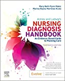
Ackley and Ladwig’s Nursing Diagnosis Handbook: An Evidence-Based Guide to Planning Care
We love this book because of its evidence-based approach to nursing interventions. This care plan handbook uses an easy, three-step system to guide you through client assessment, nursing diagnosis, and care planning. Includes step-by-step instructions showing how to implement care and evaluate outcomes, and help you build skills in diagnostic reasoning and critical thinking.
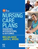
Nursing Care Plans – Nursing Diagnosis & Intervention (10th Edition)
Includes over two hundred care plans that reflect the most recent evidence-based guidelines. New to this edition are ICNP diagnoses, care plans on LGBTQ health issues, and on electrolytes and acid-base balance.
Nurse’s Pocket Guide: Diagnoses, Prioritized Interventions, and Rationales
Quick-reference tool includes all you need to identify the correct diagnoses for efficient patient care planning. The sixteenth edition includes the most recent nursing diagnoses and interventions and an alphabetized listing of nursing diagnoses covering more than 400 disorders.

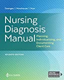
Nursing Diagnosis Manual: Planning, Individualizing, and Documenting Client Care
Identify interventions to plan, individualize, and document care for more than 800 diseases and disorders. Only in the Nursing Diagnosis Manual will you find for each diagnosis subjectively and objectively – sample clinical applications, prioritized action/interventions with rationales – a documentation section, and much more!
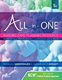
All-in-One Nursing Care Planning Resource – E-Book: Medical-Surgical, Pediatric, Maternity, and Psychiatric-Mental Health
Includes over 100 care plans for medical-surgical, maternity/OB, pediatrics, and psychiatric and mental health. Interprofessional “patient problems” focus familiarizes you with how to speak to patients.
See Also
Other recommended site resources for this nursing care plan:
- Nursing Care Plans (NCP): Ultimate Guide and Database MUST READ!
Over 150+ nursing care plans for different diseases and conditions. Includes our easy-to-follow guide on how to create nursing care plans from scratch. - Nursing Diagnosis Guide and List: All You Need to Know to Master Diagnosing
Our comprehensive guide on how to create and write diagnostic labels. Includes detailed nursing care plan guides for common nursing diagnostic labels.
Other nursing care plans related to respiratory system disorders:
- Asthma
- Aspiration Risk & Aspiration Pneumonia
- Airway Clearance Therapy & Coughing
- Bronchiolitis
- Bronchopulmonary Dysplasia (BPD)
- Chronic Obstructive Pulmonary Disease (COPD)
- Cystic Fibrosis
- Hemothorax and Pneumothorax
- Influenza (Flu)
- Ineffective Breathing Pattern (Dyspnea)
- Impairment of Gas Exchange
- Lung Cancer
- Mechanical Ventilation
- Near-Drowning
- Pleural Effusion
- Pneumonia
- Pulmonary Embolism
- Pulmonary Tuberculosis
- Tracheostomy
References and Sources
Recommended journals, books, and other interesting materials to help you learn more about pulmonary embolism nursing care plans and nursing diagnosis:
- Deaton, J. G., & Nappe, T. M. (2022, July 18). Warfarin Toxicity – StatPearls. NCBI. Retrieved January 18, 2023.
- Doenges, M. E., Moorhouse, M. F., & Murr, A. C. (2010). Nursing Care Plans: Guidelines for Individualizing Client Care Across the Life Span. F.A. Davis Company.
- Fernandes, C.J., Luppino Assad, A.P., Alves-Jr., J.L., Jardim, C., & de Souza, R. (2019). Pulmonary Embolism and Gas Exchange. Respiration, 98.
- Grzesk, G., Rogowicz, D., Wolowiec, L., Ratajczak, A., Gilewski, W., Chudzinska, M., Sinkiewicz, A., & Banach, J. (2021, August 8). The Clinical Significance of Drug–Food Interactions of Direct Oral Anticoagulants. NCBI. Retrieved January 18, 2023.
- Hanigan, S., & Barnes, G. D. (2019, October). Managing Anticoagulant-related Bleeding in Patients with Venous Thromboembolism. American College of Cardiology.
- Haukeland-Parker, S., Jervan, O., Johannessen, H. H., Gleditsch, J., Stavem, K., Steine, K., Spruit, M. A., Holst, R., Tavoly, M., Klok, F. A., & Ghanima, W. (2021, January 6). Pulmonary rehabilitation to improve physical capacity, dyspnea, and quality of life following pulmonary embolism (the PeRehab study): study protocol for a two-center randomized controlled trial – Trials. Trials Journal. Retrieved January 17, 2023.
- Haut, E. R., Owodunni, O. P., Wang, J., Shaffer, D. L., Hobson, D. B., Yenokyan, G., Kraus, P. S., Farrow, N. E., Canner, J. K., Florecki, K. L., Webster, K. L.W., Holzmueller, C. G., Aboagye, J. K., Popoola, V. O., Kia, M. V., Pronovost, P. J., Streiff, M. B., & Lau, B. D. (2022, September). Alert‐Triggered Patient Education Versus Nurse Feedback for Nonadministered Venous Thromboembolism Prophylaxis Doses: A Cluster‐Randomized Controlled Trial. Journal of the American Heart Association, 11(18).
- Olson, K. R., & Vearrier, D. (2021, October 20). Warfarin and Superwarfarin Toxicity: Practice Essentials, Etiology, Epidemiology. Medscape Reference. Retrieved January 18, 2023.
- Ouellette, D. R., & Mosenifar, Z. (2020, September 18). Pulmonary Embolism (PE): Practice Essentials, Background, Anatomy. Medscape Reference. Retrieved January 17, 2023.
- Owodunni, O. P., Lau, B. D., Wang, J., Shaffer, D. L., Kraus, P. S., Holzmueller, C. G., Aboagye, J. K., Hobson, D. B., Kia, M. V., Armocida, S., Streiff, M. B., & Haut, E. R. (2022, December). Effectiveness of Healthcare Delivery, Quality, and Safety a Patient Education Bundle on Venous Thromboembolism Prophylaxis Administration by Sex. Journal of Surgical Research, 280, 151-162.
- Sanchez, O., Caumont-Prim, A., Riant, E., Plantier, L., Dres, M., Louis, B., Collignon, M.-A., Diebold, B., Meyer, G., Peiffer, C., & Delclaux, C. (2016). Pathophysiology of dyspnoea in acute pulmonary embolism: A cross-sectional evaluation. Respirology.
- Tran, A., Redley, M., & de Wit, K. (2021, January). The psychological impact of pulmonary embolism: A mixed-methods study. Research and Practice in Thrombosis and Haemostasis, 5(2), 301-307.
- Vyas, V., & Goyal, A. (2022, August 8). Acute Pulmonary Embolism – StatPearls. NCBI. Retrieved January 17, 2023.
















Leave a Comment