Monitoring respirations and respiratory rate is an essential part of evaluating a patient’s cardiopulmonary function. Respiration reflects how effectively the body exchanges oxygen and carbon dioxide, and deviations from normal patterns can signal underlying respiratory, cardiac, or neurological issues. Because breathing is primarily an involuntary process, careful observation is necessary to avoid alerting the patient and altering their natural breathing pattern. Age, activity level, pain, and emotional state can all influence respiratory rate and effort. Accurate measurement and interpretation are crucial for timely intervention and safe patient care. This guide will provide a clear, step-by-step approach to assessing respiratory rate effectively and recognizing abnormal patterns.
Understanding Respiration
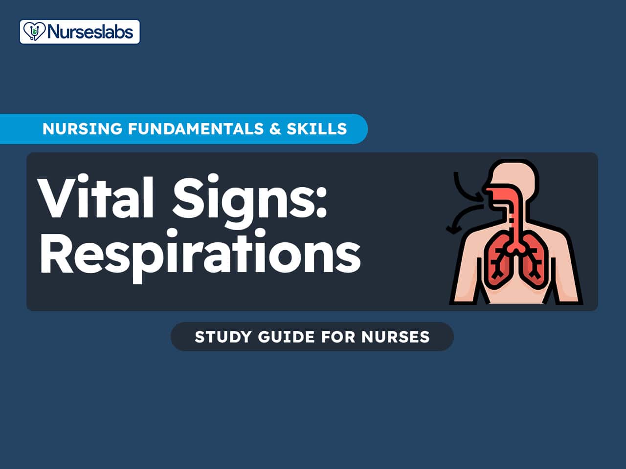
Respiration is the process by which the body supplies oxygen to its tissues and removes carbon dioxide, a waste product of metabolism. It includes two main components: external respiration, which is the gas exchange between the lungs and the bloodstream, and internal respiration, which is the exchange of gases between the blood and the body’s cells. Respiration is controlled by the brainstem and occurs automatically, though it can be influenced by voluntary effort, emotions, and physiological changes. The act of breathing involves the coordinated function of the respiratory muscles, particularly the diaphragm and intercostal muscles, which enable inhalation and exhalation.
What is Respiratory Rate?
Respiratory rate refers to the number of complete breathing cycles—one inhalation and one exhalation—that occur in one minute. It is a key clinical sign that reflects the body’s oxygen demand and overall respiratory efficiency. A normal respiratory rate varies by age and health status but typically ranges from 12 to 20 breaths per minute in a healthy adult at rest. In healthy individuals, this process is typically smooth, regular, and effortless—referred to as eupnea. Respiratory rate is just one aspect of assessment; nurses must also evaluate the depth, rhythm, quality (such as effort and sounds), and overall effectiveness of breathing. Because respiration is usually controlled subconsciously, it must be observed discreetly to avoid influencing the patient’s normal breathing pattern. Variations in any of these respiratory characteristics can signal conditions like infection, cardiac dysfunction, anxiety, or impaired oxygenation. This comprehensive approach helps determine whether gas exchange is adequate to meet the body’s metabolic demands.
Importance of Assessing Respirations
Assessing respirations is a fundamental component of patient evaluation and ongoing monitoring. Breathing patterns can provide early clues about a patient’s respiratory and overall physiological status. The following points highlight key reasons why respiratory assessment is essential in clinical practice:
1. Monitor Oxygenation and Ventilation Status
Accurate respiratory assessment helps determine if the lungs are effectively bringing in oxygen and expelling carbon dioxide. A normal respiratory rate and pattern indicate efficient gas exchange, which is vital for cellular metabolism and overall organ function. Abnormalities such as tachypnea (fast breathing), bradypnea (slow breathing), or shallow respirations may signal hypoxia, hypercapnia, or compromised lung function. Early detection allows for prompt interventions such as oxygen therapy, ventilation support, or diagnostic testing.
2. Detect Early Signs of Deterioration
Respiratory changes are often among the earliest signs of clinical decline, sometimes preceding changes in heart rate or blood pressure. For example, an increasing respiratory rate may be a compensatory response to fever, sepsis, shock, or heart failure. Observing irregular or labored breathing can also indicate neuromuscular fatigue or obstruction. Early recognition of these signs allows for timely escalation of care and may prevent respiratory arrest or the need for intensive interventions.
3. Evaluate Response to Treatment or Interventions
Continuous monitoring of respirations helps assess how a patient is responding to treatment, such as bronchodilators, antibiotics, diuretics, or pain management. A return to normal respiratory rate and effort often signals clinical improvement, while persistent or worsening abnormalities may suggest treatment failure or complications. This feedback is critical for adjusting care plans and ensuring that therapies are achieving the desired effect.
Normal Physiology of Breathing
Normal physiology of breathing is a complex, mostly involuntary process controlled by the respiratory center in the brainstem—primarily the medulla oblongata and the pons. These brain regions continuously monitor the levels of carbon dioxide (CO₂), oxygen (O₂), and pH in the blood through chemoreceptors. When CO₂ levels rise or pH drops (indicating increased acidity), the medulla stimulates the respiratory muscles to increase the rate and depth of breathing to restore balance.
The diaphragm, a dome-shaped muscle located below the lungs, is the primary muscle responsible for breathing. During inhalation, the diaphragm contracts and moves downward, creating negative pressure that pulls air into the lungs. During exhalation, it relaxes and rises back up, pushing air out of the lungs. This process occurs automatically without conscious effort but can be temporarily controlled (e.g., holding your breath). This finely tuned system ensures the body maintains proper oxygenation and acid-base balance at rest and during activity.
Normal Respiratory Rates by Age
Respiratory rates vary with age, generally being higher in infants and children and slower in adults. Newborns and infants breathe much faster to meet metabolic demands, while adults have a lower resting rate due to more efficient lung function. The table below outlines normal resting respiratory rates for different age groups:
| Age Group | Normal Respiratory Rate (breaths per minute) |
|---|---|
| Newborns (0–1 month) | 30–60 |
| Infants (1–12 months) | 30–50 |
| Toddlers (1–3 years) | 24–40 |
| Preschoolers (3–5 years) | 22–34 |
| School-age Children (6–12 years) | 18–30 |
| Adolescents (13–18 years) | 12–20 |
| Adults (18–64 years) | 12–20 |
| Older Adults (65+ years) | 12–24 |
These values represent resting respiratory rates. By adulthood, a normal resting respiratory rate is around 12–20 breaths per minute, and rates below 12 or above 20 at rest are generally considered abnormal and may indicate an underlying issue. Always compare the patient’s rate to age-appropriate norms and their baseline.
Types of Respiration
Observing the type and quality of a person’s respirations provides important insights into their respiratory and overall health status. While normal breathing (eupnea) is quiet and effortless, deviations from this pattern can indicate underlying medical conditions ranging from anxiety to life-threatening emergencies. The following are common types of respirations, each with distinct characteristics and clinical implications:
1. Eupnea (Normal Breathing)
Eupnea refers to a normal, unlabored respiratory pattern that is quiet, regular, and effortless. It typically ranges from 12 to 20 breaths per minute in a healthy adult and is essential for adequate oxygen and carbon dioxide exchange.
2. Tachypnea (Rapid Breathing)
Tachypnea is an abnormally high respiratory rate, defined as more than 20 breaths per minute in adults. It is usually shallow and may occur due to fever, anxiety, pain, respiratory infection, or metabolic acidosis.
3. Bradypnea (Slow Breathing)
Bradypnea is a slower-than-normal respiratory rate, typically less than 12 breaths per minute in adults. It can be caused by sedation, head injury, hypothyroidism, or increased intracranial pressure and may lead to inadequate oxygenation if prolonged.
4. Apnea (Absence of Breathing)
Apnea is the complete cessation of breathing for a period of time. If sustained, it can result in hypoxia, unconsciousness, and potentially death. Common examples include sleep apnea or apnea during cardiac arrest.
5. Dyspnea (Difficult or Labored Breathing)
Dyspnea is the subjective sensation of breathing discomfort or shortness of breath. It can result from heart failure, asthma, chronic obstructive pulmonary disease (COPD), or anxiety, and may require supplemental oxygen or respiratory support.
6. Orthopnea (Difficulty Breathing While Lying Flat)
Orthopnea is a form of dyspnea that worsens when the patient lies down and is relieved by sitting or standing up. It is most often associated with congestive heart failure and may require patients to sleep propped up with pillows.
7. Kussmaul Respirations
This is a deep, rapid breathing pattern often associated with metabolic acidosis, particularly diabetic ketoacidosis (DKA). It reflects the body’s effort to expel excess carbon dioxide to correct the acid-base imbalance.
8. Cheyne-Stokes Respirations
A cyclical pattern of gradual increases and decreases in respiration followed by periods of apnea. It is often seen in end-of-life care, heart failure, or neurological injuries, especially affecting the brainstem.
9. Biot’s Respirations (Ataxic Breathing)
This pattern consists of irregular breathing with variable rate and depth, followed by periods of apnea. It is typically a sign of brainstem injury or central nervous system dysfunction and is considered a medical emergency.
The table below outlines the most common types of respirations, including their defining characteristics and clinical relevance:
| Type of Respiration | Description | Clinical Significance |
|---|---|---|
| Eupnea | Normal, unlabored breathing | Indicates healthy respiratory function |
| Tachypnea | Rapid, shallow breathing (>20 breaths/min in adults) | Common in fever, anxiety, or respiratory distress |
| Bradypnea | Slow breathing (<12 breaths/min in adults) | Seen in drug overdose, head injury, or sedation |
| Apnea | Absence of breathing | Life-threatening; occurs in cardiac or respiratory arrest |
| Dyspnea | Difficult or labored breathing | Sign of heart or lung disease; patient may feel short of breath |
| Orthopnea | Difficulty breathing while lying flat | Often seen in heart failure; relieved by sitting up |
| Kussmaul | Deep, labored, rapid breathing | Associated with metabolic acidosis, especially diabetic ketoacidosis |
| Cheyne-Stokes | Cycles of deep breathing followed by apnea | Seen in end-of-life care, heart failure, or brain injury |
| Biot’s (Ataxic) | Irregular rate and depth with periods of apnea | Often indicates brainstem injury or severe neurologic condition |
Understanding these patterns is crucial. Recognizing an abnormal pattern like Cheyne–Stokes or Kussmaul’s can provide an immediate clue about the patient’s condition (for example, Cheyne–Stokes may suggest brain injury or heart failure, and Kussmaul’s strongly indicates metabolic acidosis, such as diabetic crisis). Always correlate the pattern with the patient’s clinical context and report abnormalities to the medical team promptly.
Factors Affecting Respiratory Rate
The respiratory rate is not a fixed value; it can vary depending on a range of physiological, psychological, and environmental factors. Understanding what influences respiratory rate is essential for interpreting findings accurately during patient assessments. These factors can cause temporary or long-term changes in the rate, depth, and rhythm of breathing and may reflect both normal adjustments and underlying pathology. Below is an elaboration on the most common factors that affect respiratory rate:
Age
As noted, age has a significant effect on respiratory rate. Infants and young children breathe faster to meet higher metabolic demands, whereas adults have slower rates. Elderly adults usually maintain the normal adult range, though respiratory efficiency may decrease with age due to weaker respiratory muscles or lung changes. Growth and developmental stage influence lung capacity and rate.
Exercise and Activity
Physical activity increases respiratory rate and depth to meet the body’s increased oxygen demand and CO2 removal. During strenuous exercise, breathing can become rapid and deep (hyperpnea) to supply more oxygen. In well-conditioned athletes, resting respiratory rates might be lower, but during activity their respiratory systems respond efficiently with large increases in ventilation.
Fever and Temperature
Fever raises the respiratory rate. For each 1°C rise in body temperature, the respiratory rate may increase as the body attempts to dissipate heat and meet increased metabolic needs. Conversely, in hypothermia, respiratory rate may slow. Environmental temperature can also have minor effects (e.g., breathing may accelerate in extreme heat as part of panting-like heat dissipation).
Emotions and Stress
Emotional state can significantly affect breathing. Anxiety, fear, or pain often stimulates the sympathetic nervous system, leading to faster, shallower breathing or even hyperventilation. During panic attacks, for example, people commonly develop tachypnea or hyperventilation. In contrast, relaxation or meditation can slow the respiratory rate. Nurses should be mindful that simply being in a clinical setting can make patients anxious and alter their breathing temporarily.
Body Position
Body posture influences lung expansion. A person lying flat (supine) may have more restricted chest expansion (especially if obese or pregnant), potentially leading to slightly faster, shallow breaths. Sitting up or standing allows for maximal lung expansion and may slow the rate. Patients with orthopnea or respiratory distress often instinctively assume an upright or tripod position to ease breathing. Always try to assess respiratory rate with the patient in a semi-upright, comfortable position when possible.
Medications
Many drugs affect respiratory rate and rhythm. Central nervous system depressants—such as opioids (morphine, fentanyl), sedatives, or certain anesthetics—can cause bradypnea by depressing the respiratory centers in the brain. Overdoses of these can lead to dangerous respiratory depression or apnea. On the other hand, stimulants (like amphetamines or caffeine) and some asthma medications can increase breathing rate. Always consider a patient’s medications if you notice an abnormal respiratory rate.
Illness and Health Status
A wide range of medical conditions influence respirations:
- Pulmonary diseases (e.g., asthma, chronic obstructive pulmonary disease, pneumonia) typically elevate the respiratory rate and can cause irregular rhythms or labored breathing due to impaired gas exchange. For instance, an asthmatic during an attack will have tachypnea with wheezing; a person with COPD may have an elevated rate and use accessory muscles at rest.
- Cardiac conditions. Heart failure often leads to tachypnea as the body tries to compensate for poor circulation; severe heart failure can produce Cheyne–Stokes breathing during sleep. An acute myocardial infarction (heart attack) may also present with rapid, shallow breathing due to pain and anxiety.
- Metabolic disturbances. Metabolic acidosis (as in diabetic ketoacidosis or renal failure) triggers Kussmaul’s respirations (deep, rapid breathing) as a compensatory mechanism to blow off CO2. Metabolic alkalosis might slow breathing.
- Neurological injuries. Damage to brainstem respiratory centers (from stroke, trauma, or increased intracranial pressure) can lead to abnormal rhythms like Cheyne–Stokes or Biot’s respirations, as the normal regulation of breathing is disrupted.
- Pain. Severe pain, especially thoracic or abdominal pain, can alter breathing. Patients in pain may exhibit splinting (shallow breathing to avoid pain from deep breaths, e.g., with broken ribs or post-surgery). Pain and anxiety combined can further raise the rate. Adequate pain control can help normalize breathing.
- Anemia. In anemia or low blood oxygen-carrying states, the body might increase respiratory rate to compensate for reduced oxygen delivery. Similarly, low oxygen environments (high altitude) cause increased rate and depth (hyperventilation) until the body acclimatizes.
- Smoking and airway irritants. Irritants can cause coughing and bronchoconstriction, which may alter the observed respiratory pattern and rate. Chronic smoking leads to lung changes that can elevate baseline RR over time or cause difficulty exhaling fully (as in emphysema).
Virtually any factor that affects the body’s demand for oxygen or production of carbon dioxide, or that impairs gas exchange or neural respiratory control, can influence respiratory rate. Nurses should consider these factors when interpreting RR. For example, a slight tachypnea in a febrile patient may be expected (body fighting infection), but the same tachypnea in a resting afebrile patient might signal pain, anxiety, or an impending physiological problem. Always assess the context: look at temperature, heart rate, oxygen saturation, and patient’s overall condition alongside the respiratory rate.
Step-by-Step Procedure for Assessing Respiratory Rate
Accurate assessment of respiratory rate requires careful technique and observation. Follow these steps and considerations for measuring RR:
1. Prepare yourself and gather equipment. Ensure you have a reliable timekeeping device such as a watch with a second hand or a digital timer. Wash your hands thoroughly using proper hand hygiene techniques.
Clean hands reduce the risk of cross-contamination and infection transmission. A precise timing device is critical because respiratory rate must be counted accurately, often for a full 60 seconds if irregular breathing is observed.
2. Ensure privacy and create a calm environment. Close doors, draw privacy curtains, and ensure the room is quiet and comfortable. Explain to the patient, if appropriate, that you are conducting a routine check of overall health signs—avoid directly stating you will be observing their breathing.
Respecting patient privacy upholds dignity and promotes trust. Avoiding direct mention of respiration helps prevent conscious alteration of the breathing pattern, which could skew results.
3. Position the patient appropriately. Have the patient rest in a comfortable semi-Fowler’s position (head of bed elevated 30–45°) or seated upright with arms relaxed. Wait a few minutes if the patient has recently moved, spoken, or exerted themselves.
This position facilitates full lung expansion and allows clear observation of the chest or abdominal movements. Resting ensures that the respiratory rate reflects the patient’s baseline rather than transient elevations from activity.
4. Observe without drawing attention. Begin assessing respirations discreetly—ideally right after palpating the radial pulse. Keep your fingers in place on the wrist and shift your attention to the chest or abdomen without alerting the patient.
Respiratory rate is influenced by awareness. Discreet observation ensures a more accurate assessment of involuntary, natural breathing patterns.
5. Visualize or palpate chest/abdominal movements. Observe or lightly palpate the chest or abdomen for respiratory movements. One full inspiration and expiration (rise and fall) counts as one breath. In adults, observe thoracic movement; in infants, watch the abdomen. Ensure clothing or blankets do not obstruct your view.
Proper visualization ensures each breath is counted accurately. Infants are diaphragmatic breathers, while adults use thoracic muscles—this distinction guides where to focus your observation.
6. Time and count the respirations.
- If the rhythm is regular, count for 30 seconds and multiply by 2.
- If irregular, or if the patient is an infant or critically ill, count for a full 60 seconds. Irregular or erratic breathing requires a full-minute assessment to ensure accuracy. Respiratory rate is a sensitive and early indicator of physiological distress, making timing precision essential.
7. Assess rhythm and depth.
- Rhythm. Determine whether breathing occurs at consistent intervals (regular) or varies (irregular).
- Depth. Observe whether breaths are shallow, normal, or deep.
Variations in rhythm or depth may indicate underlying medical issues. Shallow respirations may suggest pain or fatigue, while deep respirations may reflect compensation for conditions like acidosis.
8. Evaluate respiratory effort and quality. Assess for signs of labored breathing, such as nasal flaring, use of accessory muscles, intercostal retractions, or pursed-lip breathing. Listen for abnormal respiratory sounds like wheezing, gurgling, or stridor.
Increased respiratory effort and abnormal sounds often point to respiratory compromise or obstruction. Early detection of these signs allows for timely intervention and can prevent deterioration.
9. Monitor for additional signs of respiratory distress. Observe for cyanosis (bluish lips or fingertips), restlessness, altered consciousness, or changes in oxygen saturation (if pulse oximetry is available).
These signs may indicate hypoxia or serious respiratory compromise requiring urgent evaluation and intervention.
10. Document findings thoroughly. Record the following:
- Respiratory rate (e.g., “RR = 18 breaths/min”)
- Rhythm (regular/irregular)
- Depth (shallow/normal/deep)
- Quality and effort (e.g., labored, accessory muscle use, abnormal sounds)
Example. “RR = 22 breaths/min, irregular, shallow, with mild intercostal retractions.”
Clear and accurate documentation ensures that respiratory findings are communicated effectively to the healthcare team, facilitating timely care decisions and tracking of changes.
Clinical Tips and Safety Alerts
For accurate and safe respiratory assessments, keep these clinical tips in mind:
Don’t Announce the Counting
Avoid telling the patient you are counting their breaths. People tend to alter their breathing pattern if they know it’s being observed. By keeping your fingers on the pulse and subtly counting respirations afterward, you ensure a true resting respiratory rate.
Ensure Rest and Recovery
Measure respiratory rate at rest. If the patient was active (e.g., walking or just came upstairs) or visibly anxious, let them sit and rest for a few minutes before counting. Physical exertion or excitement can temporarily elevate the rate. Document if the measurement was taken during activity or distress, as this context is important.
Count for a Full Minute if in Doubt
If you note an irregular rhythm or the patient’s breathing is very shallow or very fast, count for a full 60 seconds. Irregular breathing can lead to undercounting or overcounting if a shorter interval is used. When in doubt, a full-minute count improves accuracy. This is especially important for infants (who often have irregular breathing) or any patient with an abnormal pattern.
Observe, Don’t Obstruct
Make sure nothing (clothing, bedding, your hands) is obstructing your view of the chest or abdomen movement. If you have difficulty seeing breaths, you can lightly place a hand on the patient’s upper chest or abdomen to feel the rise and fall—but be mindful not to alarm the patient or alter their pattern by doing so. In newborns, a hand on the abdomen or watching the abdomen is effective since they breathe diaphragmatically.
Look for Associated Signs
As you count, keep an eye out for any signs of respiratory distress. These include nasal flaring, cyanosis (bluish discoloration of lips or fingertips), audible gasping or wheezing, or retractions in the chest. These signs, combined with an abnormal RR, warrant prompt attention. For example, if a patient has agonal breathing (gasping, irregular breaths)—often a sign of severe brain anoxia or cardiac arrest—activate emergency response immediately (call a code/ambulance). Always prioritize patient safety: if breathing is absent or grossly inadequate, begin appropriate interventions (such as opening the airway, ventilating, or CPR if necessary) before finishing any formal counting.
Clinical Judgment with Devices
Remember that while automated monitoring devices (like bedside respiratory rate monitors or pulse oximeters with RR capability) are helpful, they can be inaccurate in certain situations. Always double-check any electronic RR reading by manually observing the respirations if the value doesn’t match the clinical picture. Never rely solely on an SpO₂ monitor’s waveform for respiratory count if the patient is critically ill—do a hands-on assessment.
Teach Breathing Awareness if Needed
If a patient is breathing very fast due to anxiety (hyperventilating), after assessing vital signs you may need to coach them in slow breathing techniques to prevent dizziness or fainting. However, perform your counting first, then intervene. For patients with known anxiety causing tachypnea, ensuring they are calm and guiding some slow breaths prior to counting can help get a measurement that reflects their baseline rather than their anxiety.
Hygiene and Comfort
If you need to be near the patient’s face (for example, to listen to breath sounds or feel airflow), practice standard precautions. For your comfort and the patient’s, be mindful of your positioning and breath (e.g., avoid directly exhaling into the patient’s face). If the patient has a communicable respiratory illness, wear appropriate PPE (mask/face shield) during close assessment.
By following these tips, nurses can ensure they obtain an accurate respiratory rate and also safeguard patient well-being. Always interpret the respiratory rate within the context of the whole clinical picture, and report concerning findings. Respiratory rate is a sensitive vital sign that often changes earlier than blood pressure or pulse in response to patient deterioration. Diligent monitoring can thus provide an early warning, allowing for timely interventions and improved patient outcomes.


Leave a Comment