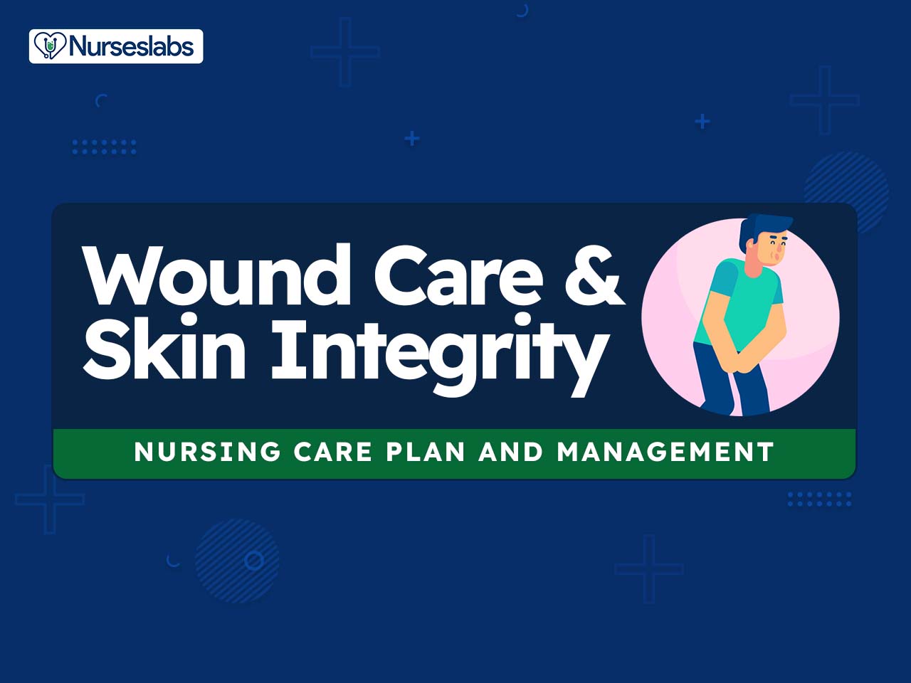
For this nursing care plan and management guide learn how to provide care for patients with wounds or break in their tissue/skin integrity. Get to know the nursing assessment, interventions, goals, and nursing diagnosis for impaired skin integrity.
What is Tissue Integrity?
What constitutes the body’s protection against external threats? Yes, it’s the integumentary system. Specifically, the skin, cornea, subcutaneous tissues, and mucous membranes are the first line of defense against threats from the external environment. In a normal setting, these defenses are adequate to defend the body from any threats. However, some factors may cause impairment or a break in this line of defense, causing impairment of tissue integrity.
The most common cause includes physical trauma (e.g., car accidents, sports injuries, cuts, blunt trauma, etc.). Other causes can be related to thermal factors (e.g., burns, frostbites), chemical injury (e.g., adverse reactions to drugs), infection, nutritional imbalances, fluid imbalances, and altered circulation (e.g., pressure injuries).
A break in tissue integrity is usually repaired by the body very well. However, there are circumstances that it doesn’t repair it at all and replaces the damaged tissue with connective tissue. When tissue integrity is left untreated, it could cause local or systemic infection and ultimately lead to necrosis.
Other factors include age, weight loss, poor nutrition and hydration, excessive moisture and dryness, smoking, and other conditions affecting blood flow. Personal hygiene behaviors such as frequent bathing, leisure activities including extended UV exposure, or unfavorable occupational environments such as wet work may compromise the skin as well (Kottner et al., 2020).
Signs and symptoms of a break in skin integrity may include unpleasant sensory feelings, inflammation, cutaneous lesions, or loss of cutaneous substance (wounds). This may not only result in reduced quality of life, itching, pain, disfigurement, and disability but also pose secondary risks including allergies and secondary infection. Overall, the global burden of skin diseases is increasing with increasing age (Kottner et al., 2020).
What is Wound Care?
A wound is a disruption of the normal structure and function of the skin and soft tissue architecture. An acute wound demonstrates normal physiology, and healing is anticipated to progress through the expected stages of wound healing, whereas a chronic wound is broadly defined as one that is physiologically impaired (Armstrong & Meyr, 2023).
Wound healing is a complex physiologic process that restores function to the skin and tissue that have been injured. The healing process is affected by several external and internal factors that either promote or inhibit healing. When providing wound care to clients, nurses, in collaboration with other members of the healthcare team, assess and manage external and internal factors to provide an optimal healing environment (Ernstmeyer & Christman, 2021).
Phases of Wound Healing
When skin is injured, there are four phases of wound healing that take place: hemostasis, inflammatory, proliferative, and maturation.
- Hemostasis. In the hemostasis phase, platelets release growth factors that alert various cells to start the repair process at the wound location. This phase lasts up to 60 minutes, depending on the severity of the injury.
- Inflammatory. After the hemostasis phase, the inflammatory phase begins, wherein vasodilation occurs so that white blood cells in the bloodstream can move into the wound to start cleaning the wound bed. This appears as edema, erythema, and exudate to the observers.
- Proliferative. The proliferative phase begins within a few days after the injury and includes four important processes: epithelialization, angiogenesis, collagen formation, and contraction.
- Epithelialization refers to the development of new epidermis and granulation tissue, which is a new connective tissue with new, fragile, thin-walled capillaries.
- Angiogenesis occurs when capillaries begin to develop within the wound 24 hours after injury. These capillaries bring more oxygen and nutrients to the wound for healing.
- Collagen is formed to provide strength and integrity to the wound. At the end of the proliferation phase, the wound begins to contract in size.
- Maturation. During this phase, collagen continues to be created to strengthen the wound. A wound typically heals within four to five weeks and often leaves behind a scar. Over time, the scar begins to soften, flatten, and become pale in about nine months (Ernstmeyer & Christman, 2021).
Types of Wound Healing
There are three types of wound healing: primary intention, secondary intention, and tertiary intention.
- Primary intention. This means that the wound is sutured, stapled, glued, or otherwise closed so the wound heals beneath the closure. This type of healing occurs with clean-edged lacerations or surgical incisions. Closed edges are referred to as approximated.
- Secondary intention. This occurs when the edges of a wound cannot be approximated or brought together, so the wound fills in from the bottom up by the production of granulation tissue. Examples of this type are pressure injuries and chainsaw injuries. These are at a higher risk of infection.
- Tertiary intention. This refers to a wound that has had to remain open or has been reopened, often due to severe infection. The wound is typically closed at a later date when the infection has resolved. These wounds have delayed healing times and increased scar tissue (Ernstmeyer & Christman, 2021).
Causes
The following are the common causes or related etiological factors for impaired skin integrity:
- Physical Trauma. Injuries such as car accidents, sports injuries, cuts, and blunt trauma that disrupt the skin’s structure.
- Thermal Factors. Exposure to extreme temperatures leading to burns or frostbites.
- Chemical Injury. Adverse reactions to drugs or contact with harmful chemicals that damage the skin.
- Infection. Presence of pathogens that invade and harm the skin tissue.
- Nutritional Imbalances. Poor intake of essential nutrients affecting skin health and repair.
- Fluid Imbalances. Dehydration or excessive fluid retention that compromises skin moisture and elasticity.
- Altered Circulation. Conditions like pressure injuries that reduce blood flow to the skin, impairing its integrity.
- Age. Increased susceptibility to skin breakdown due to thinner, less elastic skin in older adults.
- Weight Loss. Significant loss of body mass leading to reduced cushioning and support for the skin.
- Poor Nutrition and Hydration. Inadequate intake of nutrients and fluids essential for maintaining healthy skin.
- Excessive Moisture and Dryness. Imbalance in skin moisture levels causing maceration or cracking.
- Personal Hygiene Behaviors. Frequent bathing or exposure to harsh cleansing agents that strip the skin of its natural oils.
Signs and Symptoms
The following are the common signs and symptoms or defining characteristics for patients with impaired skin integrity:
- Pain. Discomfort or abnormal sensations in the affected area.
- Inflammation. Redness, swelling, heat, and pain indicating the body’s immune response to injury.
- Cutaneous Lesions. Visible abnormalities on the skin such as abrasions, lacerations, or ulcers.
- Loss of Cutaneous Substance (Wounds). Breaks in the skin that may expose underlying tissues.
- Skin and Tissue Color Changes. Alterations in skin color, including redness, purplish hues, or black discoloration.
- Swelling Around the Initial Injury. Edema surrounding the wound site indicating fluid accumulation.
- Pruritic/Itchy Skin. Persistent itching that can lead to scratching and further skin damage.
- Dry, Scaly Skin. Lack of moisture leading to rough, flaky skin surfaces.
- Thin, Fragile Skin. Increased vulnerability to tears and abrasions due to reduced skin strength.
- Exudate or Drainage. Fluid leakage from wounds, which may be clear, bloody, or purulent.
- Delayed Wound Healing. Wounds that do not progress through the normal stages of healing in an expected timeframe.
- Presence of Necrosis. Dead tissue within or around the wound site, indicating severe tissue damage.
- Erythema. Persistent redness around the wound area as a sign of inflammation.
- Edema. Swelling caused by excess fluid trapped in the body’s tissues around the wound.
Nursing Care Plans and Management
Effective wound care and the maintenance of skin integrity are vital aspects of nursing care. Nurses play a critical role in ensuring prompt and proper management of wounds to promote healing and prevent complications. A comprehensive nursing care plan is vital for addressing the unique needs of each client, taking into account their specific wound characteristics, overall health status, and individual preferences. With a dedicated focus on wound assessment, interdisciplinary collaboration, client education, and utilization of evidence-based practices, nurses are instrumental in optimizing outcomes and improving the quality of life for individuals with compromised skin integrity.
Nursing Problem Priorities
The following are the nursing priorities for clients with wounds or compromised skin integrity.
- Wound assessment. Frequent wound assessment based on the type, cause, and characteristics of the wound is necessary to help determine the type of treatment required to manage the wound effectively and promote maximal wound healing.
- Effective wound care. Quality wound care is important for rapid and uncomplicated healing through decreasing complications, repeated admissions, length of hospital stay, and costs, and enhancing client quality of life.
- Proper suture and staple removal. Proper timing and technique in suture or staple removal prevent complications that may arise from a non-healed wound.
- Drain management. The site and drain should be checked periodically throughout the shift to ensure the drain is functioning effectively and that no leaking occurs.
- Wound care for burns. Proper management of burn injuries is required to prevent wound deterioration.
- Management of pressure injuries/ulcers. Pressure injuries/ulcers cause significant pain and suffering to the client, as well as increased morbidity and mortality and higher medical costs. Prevention and effective treatment are imperative to avoid developing or worsening pressure injuries.
- Client and caregiver education. Education empowers the client and their caregivers to actively participate in the healing process, promotes adherence to treatment plans, and fosters a proactive approach to preventing complications.
Nursing Assessment
A thorough assessment of the skin or the wound must be conducted, including measurements of wound dimensions, evaluation of wound bed appearance, identification of any signs of infection, and assessment of surrounding skin integrity. This assessment guides the selection of appropriate wound dressings, cleansing agents, and adjunctive therapies.
A break in tissue integrity is characterized by the following subjective and objective data:
- The affected area is hot, and tender to touch
- Damaged or destroyed tissue (e.g., cornea, mucous membranes, integumentary, subcutaneous)
- Local pain
- Protectiveness toward site
- Skin and tissue color changes (red, purplish, black)
- Swelling around the initial injury
- Pruritic/itchy skin
- Dry, scaly skin
- Thin, fragile skin
Nursing Diagnosis
After thorough assessment, nursing diagnoses are formulated to address the challenges of impaired skin integrity, guided by the nurse’s clinical judgment and understanding of the patient’s unique condition. While nursing diagnoses help organize care, their use may vary across clinical settings. Ultimately, the nurse’s expertise and judgment shape the care plan to prioritize each patient’s needs. Here are examples of nursing diagnoses that may be useful for common concerns associated with wound care or impaired skin integrity:
- Impaired Skin Integrity related to prolonged exposure on bony prominences as evidenced by redness. and non-blanchable areas on the sacral region and heels, patient reports of pain and discomfort when repositioning secondary to CVA.
- Impaired Skin Integrity related to friction and shear from frequent repositioning.
- Impaired Skin Integrity related to insufficient nutritional intake as evidenced by delayed wound healing, dry and flaky skin.
Nursing Goals
Goals and expected outcomes may include:
- The client reports any altered sensation or pain at the site of tissue impairment.
- The client demonstrates an understanding of the plan to heal tissue and prevent injury.
- The client describes measures to protect and heal the tissue, including wound care.
- The client’s wound decreases in size and has increased granulation tissue.
Nursing Interventions and Actions
Therapeutic interventions and nursing actions for clients with impaired skin integrity include:
1. Skin and Wound Assessment
Based on observed signs, symptoms, and/or results of diagnostic tests, a medical diagnosis can be made, which guides the treatment strategy. The visual examination of the skin and the description of cutaneous lesions play a key role in diagnosing skin diseases (Kottner et al., 2020).
Determine etiology (e.g., acute or chronic wound, burn, dermatological lesion, pressure ulcer, leg ulcer).
Prior assessment of wound etiology is critical for the proper identification of nursing interventions that will guide nursing care. There are many pathological processes challenging skin integrity. Systemic diseases such as diabetes mellitus compromise skin structure and function in many ways. Foot deformity and gait problems increase callus formation with subsequent risk for foot ulceration. Dermatological diseases directly affect the cutaneous structure and function in many ways, ranging from inflammatory skin diseases to tumors (Kottner et al., 2020).
Assess the site of impaired tissue integrity and its condition.
Redness, swelling, pain, burning, and itching are indications of inflammation and the body’s immune system response to localized tissue trauma or impaired tissue integrity. Repeated irritant exposure may lead to a hardening effect that may be regarded as a protective response as well. Repeated friction leads to increased proliferation of the stratum corneum protecting the foot. These examples indicate that the skin not only adapts to the environment but also constantly compensates for risks (Kottner et al., 2020).
Assess the characteristics of the wound, including type, location, color, size (length, width, depth), drainage, and odor.
When performing an objective assessment of the wound, it is important to perform a thorough exam to check for existing wounds, as well as to evaluate their risk of skin breakdown using the Braden Scale (Ernstmeyer & Christman, 2021).
- Type
Types of wounds may include abrasions, lacerations, burns, surgical incisions, pressure injuries, skin tears, arterial ulcers, or venous ulcers. It is important to understand the type of wound present to select appropriate interventions. - Location
The location of the wound should be documented precisely. A body diagram template is helpful to demonstrate exactly where the wound is located. - Size
Wound size should be measured regularly to determine of the wound is increasing or decreasing in size. Length is measured using the head-to-toe axis, and width is measured laterally. - Degree of tissue injury
Wounds are classified as partial-thickness or full-thickness. For pressure injuries, it is important to assess the stage of the injury (see Management of Pressure Injuries). - Color of the wound base
Assess the base of the wound for the presence of healthy, pink, or red granulation tissue. Note the unhealthy appearance of dark red granulation tissue, white or yellow slough, or brown or black necrotic tissues. These determine the wound’s healing process. - Drainage
The color, consistency, and amount of exudate or drainage should be assessed and documented at every dressing change. Drainage from wounds is often described as scant, small/minimal, moderate, and large/copious amounts. The type of wound discharge should be described using serosanguinous, sanguineous, serous, or purulent (Ernstmeyer & Christman, 2021).
Assess changes in body temperature, specifically increased body temperature.
Fever is a systemic manifestation of inflammation and may indicate the presence of infection. One of the primary responses of the immune system is the release of pyrogens. Pyrogens stimulate the hypothalamus in the brain, causing it to raise the set point for normal body temperature. This results in an elevation in body temperature, leading to fever. (El-Radhi, 2019)
Assess the client’s level of pain.
Pain is part of the normal inflammatory process. The extent and depth of injury may affect pain sensations. The nurse should be aware that the degree of pain may not correlate to the extent of tissue damage. For example, skin tears are often painful because the nerve endings are exposed in the dermal layer, whereas clients with severe diabetic ulcers on their feet may experience little or no pain because of existing neuropathic damage (Ernstmeyer & Christman, 2021).
Monitor the site of impaired tissue integrity at least once daily for color changes, redness, swelling, warmth, pain, or other signs of infection.
Systematic inspection can identify impending problems early. A break in the skin allows bacteria to enter and begin to multiply. Microbial contamination of wounds can progress from localized infection to systemic infection, sepsis, and subsequent life- and limb-threatening infection (Ernstmeyer & Christman, 2021).
Monitor the status of the skin around the wound.
The skin outside the outer edges of the wound called the periwound skin, provides information related to wound development or healing. If the wound is healing by primary intention, it should be documented if the wound edges are well-approximated or if there are any signs of dehiscence (Ernstmeyer & Christman, 2021).
Monitor the client’s skin care practices, noting the type of soap or other cleansing agents used, the temperature of the water, and the frequency of skin cleansing.
An individualized plan is necessary according to the client’s skin condition, needs, and preferences. Repeated or prolonged exposure to water and other irritants such as surfactants, solvents, oils, or urine and/stool damage the skin barrier in many ways: the skin surface pH is elevated; natural moisturizing factors are removed; and the lipid bilayers are disturbed. This can also occur due to excessive bathing and cleansing habits (Kottner et al., 2020).
Assess the overall condition of the skin.
Assessment of the condition of the skin provides baseline data for possible interventions. Normal skin condition differs among individuals. Healthy skin should have good turgor (an indication of moisture), feel warm and dry to the touch, be free from impairment (cuts, wounds, abrasions, excoriation, outbreaks, and rashes), and have quick capillary refill (less than six seconds). Clients with advanced age are at high-risk risk for skin impairment because the skin is less elastic, has less moisture, and has thinning of the epidermis.
Assess for history or presence of AIDS or other immunological problems.
Skin lesions or Kaposi sarcoma are an early manifestation of diseases related to HIV. Kaposi sarcoma usually appears first as spots or lesions on the skin. The lesions can be purple, red, or brown. These lesions can be flat and not raised above the surrounding skin (patches), flat but slightly raised (plaques), or bumps (nodules). The skin lesions of Kaposi sarcoma most often develop on the legs or face, but they can also appear in other areas, such as the mucous membranes of the mouth, throat, the outside of the eye, and the inner part of the eyelids (American Cancer Society, 2018).
Assess for a history of radiation therapy.
Radiated skin becomes thin and friable, may have less blood supply, and is at higher risk for breakdown. Radiation therapy causes stasis or occlusion of small vessels and damages fibroblasts and nuclei (Nagle et al., 2022).
Evaluate the client’s strength to move (e.g., shift weight while sitting, turn over in bed, move from bed to chair).
The most significant risk factor in skin breakdown is immobility. The mobility risk factor is defined as the client’s ability to change or control their body position. Tissue damage will occur if a client is unable to reposition on their own power unless caregivers frequently change their position (Ernstmeyer & Christman, 2021).
Assess for fecal/urinary incontinence.
Stools may contain enzymes that cause skin breakdown. The urea in urine turns into ammonia within minutes and is caustic to the skin. The use of diapers and incontinence pads hastens skin breakdown. All types begin with inflammatory changes of erythema and tenderness, with progression to skin loss with prolonged exposure. Often, the normal bacterial flora is imbalanced to the point that an underlying fungal rash develops (Murphree, 2017).
Assess for edema.
Skin tightened tautly over edematous tissue is at risk for impairment. Venous stasis in the lower legs prevents diffusion of oxygen and nutrients, leading to stasis dermatitis, edema, hemosiderin deposits in the dermis, dryness, and stiffening, and a high risk for leg ulceration (Kottner et al., 2020).
Assess for environmental moisture (e.g., wound drainage, high humidity).
Moisture may contribute to skin maceration. There are four types of moisture-associated skin damage (MASD): incontinence-associated dermatitis, intertriginous dermatitis, peri-wound MASD, and peristomal MASD. prolonged or repetitive exposure to moisture is a contributing factor to MASD. (Murphree, 2017)
Assess the skin for dermatitis or exposure to chemical irritants
These conditions can cause inflammation, resulting in redness, and itching, and may cause blisters. A major “modern” problem in Western societies is repeated and extensive exposure to various skin cleansing and care activities and products. Such excessive exposure has a number of side effects such as increased skin surface pH and the destruction of corneocytes and lipid layers of the stratum corneum (Kottner et al., 2020).
Assess the skin for pruritus (itching) or mechanical trauma
Itching or mechanical traumas can result in disruptions to skin integrity and reduce its barrier function. Certain systemic diseases have long been known to cause pruritus that ranges in intensity from a mild annoyance to an intractable, disabling condition. When systemic disease underlies pruritus, clients may have normal-appearing skin or secondary lesions, such as excoriations, prurigo nodules or papules, lichen simplex chronicus, or signs of a secondary bacterial infection (Butler & James, 2023).
Observe for signs of itching and scratching.
The client who scratches the skin to alleviate extreme itching may open skin lesions and increase the risk of infection. Pruritus is most commonly associated with a primary skin disorder. However, when a primary skin condition cannot be identified as the cause, then a systemic or neuropathic cause must be sought (Butler & James, 2023).
Assess the skin for signs of long-term steroid use and other pharmacological causes of skin impairment.
Long-term steroid use may leave skin papery thin and prone to injury. Steroids decrease inflammation, inhibit epithelialization, and decrease collagen production. Hydroxyurea has been reported in multiple instances to cause non-healing ulcerations. Chemotherapeutic drugs decrease wound matric formation, lower collagen production, and inhibit wound contraction (Nagle et al., 2022).
Determine the presence of factors that can affect wound healing.
Multiple factors affect a wound’s ability to heal and are referred to as local and systemic factors. Local factors refer to factors that directly affect the wound, whereas systemic factors refer to the overall health of the client and their ability to heal. Local factors include localized blood flow and oxygenation tissue, the presence of infection or a foreign body, and venous insufficiency. Systemic factors include nutrition, mobility, stress, diabetes, age, obesity, medications, alcohol use, and smoking (Ernstmeyer & Christman, 2021).
Monitor laboratory values that affect wound healing.
When a chronic wound is not healing as expected, laboratory test results may provide additional clues regarding the causes of the delayed healing. Low hemoglobin indicates less oxygen is transported to the wound site. Increased WBC indicates infection is occurring. Elevated blood glucose and hemoglobin A1C levels indicate poor management of diabetes mellitus. Positive wound cultures indicate an infection present and provide additional information including the type and number of bacteria present, as well as identifying antibiotics to which the bacteria is susceptible (Ernstmeyer & Christman, 2021).
2. Providing Effective Skin and Wound Care
There are much more opportunities to protect and enhance skin integrity from the outside. The most important intervention is the prevention of hazards. If the skin is highly vulnerable as it occurs during severe illness or immobility, additional measures must be implemented to create a safe environment. Effective skin and wound care decreases the risk of worsening skin impairment and the development of complications as well.
Promoting skin integrity
Clean, dry, and moisturize skin, particularly bony prominences, twice daily or as indicated by incontinence or sweating. Avoid hot water. If a powder is desirable, use medical-grade cornstarch; avoid talc.
Smooth, supple skin is more resistant to injury. These measures prevent evaporation away from the skin. Avoid talc which may cause lung injury. Lipophilic leave-on products can be used to increase the epidermal moisture content and alleviate symptoms of dry skin. Empirical evidence suggests that regular application seems to be more important for dry skin treatment than the particular formulation (Kottner et al., 2020).
Massage only around the affected area.
This is to increase tissue perfusion. Massaging the actual reddened area may damage the skin further. Massage therapy can promote overall circulation and blood flow, which may help deliver nutrients and oxygen to the skin. However, it is crucial to consider the specific nature of the skin impairment and any underlying conditions. Some skin conditions or wounds may require specific treatment protocols and precautions. If there are open wounds, fragile skin, infections, or significant inflammation, massage may not be appropriate or may need to be modified to avoid further injury.
If the client is incontinent, implement an incontinence management plan.
This prevents exposure to chemicals in urine and stool that can strip or erode the skin. Methods to manage incontinence and address causative factors may include the use of absorbency and smooth pads, an improved aperture film plus a feminine pad, and a review of client toileting techniques. Some authors recommend the use of an appropriate containment or continence management system (Banharak et al., 2021).
Instruct the client to avoid rubbing and scratching. Provide gloves or clip the nails if necessary.
Rubbing and scratching can cause further injury and delay healing. Rubbing the skin vigorously or repetitively can cause abrasions. It may lead to skin breakdown, making the skin more susceptible to infection.
Assess the client’s nutritional status, including weight and weight loss, and serum albumin levels.
Inadequate nutritional intake places the client at risk for skin breakdown and compromises healing, causing impaired tissue integrity. An albumin level of less than 2.5 g/dL is a grave sign, indicating severe protein depletion and a high risk of skin breakdown. To assess the client’s nutritional status in response to therapeutic strategies, the nurse monitors the client’s hemoglobin, pre-albumin level, and body weight weekly.
Discuss the relationship between adequate nutrition consisting of fluids, protein, vitamins B and C, iron, and calories.
Nutrition plays a vital role in maintaining intact skin and in promoting wound healing. Ascorbic acid is necessary for tissue healing. Other nutrients associated with healthy skin include vitamin A, B vitamins, zinc, and sulfur.
Encourage a diet that meets nutritional needs, such as a diet of 2,000 to 3,000 kcal/day (more if increased metabolic demands).
A high-protein, high-calorie diet may be needed to promote healing. A high-protein diet with protein supplements may be helpful, and iron preparations may be necessary to raise the hemoglobin concentration. The client’s nutritional status must be adequate, and a positive nitrogen balance must be maintained because pressure injuries develop more quickly and are more resistant to treatment in clients with nutritional disorders.
Encourage adequate hydration of 2000 mL/day unless medically restricted.
Sufficient hydration help maintain skin turgor, moisture, and suppleness, which provide resilience to damage caused by pressure. Clients with a limited cardiovascular reserve may not be able to tolerate much fluid.
Collaborate with a dietician as appropriate.
The dietician can aid the client and family in food preferences to meet adequate nutritional and hydration goals. It is important to collaborate with a dietician to identify and manage nutritional deficiencies when a client is experiencing poor wound healing (Ernstmeyer & Christman, 2021).
Providing wound care
Provide wound care as needed.
Each type of wound is best treated based on its etiology. Skin wounds may be covered with wet or dry dressings, topical creams, or lubricants, hydrocolloid dressings, or vapor-permeable membrane dressings such as Tegaderm. An eye patch or hard plastic shield for corneal injury. The dressing replaces the protective function of the injured tissue during the healing process.
Perform hand hygiene prior to arranging the supplies at the bedside and after wound care.
Hand hygiene is necessary during wound care to prevent infection and cross-contamination. Touching a contaminated surface or using contaminated tools can transfer harmful microorganisms to the wound, increasing the risk of infection and delaying wound healing.
Keep a sterile dressing technique during wound care.
A sterile technique reduces the risk of infection in impaired tissue integrity. This involves the use of a sterile procedure field, sterile gloves, sterile supplies, dressing, and sterile instruments. The healthcare professional chooses the appropriate sterile technique and necessary supplies based on the clinical condition of the client, the cause of the wound, the type of dressing procedure, the goal of care, and agency policy (Doyle & McCutcheon, 2015).
Premedicate for dressing changes as necessary.
Manipulation of deep or extensive cuts or injuries may be painful. If the client experiences pain during dressing changes, it should be managed with the administration of pain medication before the scheduled dressing change (Ernstmeyer & Christman, 2021).
Cleanse the client’s wound using the appropriate cleansing solutions.
Routine cleansing should be performed at each dressing change with products that are physiologically compatible with wound tissue. Normal saline is the most gentle solution and is typically delivered using a syringe or commercial cleansers. Commercial cleansers may be used, but hydrogen peroxide, betadine, and acetic acid must be avoided because these agents can be cytotoxic (Ernstmeyer & Christman, 2021).
Identify a plan for debridement when necrotic tissue (eschar or slough) is present and if compatible with overall client management goals
Healing does not transpire in the appearance of necrotic tissue. Debridement is the removal of non-viable tissue in a wound. If necrotic tissue is present in the wound bed, it must be removed in most circumstances for the wound to heal. However, a dry eschar on a client’s heal should be left in place until the client’s vascular status is determined (Ernstmeyer & Christman, 2021).
Maintain appropriate wound moisture.
Wound dressings should maintain a moist wound environment to facilitate the development of granulation tissue. However, excessive exudate must be managed with dressings that absorb excess moisture to avoid maceration of the surrounding tissue. Dressings such as alginate or hydrofiber are used in wounds with large amounts of exudate to maintain an appropriate moisture level but also prevent the maceration of tissue (Ernstmeyer & Christman, 2021).
Pack deep wounds and wounds with tunneling with dressings appropriately.
Deep wounds and tunneling must be packed with dressings to keep the wound bed moist. Sterile gauze dressings moistened with normal saline or hydrogel-impregnated dressings are examples of packing agents used to keep the wound bed moist and eliminate dead space. They should be easy to remove from the wound base during each dressing change to avoid injuring the fragile granulation tissue (Ernstmeyer & Christman, 2021).
Provide adequate odor control for wounds with odor present.
If the odor is present in a wound, the nurse should consult with the healthcare provider about the frequency of dressing changes, wound cleansing agents, and the possible need for topical antimicrobial therapy or debridement. Room deodorants can be obtained for use after dressing changes (Ernstmeyer & Christman, 2021).
Ensure that the periwound skin is protected during wound care.
Heavily draining wounds or the improper use of moist dressings can cause maceration of the periwound skin. The nurse must apply dressings carefully to maintain wound bed moisture yet also protect the periwound skin. Skin barrier creams, skin protective wipes, or skin barrier wafers can also be used to protect the periwound skin (Ernstmeyer & Christman, 2021).
Administer antibiotics as ordered.
Although intravenous antibiotics may be indicated, wound infections may be managed well and more efficiently with topical agents. One of the primary goals of wound care is to protect the wound base from bacteria and contaminants (Ernstmeyer & Christman, 2021).
- Polyhexamethylene biguanide
When PHMB attaches to the acid membrane elements of the bacteria, the bacteria subsequently lose fluidity, causing the separation of the individual membrane lipids and dissolution of the bacterial cell. This bactericidal mechanism means there are no residual organisms left alive to facilitate resistance. PHMB has been combined in gauze and foam dressing formats. They are best utilized for healable surface wounds with exudate. - Silver
Silver is ideally suited to healable wounds with critical colonization. It is an antibacterial agent in an ionized form that can attack cell membranes, cytoplasmic organelles, and DNA, so resistance is uncommon. Silver is most often combined with calcium alginates, hydrofibers, foams, and hydrogels. - Iodine
Iodine has several antimicrobial actions including blocking bacterial cell efflux pumps, interfering with cellular respiratory processes, changing DNA structure, and denaturing cellular proteins and enzymes. Iodophors are safer-slow-release iodine delivery systems. The two most commonly used are povidone-iodine and cadexomer iodine. Clients on iodine for large wounds or extended periods should have thyroid function tests at regular intervals as hypothyroidism or hyperthyroidism can be induced by iodine wound dressings. - Methylene blue and crystal violet foam dressings
This product is a relatively non-release foam dressing with two agents, MB and CV, which produce a redox environment inhibiting the growth and survival of bacteria. The original polyvinyl alcohol foam needs to be partially hydrated to bind the surface slough and provide autolytic debridement. - Honey
Honey has been used in wound care for centuries because of its antibacterial and anti-inflammatory properties. Its acidic pH and high sugar content make the local wound environment hostile to bacteria. Honey may lose its antibacterial action when diluted with wound exudate, but this may not increase the incidence of bacterial resistance. Medical-grade honey should be used instead of honey from food sources this is because bacterial spores can persist in honey and have the potential to cause disease if activated (Sibbald et al., 2017).
Wrap blisters with gauze or apply a hydrocolloid dressing.
This prevents the skin from harmful pathogens. Sterile gauze can be used as a primary dressing or moistened wound packing. It must be changed frequently, or at least daily. A hydrocolloid dressing is used as an occlusive dressing to avoid infection in wounds (Ernstmeyer & Christman, 2021).
Prepare wound swabs for every dressing change.
If new signs of infection are present during a wound dressing change, wound swabs should be taken according to agency policy and the need for wound culture and possible antibiotic therapy must be discussed with the primary provider (Ernstmeyer & Christman, 2021).
Assist in managing a wound vac.
A wound vac refers to a device used with special foam dressings and suctioning to remove fluid and decrease air pressure around the wound to assist in healing. The nurse applies a special foam dressing over an open wound and seals it in with a thin film layer. The film has an opening that rubber tubing fits through to connect to a vacuum pump. Once connected, the vacuum pump removes fluid from the wound while also helping to pull the edges of the wound together. This device is usually worn for 24 hours a day while the wound is healing (Ernstmeyer & Christman, 2021).
Administer advanced topical wound therapy as indicated.
Wounds should proceed in an unimpeded fashion through the stages of healing. If wounds fail to progress as expected, more advanced topical therapies may be indicated. Topical wound therapies are available as topical growth factors, placental/umbilical cord tissue allograft, acellular dermal matrices, and cell-based therapies.
- Topical growth factor therapy
This is a platelet-derived growth factor that recruits and stimulates fibroblast proliferation, promoting granulation tissue formation. It is usually indicated for neuropathic diabetic ulcerations and wounds that extend to the subcutaneous tissues or deeper. This can be applied once daily to the wound bed. - Acellular extracellular matrices
These are non-living tissues derived from allogenic, xenographic, or synthetic sources that accelerate healing with minimized scar tissue formation. These agents are indicated for partial and full-thickness wounds, burn wounds, traumatic wounds, and surgical wounds. - Placental tissue allografts
These are derived from the human amnion/chorion placental membrane or the umbilical cord. They deliver exogenous growth factors to the wound bed and are antimicrobial and anti-inflammatory. These allografts are indicated for acute and chronic wounds and non-healing wounds. They can be applied directly to the wound bed. - Cell-based therapies
These deliver exogenous growth factors to the wound bed with visible cells cultured on different bioabsorbable matrices. Cell-based therapies can be epidermal, dermal, or bilayer therapies. These cells are removed from cell-based therapies including hair follicles, sweat glands, blood vessels, and immune cells. They are indicated for partial and full-thickness wounds, chronic non-healing wounds, and burn wounds (Jaffe & Wu, 2019).
3. Proper Suture and Staple Removal
Sutures are tiny threads, wire, or other materials used to sew body tissue and skin together. They may be absorbent (dissolvable) or non-absorbent (must be removed). Staples are made of stainless steel wire and provide strength for wound closure. Removal of staples requires a sterile technique and a staple extractor. Suture and staple removal are determined by how well the wound has healed and the extent of the surgery. If sutures are removed too early in the wound healing process, dehiscence can occur.
Remove the dressing and assess the wound to determine suture removal.
Sutures must be left in place long enough to establish wound closure with enough strength and support internal tissues and organs. The wound line must be observed for separations during the process of suture removal, and the procedure stopped if there are any concerns. If the wound is well-healed, all the sutures may be removed at the same time (Ernstmeyer & Christman, 2021).
Irrigate the wound before the removal of sutures.
Irrigate the wound with sterile normal saline to remove surface debris or exudate and to help prevent specimen contamination. Alternatively, commercial wound cleansers may be used. This reduces the risk of infection from microorganisms on the wound site or surrounding skin. Irrigation also loosens and removes any dried blood or crusted exudate from the sutures and wound bed (Ernstmeyer & Christman, 2021).
When removing sutures, cut under the knot as close as possible to the skin.
Never cut both ends of the knot as there will be no way to remove the suture from below the surface. Do not pull the contaminated suture (the suture on top of the skin) through the tissue as this can increase the risk of infection of the newly healed wound bed. If using a blade to cut the suture, point the blade away from the client (Ernstmeyer & Christman, 2021).
Remove remaining sutures on the incision line if indicated.
Only remove the remaining sutures if the wound is well approximated. Alternate sutures (every second suture) are typically removed first, and the remaining sutures are removed once an adequate approximation of the skin tissue is determined. The removal of the remaining sutures may be days or weeks later (Doyle & McCutcheon, 2015).
Place Steri-Strips if separation occurs or on every location of every removed staple.
Inspect the incision line while cutting the sutures. This reduces the risk of separation of incision during the procedure. If separation occurs, stop the procedure, apply Steri-Strips, and notify the healthcare provider. Then, apply a sterile dressing or leave it open to the air. This step reduces the risk of infection. Steri-Strips support wound tension across the wound and eliminate scarring. This allows the wound to heal by primary intention (Doyle & McCutcheon, 2015).
Place the lower tip of the staple extractor beneath the staple but do not pull up while depressing the handle.
The closed handle depresses the middle of the staple, causing the two ends to bend outward and out of the top layer of the skin. Close the handle, then gently remove the staple side to side to remove (Doyle & McCutcheon, 2015).
Keep the handle closed and move the staple extractor away from the skin.
When both ends of the staple are visible, move the staple extractor away from the skin and place the staple on a sterile piece of gauze by releasing the handles on the staple extractor. This avoids pulling the staple out prematurely and avoids putting pressure on the wound. It also prevents scratching the skin with the sharp staple (Doyle & McCutcheon, 2015).
4. Drain Management
Drain management systems are commonly used during post-operative surgical management to remove drainage, prevent infection, and enhance wound healing. The site and rain should be checked regularly to ensure that the drain is functioning effectively and no leakage is present (Ernstmeyer & Christman, 2021).
Monitor for proper placement of tubes, catheters, and other devices. Assess the skin and tissue affected by the tape that secures these devices.
Mechanical damage to skin and tissues due to pressure, friction, or shear is often associated with external devices. Feeding tubes and other percutaneous tubes, intravenous tubes, chest tubes, and other medical-related drain tubes cause impairment in skin integrity (Murphree, 2017).
Perform hand hygiene before and after drain management and don non-sterile gloves and goggles or face shields as appropriate.
Hand hygiene reduces the risk of infection. Personal protective equipment reduces the transmission of microorganisms and protects against accidental body fluid exposure (Ernstmeyer & Christman, 2021).
Remove the plug from the pouring spout using a sterile technique.
Opening the plug pointing away from the nurse’s face avoids an accidental splash of contaminated fluid. The plug’s sterility must be maintained to avoid cross-contamination of microorganisms from the drain or from the environment. Then, the vacuum will be broken and the reservoir or the drainage collection system will expand (Ernstmeyer & Christman, 2021).
Compress the drain after removing all contents.
Place the drainage container on the bed or a hard surface, tilt it away from the nurse’s face and compress the drain or flatten it with one hand. Gently squeezing the drain to flatten and remove all the air prior to losing the spout will establish the vacuum system (Ernstmeyer & Christman, 2021).
Secure the device and check the patency and placement of the tube.
Proper placement of the reservoir allows gravity to facilitate the wound drainage. Ensure that enough slack is present in the tubing, and that reservoir hangs lower than the wound. Providing enough slack to accommodate client movement prevents tension in the drainage system and pulling on the tubing and insertion site (Ernstmeyer & Christman, 2021).
When removing drainage tubing, firmly grasp it with one hand and remove it with a swift and steady motion.
A slight resistance may be felt when removing the drainage tubing. If there is strong resistance, stop, cover the site, and call the healthcare provider. Ensure that the drainage tip is intact after successful removal. The end of the tip should be smooth. Some agencies require that the tip be sent to the laboratory for analysis of microorganisms (Ernstmeyer & Christman, 2021).
Cleanse the old drain site and cover it with sterile dressings.
Use an aseptic technique when cleansing the old drain site. This step prevents contamination of the drain site. Then, cover the drain site with a sterile dressing to further prevent the risk of infection (Ernstmeyer & Christman, 2021).
Collaborate with a wound, ostomy, and continence nurse (WOCN).
The WOCN can assist staff, clients, and families in product selection, education, and the development of a prevention plan. If the client is experiencing delayed wound healing or has a chronic wound, it is helpful to advocate for a referral to a wound care nurse specialist (Ernstmeyer & Christman, 2021).
5. Management of Burn Wounds
Most burn wounds are small’ clients with small burns are appropriately treated in an outpatient setting if the burns do not involve critical areas such as the face, hands, genitals, or feet. In some situations, the best plan is to coordinate outpatient management with the burn unit’s team of doctors, nurses, and therapists because their expertise may facilitate attaining optimal results (Sheridan & Geibel, 2021).
Assessment of burn injuries
Evaluate the burn client using the ABCs.
Burn clients should be systematically evaluated using the methodology of the American College of Surgeons Advanced Trauma Life Support course. This evaluation is described by the primary survey. With its emphasis on support of the airway, gas exchange, and circulatory stability. First, evaluate the airway. Early recognition of impending airway compromise, followed by prompt intubation, can be lifesaving (Sheridan & Geibel, 2021).
Assess the extent and location of the burn wound.
Persons with facial burns should undergo a careful examination of the cornea prior to the development of lid swelling that can compromise examination. Large injuries are associated with a systemic response caused by a loss of the skin barrier, the release of vasoactive mediators from the wound, and subsequent infection (Sheridan & Geibel, 2021).
Obtain an accurate estimation of the burn size.
An accurate estimation of burn size is important for treatment and transfer decisions. Burn size or extent can be estimated in a number of ways. Most accurate is the age-specific chart based on the Lund-Browder diagram that compensates for the changes in body proportions with growth. A burn is drawn on a cartoon figure, and an associated age-specific table is used to calculate the body surface area involved (Sheridan & Geibel, 2021).
Utilize the rule of nines in assessing the burn wound.
An alternative in the adult assessment of the burn wound is the rule of nines. This is less accurate in children because their body proportions are different from those of adults. For areas of irregular or nonconfluent burns, the palmar surface of the client’s hand can be used. For a wide age range, the area of the palm without the fingers represents 0.5% of the body surface (Sheridan & Geibel, 2021).
Determine the depth of the burn wound.
Burn depths are routinely underestimated during the initial examination. The wound appearance changes over the days following injury. Serial examination of burn wounds can be very useful. Burn depth is classified as the first, second, third, or fourth degree.
- First-degree burns
These are usually red, dry, and painful. Burns initially termed first-degree are often actually superficial second-degree burns, with sloughing occurring the next day. - Second-degree burns
These burns are often red, wet, and very painful. Their depth, ability to heal, and propensity to form hypertrophic scars vary enormously. - Third-degree burns
These are generally leathery in consistency, dry, insensate, and waxy. These wounds will not heal, except by contraction and limited epithelial migration, with resulting hypertrophic and unstable cover. - Fourth-degree burns
These burns involve underlying subcutaneous tissue, tendon, or bone. Usually, even an experienced examiner has difficulty accurately determining burn depth during the early examination (Sheridan & Geibel, 2021).
Assess for signs of infection in the burn wound.
An ability to make the diagnosis of burn wound infection is important. Local signs of burn wound infection include the conversion of a partial-thickness injury to a full-thickness wound, worsening cellulitis of surrounding tissue, eschar separation, and tissue necrosis. Burn cellulitis may manifest as erythema, induration, warmth, and tenderness in the tissue surrounding the burn wound or wound eschar (Animalu & Chandrasekar, 2022).
Emergency burn management
Remove the source of flames.
When clothing catches fire, the flames can be extinguished if the client drops to the floor or ground and rolls; anything available to smother the flames, such as a blanket, rug, or coat, may be used. The older adult or a person with impaired mobility could be instructed to “stop, sit, and pat” to prevent musculoskeletal injuries.
Provide cooling methods to the burn.
After the flames are extinguished, the burned area and adherent clothing are soaked with cool water, briefly, to cool the wounded and halt the burning process. However, never apply ice directly to the burn, never wrap the person in ice, and never use cold soaks or dressings for longer than several minutes. It may worsen tissue damage and lead to hypothermia.
Remove restrictive objects.
If possible, remove the clothing immediately. Adherent clothing may be left in place once cooled. Other clothing and all jewelry, including piercings, should be removed to allow for assessment and to prevent constriction secondary to rapidly developing edema.
Cover the wound.
The burn should be covered as quickly as possible to minimize bacterial contamination, maintain body temperature, and decrease pain by preventing air currents from coming into contact with the injured surface. Any clean, dry cloth can be used as an emergency dressing. Ointments and salves should not be used. No medication or material should be applied to the burn wound other than the dressing.
Irrigate chemical burns.
Chemical burns resulting from contact with a corrosive material are irrigated immediately. If such an injury occurs at home, brush off the chemical agents, remove the clothes immediately, and rinse all areas of the body that have come into contact with the chemical. Rinsing can occur in the shower or any other source of continuous running water.
Wound cleaning and dressing
Cleanse and dress the wound gently and regularly.
Gently clean the wound of debris and exudate on a regular basis. This usually requires daily removal of accumulated exudate and topical medications. The client may clean a small burn with lukewarm tap water and mild soap. Soaking the dressings in lukewarm tap water may decrease the pain associated with removal. Most small burns are managed adequately with daily cleansing and dressing (Sheridan & Geibel, 2021).
Determine the proper wound dressing materials for the client.
Most topical dressings for outpatient use have a viscous carrier that prevents wound desiccation and a broader antibacterial spectrum that reduces wound colonization. The addition of a gauze wrap minimizes the soiling of both clothing and unburned skin and protects the wound from the external environment (Sheridan & Geibel, 2021).
Provide adequate pain management for every dressing change as appropriate.
For most clients, an oral narcotic medication administered 30 to 60 minutes prior to a planned dressing change provides adequate pain control. Because most dressings are occlusive, pain control between dressing changes tends to be managed adequately without narcotics in most clients (Sheridan & Geibel, 2021).
Wet the dressings thoroughly with sterile normal saline solution before removal.
Saturating dressings will ease the removal by loosening adherents and decreasing pain, especially with burns. Soaking dressings in lukewarm water may decrease the pain associated with dressing removal. Gently cleanse the wound with a gauze or clean cloth, inspect for signs of infection, pat dry with a clean towel, and redress the client (Sheridan & Geibel, 2021).
Modify dressings to accommodate splints or other positioning devices.
After the prescribed topical agents are applied, the wound is covered with several layers of dry dressings with lighter dressing over joints to allow for mobility. Circumferential dressings should always be applied distally to proximally in order to promote the return of excess fluid to the central circulation.
Assist in excision and grafting for clients with full-thickness burn wounds.
Early excision and closure of full-thickness wounds change the natural history of burn injury, avoiding the otherwise common occurrence of wound sepsis. If wounds involve more than 40% TBSA, this may require staged procedures. When autograft material is exhausted, temporary biologic closure is achieved with human allograft or other temporary wound closure material (Sheridan & Geibel, 2021).
Medication administration
Apply topical antibacterials to burn wounds as indicated.
The goal of topical therapy is to provide a dressing that is effective against gram-positive and gram-negative organisms and fungi, penetrates eschar but is not systematically toxic; cost-effective, available, and acceptable to the client; easy to apply and remove, and decreases the frequency of dressing changes, decrease pain, and minimizes nursing time.
- Silver. Silver has a broad antibacterial spectrum and is painless upon application.
- Aqueous 0.5% silver nitrate. This has broad-spectrum coverage, including fungi; however, it leeches off electrolytes.
- Mafenide acetate. This agent has also broad antibacterial spectrum properties and can best penetrate eschars.
- Petrolatum. This is a bland and nontoxic topical material.
- Debriding enzymes. These enzymes are useful in selected partial-thickness wounds.
Assist in the application of membranes as appropriate.
Wound membranes are different from medications and dressings in that they provide transient physiologic wound closure. Thai implies a degree of protection from mechanical trauma, vapor transmission characteristics similar to skin, and a physical barrier to bacteria (Sheridan & Geibel, 2021).
- Porcine xenograft
This adheres to the wound coagulum and provides excellent pain control. - Split-thickness allograft
This graft vascularizes and provides durable temporary closure of the wounds. - Hydrocolloid dressings
These provide a vapor and bacteria barrier while absorbing wound exudate. - Impregnated gauzes
These provide a vapor and bacteria barrier while allowing drainage. - Acticoat
This is a nonadherent wound dressing that delivers a low concentration of silver for antisepsis. - Biobrane
A synthetic bilaminate that facilitates fibrovascular tissue growth into the inner layer and provides a temporary vapor and bacteria barrier. - Transcyte
A synthetic bilaminate that facilitates fibrovascular tissue growth into the inner layer populated with allogenic fibroblasts and an overlying layer that provides a temporary vapor and bacteria barrier. - Alloderm R
This consists of cell-free allogenic human dermis and requires an immediate thin overlying autograft. - Integra R
This provides a scaffold for the neodermis and requires delayed thin autograft (Sheridan & Geibel, 2021).
Surgical management of burns
Assist in wound debridement as indicated.
The goals of debridement are the removal of devitalized tissue or burn eschar in preparation for grafting and wound healing, and the removal of tissue contaminated by bacteria and foreign bodies. There are four types of debridement:
- Natural debridement
The devitalized tissue separates from the underlying viable tissue spontaneously. Bacteria present at the interface of the burned tissue and the viable tissue gradually liquefy the fibrils of collagen that hold the eschar in place. The process may take weeks to months to occur. - Mechanical debridement
This involves the use of surgical tools to separate and remove the eschar. Dressing changes and wound cleaning aid in the removal of wound debris. Wet-to-dry dressings are not advocated in burn care because of the possibility of removing viable cells along with necrotic tissue. - Chemical debridement
Topical enzymatic agents are available to promote the debridement of burn wounds. Because such agents usually do not have antimicrobial properties, they may be used together with topical antibacterial therapy to protect the client from bacterial invasion. - Surgical debridement
Early surgical excision to remove devitalized tissue along with early burn wound closure is now recognized as one of the most important factors contributing to the survival of a client with a major burn injury. This may be performed as soon as possible after the burn, once the client is hemodynamically stable and edema has decreased. Ideally, the wound is then covered immediately with a skin graft and a dressing.
6. Management of Pressure Injuries
Once a pressure injury has developed, immediate treatment is required. Commonly used treatments over the years have included innovative mattresses, ointments, creams, solutions, dressings, ultrasonography, ultraviolet heat lamps, sugar, and surgery. In choosing the treatment strategy, consideration should be given to the stage of the wound and the purpose of treatment (Kirman & Geibel, 2022).
Assessment and classification of pressure injuries
Classify pressure ulcers by assessing the extent of tissue damage.
According to the National Pressure Injury Advisory Panel (NPIAP), wound assessment is more reliable when classified in such a manner. The following are the stages of pressure ulcers:
- Stage I. Nonblanchable erythema signaling potential ulceration.
- Stage II. Partial-thickness skin loss (abrasion, blister, or a shallow crater) involving the epidermis and may extend through the dermis.
- Stage III. Full-thickness skin loss involving damage to or necrosis of subcutaneous tissue that may extend down to but not through underlying fascia; ulcer appears as a deep crater with or without undermining of adjacent tissue.
- Stage IV. Full-thickness skin loss with extensive destruction; tissue necrosis; or damage to muscle, bone, or supporting structures (e.g., tendons, joint capsules).
- Unstageable pressure injury. Full-thickness skin and tissue loss in which the extent of tissue damage within the ulcer cannot be confirmed because it is obscured by slough or eschar.
- Deep tissue pressure injury. Intact or non-intact skin with localized area of persistent nonblanchable deep red, maroon, purple discoloration or epidermal separation revealing a dark wound bed or blood-filled blister.
Check on bony prominences such as the sacrum, trochanters, scapulae, elbows, heels, inner and outer malleolus, inner and outer knees, and back of the head).
Specific areas where the skin is stretched tautly are at higher risk for the breakdown because the possibility of ischemia to the skin is high as a result of compression of skin capillaries between a hard surface (e.g., mattress, chair, or table) and the bone. For lightly pigmented skin, pressure areas appear to be red. For darker skin tones, these areas appear to be red, blue, or purple hue spots.
Evaluate the client’s awareness of the sensation of pressure.
Usually, individuals change position off pressure areas every few minutes; these occur automatically even during sleep. Clients who are unaware of sensation tend to do nothing thus resulting in prolonged pressure on skin capillaries and eventually in skin ischemia. Conscious or unconscious feedback from the areas of compression leads them to change position, thereby shifting the pressure from one area to another long before any irreversible ischemic damage occurs (Kirman & Geibel, 2022).
Use an objective tool for pressure injury/ulcer risk assessment.
These are validated tools for risk assessment.
- Acute care: Assessment should be every 24 to 48 hours or sooner if the client’s condition changes. The size and dimensions must be measured and documented. Accurate measurements help track changes in the wound’s size over time and guide appropriate treatment.
- Long-term care: Assess on admission, weekly for four weeks, and then quarterly and whenever the resident’s condition changes.
- Braden Scale
This is a widely used scale. It consists of six subscales: sensory, perception, moisture, activity and mobility, nutrition, and friction/shear. These factors are rated on a scale from 1 to 4 with 1 being “completely limited” to 4 being “no impairment” (Ernstmeyer & Christman, 2021). - Norton scale
This system remains popular due to its ease of use. It includes the assessment of physical condition, mental condition, activity, mobility, and incontinence. The five subscale scores of the scale are added together for a total score that ranges from 5 to 20. A lower Norton score indicates higher levels of risk for pressure injury development (Agency for Healthcare Research and Quality, 2014).
Assess the amount of shear (pressure exerted laterally) and friction (rubbing) on the client’s skin.
A typical cause of shear is elevating the head of the client’s bed: the body’s weight is displaced downward onto the client’s sacrum. Typical causes of friction include the client rubbing heels or elbows against bed linen, and moving the client up in bed without the use of a lift sheet. Shear occurs when tissue layers move over the top of each other, causing blood vessels to stretch and break as they pass through the subcutaneous tissue (Ernstmeyer & Christman, 2021).
Assess the surface that the client consumes most of their time on (e.g., a mattress for a bedridden client, or a cushion for people in wheelchairs).
Clients who spend the majority of their time on one surface require a pressure reduction or pressure relief device to distribute pressure more evenly and reduce the risk of breakdown. The first step in healing a pressure injury is the determination of the cause, such as pressure, friction, or shear. Pressure reduction may be achieved through the use of specialized support surfaces for bedding and wheelchairs that can keep tissue pressures below 32 mm Hg (Kirman & Geibel, 2022).
For clients with limited mobility, use a risk assessment tool to assess immobility-related risk factors systematically.
This identifies the client’s risk for immobility-related skin breakdown.
- Level 4. No limitations; the client makes major and frequent changes in position without assistance.
- Level 3. Slightly limited; the client makes frequent though slight changes in body or extremity position independently.
- Level 2. Very limited; the client makes occasional slight changes in body or extremity position but is unable to make frequent or significant changes independently.
- Level 1. Completely immobile; the client does not make even slight changes in body or extremity position without assistance. (Ernstmeyer & Christman, 2021)
Monitor the client’s continence status and minimize exposure of skin impairment site and other areas of moisture from incontinence, perspiration, or wound drainage.
This prevents exposure to chemicals in urine and stool that can strip or erode the skin causing further impaired tissue integrity. When exposed to urine and fecal matter, the skin becomes erythematous with the potential of developing dermal and epidermal erosion. The combination of an alkaline pH and noxious chemical irritants can stimulate the skin to erode. Damage to the skin can occur within 10 to 15 minutes following contact with moisture from stool or urine (Banharak et al., 2021).
Pay special attention to all high-risk areas such as bony prominences, skin folds, sacrum, and heels.
Systematic inspection can identify impending problems early and provide early treatment. Pressure is not equally distributed from the bony surface to the overlying skin; it is greatest at the bony prominence, decreasing gradually toward the periphery (Kirman & Geibel, 2022).
Assess for the presence of risk factors.
Immobility, impaired sensory perception or cognition, decreased tissue perfusion, decreased nutritional status, friction and shear forces, increased moisture, and age-related skin changes all contribute to the development of pressure injuries.
Provide relief from pressure
Check every two (2) hours for proper placement of footboards, restraints, traction, casts, or other devices, and assess skin and tissue integrity.
Mechanical damage to skin and tissues (pressure, friction, or shear) is often associated with external devices. In addition to regular turning, small shifts of boy weight, such as repositioning of an ankle, elbow, or shoulder, are necessary. The skin is inspected at each position change and assessed for temperature elevation. If redness or heat is noted or if the client complains of discomfort, pressure on the area must be relieved.
Encourage the use of pillows, foam wedges, and pressure-reducing devices.
These measures help redistribute and relieve pressure and prevent pressure injury. A pillow or commercial heel protector may be used to support the heels off the bed when the client is supine. Placing pillows superior and inferior to the sacrum relieves sacral pressure. Placing a small rolled towel or sheepskin under a shoulder or hip allows a return of blood flow to the skin in the area on which the client is sitting or lying.
Maintain the head of the bed at the lowest degree of elevation possible.
This reduces shear and friction. The recumbent position is preferred to the semi-Fowler position because of the increased supporting body surface area in this position. Attention should be paid to client migration or moving down the bed in those on bed rest, particularly when the head of the bed is elevated.
Encourage the implementation of pressure-relieving devices commensurate with the degree of risk for skin impairment:
- For low-risk clients: good-quality (dense, at least five inches thick) foam mattress overlay
Egg crate-type mattresses less than four to five inches thick do not relieve pressure. Because they are made of foam, moisture can be trapped. A false sense of security with the use of these mattresses can delay the initiation of devices useful in relieving pressure. Foam mattresses are lightweight and require no maintenance; however, they can retain moisture and heat (Kirman & Geibel, 2022). - For moderate-risk clients: water mattress, static or dynamic air mattress
Dynamic devices electronically alternate inflation and deflation of the device. Static devices consist of gel, foam, water, or air that remains in a constant state of inflation. In the home, a waterbed is a good alternative. Water mattresses are readily available in the community and are easy to clean, however, they can leak, are heavy, and are difficult to maintain. Gel mattresses are low maintenance and can resist puncture; however, they are expensive (Kirman & Geibel, 2022). - For high-risk clients or those with existing stage III or IV pressure injuries/ulcers (or with stage II pressure injuries/ulcers and multiple risk factors): low-air-loss beds or air-fluidized therapy
Low-air-loss beds allow the elevated head of the bed and client transfer. These should be used when pulmonary concerns necessitate elevating the head of the bed or when getting the client up is feasible. However, they are noisy, expensive, make transfers difficult, and restrict mobility. Air-fluidized therapy supports the client’s weight at well below capillary closing pressure but restricts getting the client out of bed easily.
Encourage ambulation if the client is able.
Ambulation reduces pressure on the skin from immobility thus lessening the factors that may result in impaired skin integrity. After successful wound closure, ambulatory clients should be out of bed with assistance as soon as possible. However, more strenuous physical activity should be delayed for approximately six weeks (Kirman & Geibel, 2022).
Improve the client’s sensory perception.
Strategies to improve cognition and sensory perception may include stimulating the client to increase awareness of self in the environment, encouraging the client to participate ins elf-care, or supporting the client’s efforts toward active compensation for loss of sensation.
Proper client positioning
Do not position the client on the site of impaired tissue integrity. If ordered, turn and position the client at least every two (2) hours and carefully transfer the client.
This is to avoid the adverse effects of external mechanical forces (pressure, friction, and shear). Turning and repositioning the client remains the cornerstone of prevention and treatment through pressure relief. Repositioning should be performed every two hours, even in the presence of a specialty surface or bed (Kirman & Geibel, 2022).
Encourage the client to change position every 15 minutes and change chair-bound positions every hour.
During sitting, the pressure over the sacrum may exceed 100 mm Hg. The pressure needed to close capillaries is around 32 mm Hg; any pressure above 32 mm Hg leads to ischemia. The client may push down on the armrests and raise their buttocks off the seat of the chair, or they may repeat the push-up on the right side and then the left, pushing up on one side by pushing down on the armrest. The client may also move from one side to the other while sitting on the chair.
Encourage the implementation of a turning schedule, restricting time in one position to two hours or less, if the client is restricted to the bed.
Turning every two hours is the key to preventing breakdown. The head of the bed should be kept at 30 degrees or less to avoid sliding down on the bed. The degree of ability to move independently- the comfort, fatigue, loss of sensation, overall physical and mental status, and specific disorder- influences the plans for changing position. The client should be positioned laterally, prone, and dorsally in sequence unless a position is not tolerated or is contraindicated.
Promote interventions to improve tissue perfusion.
Activity, exercise, and repositioning improve tissue perfusion. Massage of the erythematous area is avoided because damage to the capillaries and deep tissue may occur. Positioning and elevation of the edematous body part to promote venous return and diminish congestion improve tissue perfusion.
Medical management of pressure injuries
Assist in preparing the client for hyperbaric oxygen therapy.
Hyperbaric oxygen therapy involves either the application of topical oxygen at increased pressure directly to the wound or placing the client into a hyperbaric oxygen chamber. Both methods of hyperbaric oxygen therapy promote wound healing by stimulating new vascular growth and aiding in the preservation of damaged tissue.
Administer muscle relaxants for spasticity or prepare the client for surgery.
Spasticity should be controlled pharmacologically with medications such as diazepam, baclofen, or dantrolene sodium. Clients with spasticity refractory to medication may be candidates for neurosurgical ablation. Flexion contractures may also be relieved surgically (Kirman & Geibel, 2022).
Provide oral nutritional supplementation as recommended.
Malnutrition is one of the few reversible contributing factors for pressure injuries. In a review of size clinical studies aimed at examining the effect of oral nutritional supplementation enriched with arginine, vitamin C, and zinc in pressure ulcer care, ONS was found to have positive effects on pressure injury healing and potentially reduce the risk of developing these injuries (Kirman & Geibel, 2022).
Surgical management of pressure injuries
Prepare the client for surgical procedures as indicated.
Surgical intervention is necessary when the injury is extensive when complications exist, and when the ulcer does not respond to treatment. Surgical procedures include debridement, incision and drainage, bone resection, and skin grafting. Osteomyelitis is a common complication of wounds of stage IV depth.
Assist during urinary or fecal diversion procedures as indicated.
Urinary or fecal diversion may be necessary to optimize wound healing. Many of these clients are incontinent and their wounds are contaminated with urine and feces daily (Kirman & Geibel, 2022).
Prepare the client for the reconstruction of pressure injury as indicated.
Reconstruction of a pressure injury is aimed at the improvement of client hygiene and appearance, prevention or the resolution of osteomyelitis and sepsis, reduction of fluid and protein loss through the wound, and prevention of future malignancy. Stage 3 and 4 pressure injuries tend to require flap reconstruction (Kirman & Geibel, 2022).
7. Client and Caregiver Education
Educating clients and caregivers about wound care and skin integrity empowers them to take an active role in their own care. By providing knowledge bout proper wound cleaning, dressing changes, and preventive measures, individuals can confidently perform self-care activities and actively contribute to their healing process.
Educate the client about proper nutrition, hydration, and methods to maintain tissue integrity.
The client needs proper knowledge of their condition to prevent impaired tissue integrity. Malnutrition is one of the few reversible contributing factors for pressure injuries, and establishing adequate caloric intake has been shown to improve the healing of these lesions (Kirman & Geibel, 2022).
Teach the client and caregiver about skin and wound assessment and ways to monitor for signs and symptoms of infection, complications, and healing.
Early assessment and intervention help prevent the development of serious problems. Signs of a localized wound infection include redness, warmth, and tenderness around the wound. Purulent and malodorous drainage may also be present (Ernstmeyer & Christman, 2021).
Instruct client, significant others, and family in the proper care of the wound, including handwashing, wound cleansing, dressing changes, and application of topical medications.
Accurate information increases the client’s ability to manage therapy independently and reduces the risk of infection. Nursing management for clients who must perform self-care for skin problems, such as applying medications and dressings, focuses on educating the client about how to cleanse the affected area and pat it dry, apply medication to the lesion while the skin is moist, cover the area with plastic if recommended, and cover with an elastic bandage, dressing, or paper tape to seal the edges.
Educate the client on the need to notify the healthcare provider or nurse.
This is to prevent further impaired tissue integrity complications. New signs of infection should be reported to the healthcare provider with an anticipated order for a wound culture. Concerns about the healing of a chronic wound or the dressings should be communicated to the healthcare provider (Ernstmeyer & Christman, 2021).
Inform the client or caregiver against elevating the head of the bed repeatedly. Encourage the use of lifting devices like trapeze or bed linen to move the client in bed.
The common cause of impaired skin integrity is friction which involves rubbing heels or elbows toward bed linen and moving the client up in bed without the use of a lift sheet. A common cause of shear is elevating the head of the client’s bed: the body’s weight is shifted downward onto the client’s sacrum.
Educate the client and caregiver about the causes of pressure.
This information can assist the client or caregiver in finding methods to prevent skin breakdown. The development of pressure injuries is directly related to the duration of immobility: if the pressure continues long enough, small vessel thrombosis and tissue necrosis occur, and a pressure injury results.
Reinforce the importance of turning, mobility, and ambulation.
These will enhance their sense of efficacy and can improve compliance with the prescribed interventions. When a person is immobile and inactive, pressure is exerted on the skin and subcutaneous tissue by objects on which the person rests, such as a mattress, chair seat, or cast.
Educate clients and caregivers about proper skin care.
Educating clients and caregivers on methods to maintain skin integrity enhances their sense of self-efficacy and prevents skin breakdown. Continuous moisture on the skin must be prevented by meticulous hygiene measures. It is important to pay special attention to skin folds, including areas under the breasts, arms, and groin, and between the toes. The skin may be lubricated with a bland lotion to keep it soft and pliable. Drying agents and powders are avoided.
Educate the client to inspect pressure areas routinely.
When decreased sensory perception exists, the client and caregivers are taught to inspect potential pressure areas visually every morning and evening, using a mirror if necessary, for evidence of pressure injury development.
Reinforce the importance of regularly checking diapers and incontinence pads in immobile clients.
Diapers and incontinence pads may be useful in absorbing moisture away from the surface of the skin, provided that they are checked regularly and changed when soiled. If used inappropriately, these products may actually aggravate maceration and result in dermatitis (Kirman & Geibel, 2022).
Alert the client’s caregivers to observe for environmental factors that contribute to pressure injuries.
The nurse must instruct the family to be alert for environmental factors that may contribute to pressure on the skin and diminished circulation such as wrinkles in sheets or pressure of tubes on the skin, and remove the source of pressure.
Ensure that the client has a strong support system at home.
Clients and their support system must realize that it is their responsibility to avoid recurrent and new ulceration and that this is a lifelong process. Education on the proper avoidance of pressure should begin in the hospital and continue into the home (Kirman & Geibel, 2022).
Recommended Resources
Recommended nursing diagnosis and nursing care plan books and resources.
Disclosure: Included below are affiliate links from Amazon at no additional cost from you. We may earn a small commission from your purchase. For more information, check out our privacy policy.
Ackley and Ladwig’s Nursing Diagnosis Handbook: An Evidence-Based Guide to Planning Care
We love this book because of its evidence-based approach to nursing interventions. This care plan handbook uses an easy, three-step system to guide you through client assessment, nursing diagnosis, and care planning. Includes step-by-step instructions showing how to implement care and evaluate outcomes, and help you build skills in diagnostic reasoning and critical thinking.

Nursing Care Plans – Nursing Diagnosis & Intervention (10th Edition)
Includes over two hundred care plans that reflect the most recent evidence-based guidelines. New to this edition are ICNP diagnoses, care plans on LGBTQ health issues, and on electrolytes and acid-base balance.

Nurse’s Pocket Guide: Diagnoses, Prioritized Interventions, and Rationales
Quick-reference tool includes all you need to identify the correct diagnoses for efficient patient care planning. The sixteenth edition includes the most recent nursing diagnoses and interventions and an alphabetized listing of nursing diagnoses covering more than 400 disorders.

Nursing Diagnosis Manual: Planning, Individualizing, and Documenting Client Care
Identify interventions to plan, individualize, and document care for more than 800 diseases and disorders. Only in the Nursing Diagnosis Manual will you find for each diagnosis subjectively and objectively – sample clinical applications, prioritized action/interventions with rationales – a documentation section, and much more!

All-in-One Nursing Care Planning Resource – E-Book: Medical-Surgical, Pediatric, Maternity, and Psychiatric-Mental Health
Includes over 100 care plans for medical-surgical, maternity/OB, pediatrics, and psychiatric and mental health. Interprofessional “patient problems” focus familiarizes you with how to speak to patients.

See also
Other recommended site resources for this nursing care plan:
- Nursing Care Plans (NCP): Ultimate Guide and Database MUST READ!
Over 150+ nursing care plans for different diseases and conditions. Includes our easy-to-follow guide on how to create nursing care plans from scratch. - Nursing Diagnosis Guide and List: All You Need to Know to Master Diagnosing
Our comprehensive guide on how to create and write diagnostic labels. Includes detailed nursing care plan guides for common nursing diagnostic labels.
References and Sources
Recommended resources for the wound care and tissue integrity nursing care plan:
- Agency for Healthcare Research and Quality. (2014). Section 7. Tools and Resources (continued). AHRQ.
- American Cancer Society. (2018). Kaposi Sarcoma Early Detection, Diagnosis, and Staging. American Cancer Society.
- Animalu, C. N., & Chandrasekar, P. H. (2022, June 14). Burn Wound Infections: Background, Pathophysiology, Epidemiology. Medscape Reference.
- Armstrong, D. G., & Meyr, A. J. (2023). Basic principles of wound management. UpToDate.
- Banharak, S., Panpanit, L., Subindee, S., Narongsanoi, P., Sanun-aur, P., Kulwong, W., Songtin, P., & Khemphimai, W. (2021). Journal of Multidisciplinary Healthcare. 10.2147/JMDH.S329672
- Baranoski, S., & Ayello, E. A. (2008). Wound care essentials: Practice principles. Lippincott Williams & Wilkins.
- Berman, A., Snyder, S. J., Kozier, B., Erb, G. L., Levett-Jones, T., Dwyer, T., … & Parker, B. (2014). Kozier & Erb’s Fundamentals of Nursing Australian Edition (Vol. 3). Pearson Higher Education AU.
- Butler, D. F., & James, W. D. (2023, March 21). Pruritus and Systemic Disease: Background, Pathophysiology, Etiology. Medscape Reference.
- Corbett, L. Q. (2012). Wound care nursing: professional issues and opportunities. Advances in wound care, 1(5), 189-193.
- Daley, B. J., & Panthaki, Z. J. (2020, April 24). Wound Care: Background, Epidemiology, Etiology. Medscape.
- Doyle, G. R., & McCutcheon, J. A. (2015). Clinical Procedures for Safer Patient Care. BC Open Textbook Project.
- El-Radhi, A. S. (2019, January 2). Pathogenesis of Fever – PMC. NCBI.
- Ernstmeyer, K., & Christman, E. (2021). Nursing Fundamentals. Open Resources.
- Ernstmeyer, K., & Christman, E. (2021). Nursing Skills. Open RN.
- Hardy, M. A. (1990). A pilot study of the diagnosis and treatment of impaired skin integrity: dry skin in older persons. International Journal of Nursing Terminologies and Classifications, 1(2), 57-63.
- Hinkle, J. L., & Cheever, K. H. (2018). Brunner & Suddarth’s Textbook of Medical-surgical Nursing. Wolters Kluwer.
- Iverson-Carpenter, M. S. (1988). Impaired skin integrity. Journal of geron
- Jaffe, L., & Wu, S. C. (2019). Dressings, Topical Therapy, and Negative Pressure Wound Therapy. Clinics in Podiatric Medicine & Surgery, 36(3).
- Kent, D. J., Scardillo, J. N., Dale, B., & Pike, C. (2018). Does the use of clean or sterile dressing technique affect the incidence of wound infection?. Journal of Wound, Ostomy and Continence Nursing, 45(3), 265-269.
- Khan, T., & Khardori, R. (2020, October 15). Diabetic Foot Ulcers: Practice Essentials, Pathophysiology, Etiology. Medscape Reference.
- Kirman, C. N., & Geibel, J. (2022, April 29). Pressure Injuries (Pressure Ulcers) and Wound Care Treatment & Management: Approach Considerations, General Measures for Optimizing Medical Status, Pressure Reduction. Medscape Reference.
- Kottner, J., Beeckman, D., Vogt, A., & Blume-Peytavi, U. (2020). Skin health and integrity. Innovations and Emerging Technologies in Wound Care.
- Malaquias, S. G., Bachion, M. M., Martins, M. A., Nunes, C. A. D. B., Torres, G. D. V., & Pereira, L. V. (2014). Impaired tissue integrity, related factors and defining characteristics in persons with vascular ulcers. Texto & Contexto-Enfermagem, 23(2), 434-442.
- Mendham, J. E. (2004). Gabapentin for the treatment of itching produced by burns and wound healing in children: a pilot study. Burns, 30(8), 851-853.
- Menna Barreto, L. N., Silva, M. B. D., Engelman, B., Figueiredo, M. S., Rodríguez‐Acelas, A. L., Cañon‐Montañez, W., & Almeida, M. D. A. (2019). Evaluation of surgical wound healing in orthopedic patients with impaired tissue integrity according to nursing outcomes classification. International journal of nursing knowledge, 30(4), 228-233.
- Menna Barreto, L. N., Swanson, E. A., & de Abreu Almeida, M. (2016). Nursing outcomes for the diagnosis impaired tissue integrity (00044) in adults with pressure ulcer. International journal of nursing knowledge, 27(2), 104-110.
- Murphy, J. V., Banwell, P. E., Roberts, A. H., & McGrouther, D. A. (2000). Frostbite: pathogenesis and treatment. Journal of Trauma and Acute Care Surgery, 48(1), 171.
- Nagle, S. M., Stevens, K. A., & Wilbraham, S. C. (2022). Wound Assessment – StatPearls. NCBI.
- Pezzi, M. V., Rabelo‐Silva, E. R., Paganin, A., & de Souza, E. N. (2016). Nursing Interventions and Outcomes for the Diagnosis of Impaired Tissue Integrity in Patients After Cardiac Catheterization: Survey. International journal of nursing knowledge, 27(4), 215-219.
- Piloian, B. B. (1992). Defining characteristics of the nursing diagnosis” high risk for impaired skin integrity”. Decubitus, 5(5), 32-4.
- Ratliff, C. (1990). Impaired skin integrity related to radiation therapy. Journal of enterostomal therapy, 17(5), 193.
- Sheridan, R. L., & Geibel, J. (2021, June 8). Initial Evaluation and Management of the Burn Patient: Overview, Initial Evaluation and Resuscitation, Evaluation of the Burn Wound. Medscape Reference.
- Sibbald, R. G., Elliott, J. A., Verma, L., Brandon, A., Persaud, R., & Ayello, E. (2017). Update: Topical Antimicrobial Agents for Chronic Wounds. Advances in Skin & Wound Care, 30(10).
- Tegegne, B., Yimam, F., Yalew, Z. M., Wuhib, M., Mekonnen, L., Yitayew, Y. A., Ademe, S., Tsegaye, D., Eshetie, Y., & Chanie, M. (2022). Knowledge and Practice of Wound Care and Associated Factors among Nurses Working in South Wollo Zone Government Hospitals, Ethiopia. Chronic Wound Care Management and Research, 2022.


Leave a Comment