Understanding the fundamental aspects of skin, skin integrity, and wound care is essential for healthcare professionals, especially those in nursing. Proper cleaning and dressing of wounds are essential to reduce the risk of infection, manage exudate, and ensure optimal healing conditions. This comprehensive guide outlines the best practices for cleaning and dressing wounds in a nursing context.
Structure of the Skin
The skin is the body’s largest organ, serving as a critical barrier against environmental hazards, regulating temperature, and providing sensory information. It serves multiple critical functions, such as protection, regulation, and sensation. The outermost layer, the epidermis, acts as a barrier against environmental hazards, while the dermis, located beneath the epidermis, houses blood vessels, nerves, and hair follicles, supporting the skin’s structure and function. Below the dermis lies the hypodermis, composed of fat and connective tissue, providing insulation and cushioning to internal organs.
Functions of the Skin
Understanding the functions of the skin helps healthcare professionals recognize its crucial role and the importance of diligent care to maintain skin integrity. Here are the key functions of the skin:
1. Protection
- Barrier Against Pathogens. The skin acts as a physical barrier that prevents the entry of harmful microorganisms such as bacteria, viruses, and fungi.
- Chemical Protection. It shields the body from harmful chemicals and toxins present in the environment.
- UV Radiation Protection. The skin contains melanin, a pigment that absorbs and dissipates ultraviolet (UV) radiation, reducing the risk of DNA damage and skin cancer.
2. Regulation
- Temperature Regulation. The skin helps regulate body temperature through sweating and vasodilation (widening of blood vessels) to release heat, and vasoconstriction (narrowing of blood vessels) to retain heat.
- Water Balance. The skin prevents excessive water loss from the body through a process called transepidermal water loss (TEWL), maintaining hydration and fluid balance.
3. Sensation
- Touch and Pressure. The skin contains various types of sensory receptors that detect touch, pressure, and vibration, allowing us to interact with and respond to our environment.
- Pain. Nociceptors in the skin detect painful stimuli, helping to protect the body from injury by signaling potential harm.
- Temperature. Thermoreceptors in the skin detect changes in temperature, providing essential feedback for maintaining homeostasis.
4. Metabolic Functions
- Vitamin D Synthesis. When exposed to sunlight, the skin synthesizes vitamin D, which is crucial for calcium absorption and bone health.
- Fat Storage. The hypodermis, the deepest layer of the skin, stores fat, providing insulation, energy reserves, and cushioning to protect internal organs.
5. Immune Defense
- Langerhans Cells. These immune cells are present in the epidermis and play a crucial role in recognizing and presenting antigens to the immune system, initiating an immune response.
- Antimicrobial Peptides. The skin produces antimicrobial peptides that inhibit the growth of pathogens and maintain a healthy skin microbiome.
6. Excretion
- Sweat Production. Sweat glands in the skin excrete waste products such as urea, salts, and ammonia through perspiration, helping to detoxify the body.
What is Skin Integrity
Skin integrity refers to the state of the skin being whole, intact, and undamaged. It is a crucial aspect of overall health, as the skin serves as the body’s first line of defense against environmental hazards such as bacteria, viruses, chemicals, and physical injuries. Maintaining skin integrity ensures that the skin can effectively perform its protective functions. Maintaining skin integrity is essential for overall health, as breaches in this barrier can lead to infections and other complications.
Factors Affecting Skin Integrity
- Age. Skin becomes thinner and less elastic with age, making it more susceptible to damage.
- Nutrition. Adequate nutrition, including vitamins and minerals, is essential for skin health.
- Hydration. Proper hydration helps maintain skin elasticity and resilience.
- Mobility. Immobility can lead to pressure ulcers and other skin issues.
- Hygiene. Regular cleansing helps prevent infections and maintain skin health.
Threats to Skin Integrity
Pressure ulcers, also known as bedsores, are injuries that occur due to prolonged pressure on the skin, commonly affecting bedridden patients. Prolonged exposure to moisture can cause maceration, where the skin becomes overly soft and breaks down. Excoriation, which refers to the abrasion of the skin caused by scratching or mechanical damage, and skin tears, resulting from shear, friction, or blunt force, are other common threats to skin integrity.
Maintaining Skin Integrity
Maintaining skin integrity is crucial for overall health, as breaches in this barrier can lead to severe complications.
- Regular Assessment. Frequent examination of the skin, especially in high-risk areas, to identify and address issues early.
- Moisturization. Using creams and lotions to keep the skin hydrated and prevent dryness.
- Pressure Relief. Repositioning patients regularly to avoid prolonged pressure on any one area.
- Proper Nutrition and Hydration. Ensuring an adequate intake of nutrients and fluids to support skin health.
- Infection Control. Using appropriate hygiene practices and treatments to prevent and manage infections.
Wound Care
Effective wound care is a vital component of maintaining skin integrity and promoting healing. A wound is any break in the skin or underlying tissues caused by injury or surgery. While acute wounds typically heal through the normal stages of wound healing, chronic wounds do not heal within the expected timeframe due to underlying conditions like diabetes or vascular disease. Debridement, the removal of dead or infected tissue, is often necessary to promote a healthy wound bed and facilitate healing.
Exudate, the fluid that oozes from a wound, is a normal part of the healing process but must be managed properly to prevent infection and maceration. The presence of granulation tissue, a red, bumpy tissue that forms during healing, indicates progress. Epithelialization, where new epithelial tissue forms over the wound, is the final stage of healing.
Maintaining a healthy wound bed is essential, and various wound dressings are used to protect the wound, manage exudate, and create an optimal healing environment. Types of dressings include hydrocolloids, hydrogels, foams, and alginates, each offering specific benefits depending on the wound’s condition. Infection control measures, such as using sterile techniques and appropriate wound cleansers, are critical in preventing infections and promoting healing.
Type of Wounds
Wounds to the body can be categorized as either intentional or unintentional. Intentional wounds occur during therapeutic procedures such as surgeries or venipunctures, where tissue trauma is necessary for treatment, such as tumor removal. Unintentional wounds, on the other hand, result from accidents, such as fractures sustained in automobile collisions. A wound is considered closed if tissues are traumatized without breaking the skin, whereas it is classified as open when there is a break in the skin or mucous membrane surface.
Wounds can be classified based on their acquisition and the likelihood of contamination:
- Clean Wounds. These are uninfected wounds with minimal inflammation, where there has been no entry into respiratory, gastrointestinal, genital, or urinary tracts. Clean wounds are typically closed wounds.
- Clean-Contaminated Wounds. Surgical wounds where the respiratory, gastrointestinal, genital, or urinary tract has been entered, but there is no evidence of infection present.
- Contaminated Wounds. These include open, fresh accidental wounds or surgical wounds where there has been a significant breach in sterile technique or exposure to a large amount of gastrointestinal spillage. Contaminated wounds show signs of inflammation.
- Dirty or Infected Wounds. These wounds contain dead tissue or demonstrate clinical infection, such as purulent drainage. They require specific treatment to manage infection and promote healing.
Wound Assessment
Wound assessment plays a critical role in effective wound management and patient care. By prioritizing thorough assessment, healthcare teams can optimize outcomes and promote the best possible recovery for patients with wounds. Here is a list of wound assessment parameters:
- Size and Dimensions. Assessing the length, width, and depth of the wound provides a baseline measurement to track healing progress or identify deterioration.
- Depth. Determining the depth of the wound helps assess involvement of underlying tissues and guides appropriate wound care interventions.
- Wound Bed Appearance (e.g., color, presence of granulation tissue). The color of the wound bed indicates tissue perfusion and healing progress. Granulation tissue signifies healthy wound healing.
- Exudate (amount, color, consistency). Assessing exudate helps monitor infection, inflammation, and the effectiveness of wound management. Excessive or abnormal exudate may indicate complications.
- Periwound Skin Condition (integrity, color, temperature). Evaluating the skin around the wound helps detect signs of maceration, irritation, infection, or impaired healing.
- Edges of the Wound (regularity, presence of undermining or tunneling). Examining wound edges helps determine wound healing progression. Irregular edges, undermining, or tunneling may indicate delayed healing or infection.
- Pain Assessment (location, intensity). Assessing pain levels helps evaluate wound healing progress and guides pain management interventions.
- Odor. Odor may indicate infection or necrotic tissue, influencing wound management decisions.
- Presence of Foreign Bodies. Identifying and removing foreign bodies reduces the risk of infection and promotes wound healing.
- Patient’s Overall Condition (e.g., vital signs, systemic symptoms). Systemic symptoms like fever or increased heart rate may indicate systemic infection or complications affecting wound healing.
- Previous Wound Care Interventions. Understanding prior treatments helps assess their effectiveness and informs future care decisions.
- Patient’s Understanding and Compliance with wound care instructions. Ensuring patient understanding and compliance promotes effective wound healing and reduces the risk of complications.
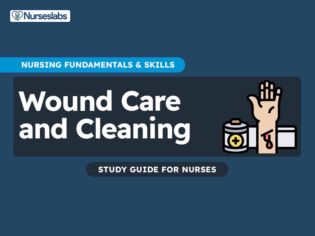
Cleaning and Dressing Wounds
In nursing, the meticulous process of cleaning and dressing wounds is vital for promoting healing and preventing complications. This structured approach involves systematic steps that ensure the wound is properly cleansed, assessed, and dressed with appropriate materials, tailored to the specific needs of each patient. By following these steps, healthcare providers can effectively manage wounds, enhance patient comfort, and support optimal recovery outcomes.
1. Collect all necessary supplies such as gloves, sterile gauze, wound cleanser, dressings, and any prescribed medications.
Ensures that all needed materials are readily available, reducing interruptions and maintaining a sterile environment to prevent infection.
2. Wash hands thoroughly with soap and water or use an alcohol-based hand sanitizer before and after the procedure.
Reduces the risk of introducing contaminants to the wound and protects both the patient and healthcare provider from infection.
3. Explain the procedure to the patient to obtain their consent and cooperation. Position the patient comfortably, ensuring the wound area is accessible.
Informs the patient and reduces anxiety, facilitating cooperation. Proper positioning helps ensure thorough and effective wound care.
4. Wear gloves, and if necessary, additional PPE such as a gown or mask.
Protects both the patient and the healthcare provider from potential contamination and infection.
5. Carefully remove the old dressing (if any), noting the type and amount of drainage. Dispose of the old dressing in a biohazard bag.
Prevents contamination of the wound and allows for assessment of wound exudate, which can indicate the wound’s healing progress or the presence of infection.
6. Assess the wound for size, depth, color, odor, and signs of infection or other complications.
Provides essential information to guide treatment decisions and monitor healing progress.
7. Use a sterile wound cleanser or saline solution to gently irrigate the wound. Clean from the least contaminated area (the wound itself) to the most contaminated area (the surrounding skin).
Removes debris, bacteria, and exudate, reducing the risk of infection and creating a clean environment for healing.
8. Pat the wound and surrounding skin dry with sterile gauze.
Prevents maceration of the skin, which can occur if the area remains too moist, and prepares the wound bed for dressing application.
9. Apply any prescribed topical medications or ointments to the wound.
Delivers targeted treatment to promote healing, reduce infection risk, or address other specific wound needs.
10. Place a new, sterile dressing over the wound, ensuring it covers the wound completely and adheres properly.
Protects the wound from contamination, absorbs exudate, and maintains an optimal moist environment for healing.
11. Use tape, bandages, or other securing methods to keep the dressing in place without causing undue pressure or restricting circulation.
Ensures the dressing stays in place, providing continuous protection and support to the wound.
12. Dispose of used supplies and PPE appropriately. Clean the surrounding area if necessary.
Maintains a clean and safe environment, reducing the risk of cross-contamination or infection.
13. Wash hands thoroughly with soap and water or use an alcohol-based hand sanitizer.
Completes the infection control process, ensuring that any potential contaminants picked up during the procedure are removed.
14. Record the wound care provided, including the wound assessment findings, the type of dressing applied, and any relevant observations.
Ensures continuity of care by providing accurate and up-to-date information for other healthcare providers and informs clinical decisions.
15. Educate the patient and caregivers on wound care instructions, signs of complications, and follow-up care.
Empowers patients to participate in their healing process, promotes adherence to treatment plans, and enhances overall patient outcomes.
Negative Pressure Wound Therapy (NPWT)
Negative Pressure Wound Therapy (NPWT) is a specialized treatment used to promote wound healing by applying negative pressure to the wound bed. Following these steps with careful consideration of rationale ensures safe and effective NPWT application in nursing practice. Here are the essential steps involved in NPWT:
1. Conduct a thorough assessment of the wound to determine its size, depth, exudate levels, and overall condition.
Proper assessment ensures that NPWT is suitable for the wound type and helps establish baseline measurements to monitor healing progress.
2. Collect NPWT equipment including the vacuum-assisted closure (VAC) device, sterile foam or gauze dressing, transparent film, tubing, and appropriate dressings for the wound.
Having all necessary supplies ready ensures smooth application of NPWT and minimizes interruptions during the procedure.
3. Explain the NPWT procedure to the patient, addressing any concerns and obtaining informed consent.
Fosters patient understanding and cooperation, promoting comfort and reducing anxiety during treatment.
4. Debride the wound as necessary to remove necrotic tissue, foreign debris, and excess exudate.
Promotes a clean wound bed, enhances effectiveness of NPWT, and reduces the risk of infection.
5. Place sterile foam or gauze dressing into the wound bed, ensuring it covers the entire wound area. Seal the dressing with a transparent film and connect it to the NPWT device via tubing.
Creates an airtight seal around the wound, allowing for controlled negative pressure application and promoting wound healing through enhanced circulation and removal of exudate.
6. Program the NPWT device to the prescribed negative pressure level based on wound characteristics and healthcare provider’s recommendations.
Optimal negative pressure settings facilitate effective wound healing by promoting granulation tissue formation, reducing edema, and enhancing tissue perfusion.
7. Regularly assess the NPWT system for proper functioning, ensuring adequate suction and seal integrity. Monitor the wound site for signs of discomfort, excessive bleeding, or other complications.
Continuous monitoring helps maintain treatment efficacy, prevents potential complications, and ensures patient safety throughout NPWT.
8. Educate the patient and caregivers on NPWT, including device operation, dressing changes, signs of complications, and follow-up care instructions.
Empowers patients to actively participate in their wound care, promotes adherence to treatment protocols, and enhances overall treatment outcomes.
9. Record detailed documentation of NPWT application, including initial wound assessment findings, treatment parameters, wound measurements, and patient response.
Provides a comprehensive record of care delivered, supports continuity of care among healthcare providers, facilitates treatment evaluation, and ensures accountability.
References
- Balsa, I. M., & Culp, W. T. N. (2015). Wound care. Veterinary Clinics of North America: Small Animal Practice. Advance online publication.
- Lei, J., Sun, L., Li, P., Zhu, C., Lin, Z., Mackey, V., … & He, Q. (2019). The wound dressings and their applications in wound healing and management. Health Science Journal, 13(4), 1-8.
- Rezvani Ghomi, E., Khalili, S., Nouri Khorasani, S., Esmaeely Neisiany, R., & Ramakrishna, S. (2019). Wound dressings: Current advances and future directions. Journal of Applied Polymer Science, 136(43), Article 47738.
- Wilkins, R. G., & Unverdorben, M. (2013). Wound cleaning and wound healing. Advances in Skin & Wound Care, 26(4), 160–163.


Leave a Comment