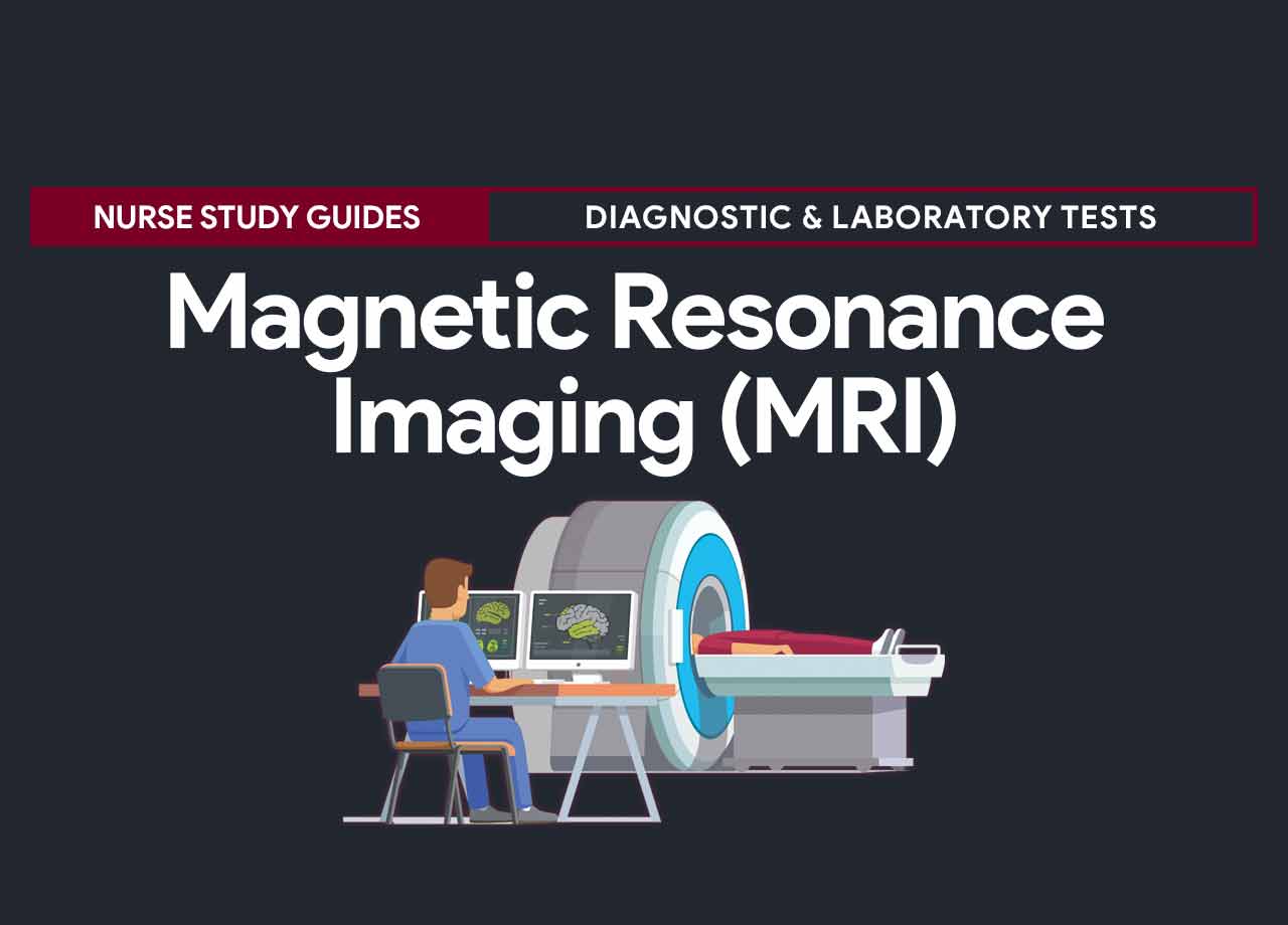
Magnetic resonance imaging (MRI) has the ability to see it through bone and to delineate fluid-filled soft tissue in great detail and produces images of organs and vessels in motion. In this noninvasive procedure, the patient is placed in a magnetic field into which a radiofrequency beam is introduced. Resulting energy changes are measured and used by the MRI computer to generate images on a monitor. Cross-sectional images of the anatomy are viewed in multiple planes and recorded for the permanent record.
Because the magnetic fields and radiofrequency waves used are imperceptible by the patient, no harmful effects have been documented. Research continues on the optimal magnetic fields and radiofrequency waves for various tissue types.
Procedure
The protocol and procedure for magnetic resonance imaging (MRI) differs per area but generally, the following steps are followed.
- If indicated, an IV line is started to administer a contrast medium before the procedure begins.
- The patient is placed in a supine position on a narrow, padded, nonmetallic bed that slides to the desired position inside the scanner.
- The patient is asked to remain still.
- Radiofrequency energy is directed at the area being tested. The radiologist may vary the waves and use the computer to manipulate and enhance the images.
- The resulting images are displayed on a monitor and recorded on film or magnetic tape for permanent storage.
- The patient is advised to keep his eyes closed to promote relaxation and prevent a closed-in-feeling.
- If nausea occurs because of claustrophobia, the patient is encouraged to take deep breaths.
- If the test is prolonged with the patient lying flat, monitor him for orthostatic hypotension.
Types
The following are the different types of magnetic resonance imaging (MRI):
Abdominal and pelvic
| Indication | Abnormal Results |
Blood Vessels (magnetic resonance angiography)
| Indication | Abnormal Results |
|
|
Breast
| Indication | Abnormal Results |
|
|
Cardiac
| Indication | Abnormal Results |
|
|
Intracranial
| Indication | Abnormal Results |
|
|
Musculoskeletal
| Indication | Abnormal Results |
|
Spinal
| Indication | Abnormal Results |
|
Urinary tract
| Indication | Abnormal Results |
|
|
Contraindication of MRI
Magnetic resonance imaging is contraindicated in:
- Patients with severe obesity (usually more than 300 pounds)
- Patients with claustrophobia
- Patients who are confused or agitated
- Patients who are unstable and require continuous life-support equipment, because most monitoring equipment cannot be used inside the scanner room. Magnet adaptive equipment is becoming available for use in the MRI scanner room.
- Patients with implantable metal objects such as pacemakers, infusion pumps, aneurysm clips, inner ear implants, and metal fragments in one or both eyes, because the magnet may move the object within the body and injure the patient.
Interfering Factors
- Patients inability to remain still while the procedure is ongoing
- Patients inability to fit into the scanner
Nursing Responsibilities for CT Scan
The following are the nursing interventions and nursing care considerations for a patient undergoing magnetic resonance imaging:
Before the procedure
The following are the nursing interventions before magnetic resonance imaging:
- Explain to the patient the purpose of the test. Tell him who will perform the test and where it will take place.
- Inform the patient that he’ll need to lie flat on a narrow bed, which slides into a large cylinder that houses the MRI magnets. Tell him that the scanner will make clicking, whirring, and thumping noises as it moves inside its housing and that he may receive earplugs.
- Explain to the patient that MRI is painless and involves no exposure to radiation from the scanner. A radioactive contrast dye may be used, depending on the tissue being studied.
- For MRI of the urinary tract, advise the patient to avoid alcohol, caffeine-containing beverages, and smoking for at least 2 hours and food for at least 1 hour before the test. Explain to the patient that he can continue taking medications, except for iron, which interferes with the imaging.
- Advise the patient that he’ll have to remain still for the entire procedure.
- Explain to the patient who’s claustrophobic or anxious about the test’s duration that he’ll receive a mild sedative to reduce his anxiety or that he may need to be scanned in an open MRI scanner, which may take longer but is less confining. Tell him that he’ll be able to communicate with the technician at all times and that the procedure will be stopped if he feels claustrophobic.
- If contrast media will be used, obtain a history of allergies or hypersensitivity to these agents. Mark any sensitivities on the chart and notify the practitioner.
- Instruct the patient to remove all metallic objects, including jewelry, hairpins, and watches.
- Ask the patient if he has any implanted metal devices or prostheses, such as vascular clips, shrapnel, pacemakers, joint implants, filters, and intrauterine devices. If so, the test may not be able to be performed.
- Make sure that the patient or a responsible family member has signed an informed consent form.
- Administer the prescribed sedative if ordered.
- At the scanner room door, recheck the patient one last time for metal objects.
- Just before the procedure, have the patient urinate.
During the procedure
- Remind the patient to remain still throughout the procedure.
- Assess how the patient responds to the enclosed environment. Provide reassurance if necessary.
- Monitor the cardiac function for signs of ischemia (chest pressure, shortness of breath, or changes in hemodynamic status).
- If the patient is unstable, make sure an IV line with no metal components is in place and that all equipment is compatible with MRI. If necessary, monitor the patient’s oxygen saturation, cardiac rhythm, and respiratory status during the test. An anesthesiologist may be needed to monitor a heavily sedated patients.
After the procedure
The nurse should be aware of these post-procedure nursing interventions after magnetic resonance imaging:
- Tell the patient that he may resume his usual activities as ordered.
- If the test took a long time and the patient was lying flat for an extended period, observe him for orthostatic hypotension.
- Provide comfort measures and pain medication as needed and ordered because of prolonged positioning the scanner.
- Monitor the patient for the adverse reaction to the contrast medium (flushing, nausea, urticaria, and sneezing).
Normal Results
The following are the expected normal results of magnetic resonance imaging:
- Results dependent on the specific type of MRI
- Structure and function of the studied organ within normal parameters for the patient
Abnormal Results
The abnormal results of a magnetic resonance imaging varies per area.
- Results dependent on a specific type of MRI.
- Abnormalities dependent on the particular organ studied (see abnormal results above).
Gallery
Images related to magnetic resonance imaging (MRI):
References
Additional resources and references for this guide:
- Alghamdi, A., Alghamdi, M., Alamri, S., Alshehri, M., Alatawi, I., Alzahrani, S., … & Alali, N. (2021). Assessment of Saudi Arabian Nurses’ Knowledge and Attitudes Toward Magnetic Resonance Imaging Safety. Journal of Radiology Nursing.
- Kanal, E. (1994). Pregnancy and the safety of magnetic resonance imaging. Magnetic resonance imaging clinics of North America, 2(2), 309-317.
- Smart, G. (1997). Helping children relax during magnetic resonance imaging. MCN: The American Journal of Maternal/Child Nursing, 22(5), 237-241.
- Westbrook, C., & Talbot, J. (2018). MRI in Practice. John Wiley & Sons.
- Wolters Kluwer Health/Lippincott Williams & Wilkins. (2009). Critical care nursing in a flash. Philadelphia.


Leave a Comment