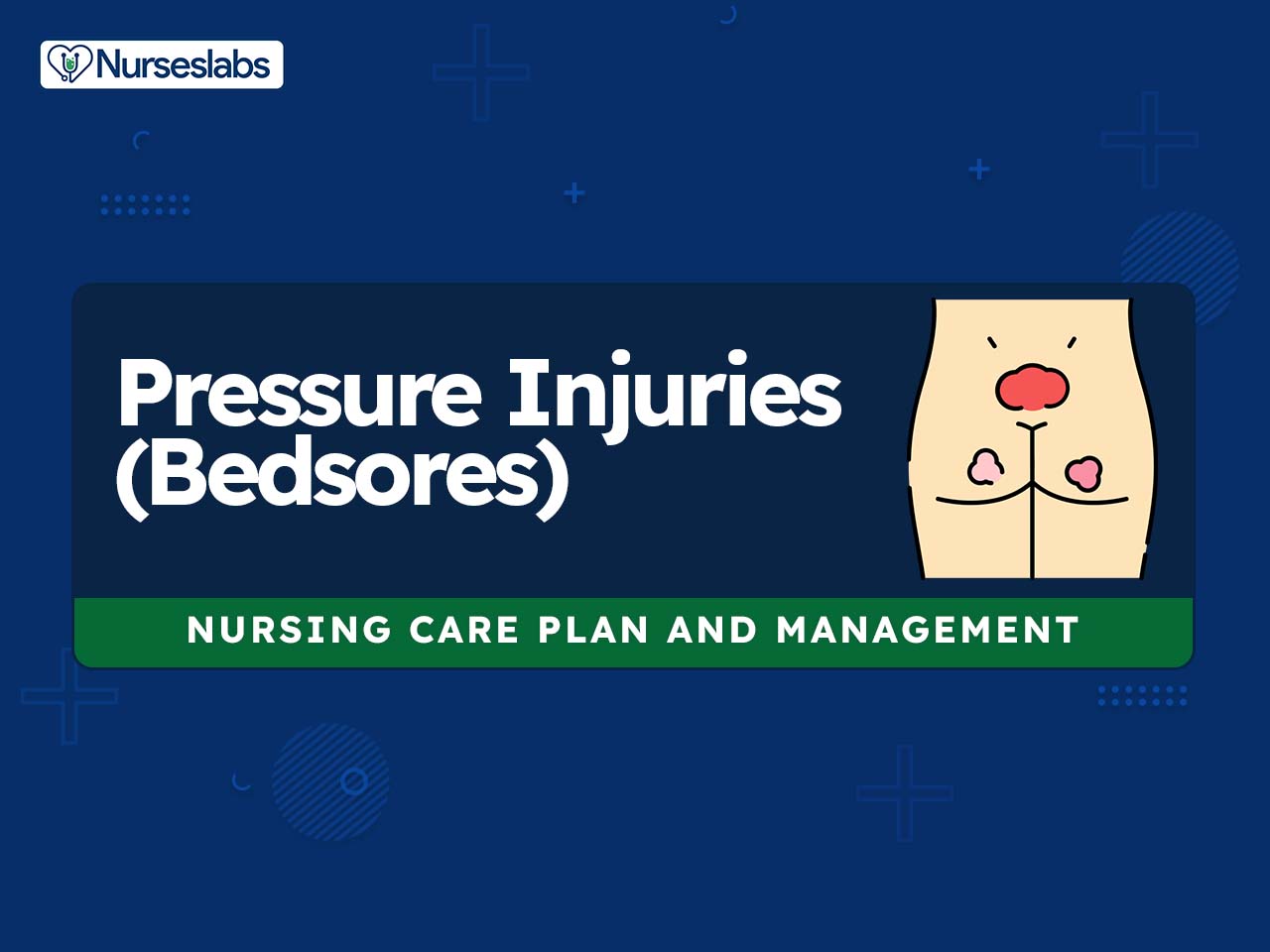
Pressure injuries, also known as bedsores or pressure ulcers, are a common problem in the healthcare industry. They occur when an area of the skin is subjected to pressure and is not relieved for a prolonged period. Currently, the National Pressure Injury Advisory Panel (NPIAP) defines a pressure injury as localized damage to the skin and underlying soft tissue, usually over a bony prominence or related to medical or other devices (Kirman & Geibel, 2022). This can happen in bedridden individuals, those using a wheelchair or having limited mobility. Pressure injuries can cause significant pain, and discomfort, and even lead to serious complications such as infections, sepsis, and even death.
Nursing care plans are essential for the treatment of pressure injuries. These care plans include a nursing diagnosis, assessment, interventions, and evaluation.
What are Pressure Injuries?
A pressure injury (also known as bedsores, pressure ulcers, or decubitus ulcers) is a localized skin injury where tissues are compressed between bony prominences and hard surfaces such as a mattress. They are caused by pressure in combination with friction, shearing forces, and moisture. The pressure compresses small blood vessels and leads to impaired tissue perfusion. The reduction of blood flow causes tissue hypoxia leading to cellular death.
Although the terms decubitus ulcer, pressure sore, and pressure ulcer have often been used interchangeably, the NPIAP has stated that pressure injury is the best term to use, given that open ulceration does not always occur. They may present as intact skin or an open ulcer and may be painful (Kirman & Geibel, 2022).
It is helpful to stage the pressure injury according to the system promulgated by the NPIAP.
- Stage 1 pressure injury – Nonblanchable erythema of intact skin
- Stage 2 pressure injury – Partial-thickness skin loss with exposed dermis
- Stage 3 pressure injury – Full-thickness skin loss
- Stage 4 pressure injury – Full-thickness skin and tissue loss
- Unstageable pressure injury – obscured full-thickness skin and tissue loss
- Deep pressure injury – persistent non-blanchable deep red, maroon, or purple discoloration
Pressure is exerted on the skin, soft tissue, muscle, and bone by the weight of an individual against a surface beneath. These pressures often exceed the capillary filling pressure of 32 mm Hg. Pressure injuries are common among clients hospitalized in acute- and chronic care facilities. It has been estimated that about one million pressure injuries occur in the United States. Older adult clients admitted to acute care hospitals for nonelective orthopedic procedures are at even greater risk for pressure injuries than other hospitalized clients. Each year, approximately 60,000 people die of complications of pressure injuries (Kirman & Geibel, 2022).
Nursing Care Plans and Management
Pressure injuries in stages I through III can be managed with aggressive local wound treatment and proper nutritional support, while stage IV pressure ulcers usually require surgical intervention. Nursing care planning goals for clients experiencing pressure injuries (bedsores) include assessing the contributing factors leading to a lack of tissue perfusion, assessing the extent of the injury, promoting compliance with the medication regimen, and preventing further injury.
Nursing Problem Priorities
The following are the nursing priorities for patients with pressure injuries:
- Assess and stage pressure injuries accurately.
- Implement effective pressure relief and redistribution strategies.
- Optimize wound care and promote healing.
- Manage pain and discomfort associated with pressure injuries.
- Prevent infection through proper wound hygiene and antimicrobial treatments.
- Provide education on self-care and prevention measures.
- Address underlying factors contributing to pressure injuries, such as immobility or poor nutrition.
- Monitor and manage complications, such as cellulitis or deep tissue damage.
- Collaborate with a multidisciplinary team for comprehensive care.
- Conduct regular reassessment and documentation of pressure injuries.
Nursing Assessment
Assess for the following subjective and objective data:
- Destruction of skin layers
- Disruption of skin surfaces
- Drainage of pus
- Invasion of body structures
- Pressure ulcer stages:
- Deep tissue injury (new stage):
- A purple or maroon localized area of intact skin or blood-filled blister resulting from pressure damage of underlying soft tissue.
- Stage I:
- Stage II:
- Partial-thickness skin loss involving the epidermis, dermis, or both. The ulcer is superficial and presents clinically as an abrasion or blister.
- Stage III:
- Full-thickness skin loss involving damage to or necrosis of subcutaneous tissue that may extend down to, but not through, underlying fascia.
- Slough may be present; may include undermining and tunneling.
- Stage IV:
- Extensive destruction, tissue necrosis, or damage to muscle, bone, or supporting structures, with or without full-thickness skin loss.
- Undermining and tunneling may develop.
- Unstageable:
- Full-thickness tissue loss in which the actual depth of the ulcer is completely obstructed by slough or eschar in the wound bed.
- Deep tissue injury (new stage):
Nursing Diagnosis
Following a thorough assessment, a nursing diagnosis is formulated to specifically address the challenges associated with pressure injury based on the nurse’s clinical judgement and understanding of the patient’s unique health condition. While nursing diagnoses serve as a framework for organizing care, their usefulness may vary in different clinical situations. In real-life clinical settings, it is important to note that the use of specific nursing diagnostic labels may not be as prominent or commonly utilized as other components of the care plan. It is ultimately the nurse’s clinical expertise and judgment that shape the care plan to meet the unique needs of each patient, prioritizing their health concerns and priorities.
Nursing Goals
Goals and expected outcomes may include:
- The client will receive stage-appropriate wound care and has controlled risk factors for the prevention of additional ulcers.
- The client will experience the healing of pressure injuries and experience pressure reduction.
- The client and caregiver will verbalize understanding of the following aspects of home care: nutrition, pressure relief, wound care, and incontinence management.
- The client and caregiver will verbalize their ability to cope adequately with the existing situation and provide support/monitoring as indicated.
- The client will demonstrate stable weight or progressive weight gain toward the goal.
- The client will be free of malnutrition.
- The client will verbalize understanding of individual interferences to adequate intake.
- The client will verbalize understanding of the impact of malnutrition on the development of pressure injuries.
- The client will participate in specific interventions to stimulate appetite and increase dietary intake.
- The client will avoid further development of preventable pressure injuries.
Nursing Interventions and Actions
Therapeutic interventions and nursing actions for patients with pressure injury may include:
1. Assessing and Staging Pressure Injuries
1. Assess the specific risk factors for pressure injuries.
Even clients with existing pressure injuries continue to be at risk for further injury. Nurses should consider all potential risk factors for pressure injury development. The pressure of an individual’s body weight or pressure from a medical device above a certain threshold for a prolonged period is thought to be the cause of pressure injuries. Many factors are identified as contributing factors to pressure injuries and injury formation, such as increased arteriole pressure, shearing forces, friction, moisture, and nutrition status (Mondragon & Zito, 2022).
2. Determine the client’s age and general condition of the skin.
Older adult clients have less elastic skin, less moisture, less padding, and thinning of the epidermis, making it more prone to skin impairment. The quality of the skin influences whether pressure leads to ulceration. Paralysis, insensibility, and aging lead to atrophy of the skin with thinning of this protective barrier. A decrease in epidermal turnover, a flattening of the dermal-epidermal junction, and loss of vascularity occur with advanced age (Kirman & Geibel, 2022).
3. Assess the client’s nutritional status, including weight, weight loss, and serum albumin levels, if indicated.
A severe protein depletion has an albumin level of less than 2.5 g/dL. Clients with pressure injuries lose big amounts of protein in wound exudates and may require 4000 kcal/day or more to remain anabolic. Malnutrition, hypoproteinemia, and anemia reflect the overall status of the client and can contribute to tissue vulnerability to trauma as well as cause delayed wound healing (Kirman & Geibel, 2022).
4. Assess for a history of preexisting chronic diseases (e.g., diabetes mellitus, acquired immune deficiency syndrome, Guillain-Barré syndrome, peripheral and/or cardiovascular disease).
Clients with chronic diseases typically exhibit multiple risk factors that predispose them to pressure injuries. These include poor nutrition, poor hydration, incontinence, and immobility. In clients with sensory deficits, an absent pressure feedback response may result in sustained pressure for a prolonged period, leading to tissue injury (Mondragon & Zito, 2022).
5. Assess the skin on admission and daily for an increasing number of risk factors.
The incidence of skin breakdown is directly related to the number of risk factor present. Prevention is the key to managing pressure injuries, and it begins with a complete medical and nursing history, a risk assessment, and a skin examination when the client is admitted (Kirman & Geibel, 2022).
6. Assess for a history of radiation therapy.
Irradiated skin becomes thin and brittle, may have less blood supply, and is at a higher risk for skin breakdown. A constant state of skin renewal that occurs approximately every 26 days makes the skin more vulnerable than any other organ to ionizing radiation injury. Ionizing radiation creates free radicals and reactive oxygen intermediates that lead to DNA and protein damage, as well as damage to cellular membranes of rapidly proliferating tissue (Manna & Cooper, 2022).
7. Assess the client’s awareness of the sensation of pressure.
Usually, people shift their weight off pressure areas every few minutes; this occurs more or less automatically, even during sleep. Clients with decreased sensation are unaware of unpleasant stimuli and do not shift weight, thereby exposing the skin to excessive pressure. In clients with normal sensitivity, mobility, and mental faculties, pressure injuries are unlikely. Conscious or unconscious feedback from the areas of compression leads them to change position, thereby shifting the pressure from one area to another long before any irreversible ischemic damage occurs (Kirman & Geibel, 2022).
8. Assess for fecal and urinary incontinence.
The urea in urine turns into ammonia within minutes and is erosive to the skin. While the stool may contain enzymes that cause skin breakdown. Diapers and incontinence pads with plastic liners trap moisture and speed up breakdown. These conditions cause the skin to be continually moist, thus leading to maceration. Additionally, frequent soiling has the effect of regularly introducing bacteria into an open wound (Kirman & Geibel, 2022).
9. Assess the client’s ability to move (shift weight while sitting, turn over in bed, move from the bed to a chair).
Immobility is a huge risk factor for pressure injury development among adult hospitalized clients. This situation may be present in clients who are neurologically impaired, heavily sedated or anesthetized, restrained, demented, or recovering from a traumatic injury. These clients cannot alter their position far enough or often enough to relieve the pressure (Kirman & Geibel, 2022).
10. Assess for environmental moisture (excessive perspiration, high humidity, wound drainage).
Moisture may contribute to skin maceration. Incontinence or the presence of a fistula contributes to ulceration in several ways. These conditions cause the skin to be continually moist, thus leading to maceration (Kirman & Geibel, 2022).
11. Assess the amount of shear (pressure exerted laterally) and friction (rubbing) on the client’s skin.
Shearing forces are most commonly noted on the sacrum, scapulae, heels, and elbows from skin-sheet friction, from semi-Fowler’s position and repositioning, and lift sheets. Trauma that causes deepithelialization or skin tears removes the barrier to bacterial contamination and leads to transdermal water loss, creating maceration and causing the skin to adhere to clothing and bedding (Kirman & Geibel, 2022).
12. Assess the surface that the clients spend the majority of time on (mattress for bedridden clients, cushion for clients in wheelchairs).
Clients who spend the majority of their time on one surface need a pressure reduction or pressure relief device to reduce the risk of skin breakdown. In theory, the reduction of tissue pressures below capillary filling pressures should allow adequate tissue perfusion. Selection of a support surface should be based on the client’s management plan, their risk factors for developing pressure injuries, and the cost of obtaining and servicing the device (Kirman & Geibel, 2022).
13. Assess the skin over bony prominences (sacrum, trochanters, scapulae, elbows, heels, inner and outer malleolus, inner and outer knees, back of the head).
These areas are at the highest risk for breakdown resulting from tissue ischemia from compression against a hard surface. The points of highest pressure with the client supine include the sacrum, heel, and occiput (40 to 60 mm Hg). With the client prone, the chest and knees absorb the highest pressure (50 mm Hg). when the client is sitting, the ischial tuberosities are under the most pressure (100 mm Hg). these pressures are greater than the end capillary pressure, which is why these are the areas where pressure injuries are most common (Kirman & Geibel, 2022).
14. Use an objective tool for pressure ulcer risk assessment: Braden scale, Norton scale, and Waterlow scale.
The Braden scale is the most widely used risk assessment. It consists of six subscales namely: activity, mobility, moisture, nutrition, sensory perception, and friction. The Norton scale includes the following items: physical condition, mental state, activity, mobility, and incontinence. Lower scores indicate a greater risk of pressure injuries. The Waterlow Scale for pressure injury risk assessment is commonly used for assessing the risk of pressure injuries in the United Kingdom and Ireland. It includes the following subscales: weight/height (body mass index), skin type visual risk, sex/age, continence, mobility, appetite, and medications. The scale includes special risk factors such as malnutrition, neurological defect, and major surgery/trauma (Satekova et al., 2016).
15. Assess the client’s level of pain, especially related to dressing changes and procedures.
Prophylactic pain medication may be indicated. However, although pain may be present at the injury site, it is more commonly absent because the client either is paraplegic in critical condition and unable to acknowledge the pain. An adequate examination of the wound may necessitate the administration of IV or oral pain medications to ensure client comfort. Chronic pain may be present among these clients and may be exacerbated by examination ulcers (Kirman & Geibel, 2022).
16. Assess and stage pressure injuries.
Staging is essential because it determines the treatment plan. Staging should be assessed at each dressing stage. It reflects whether the epidermis, dermis, fat, muscle, bone, or joint is exposed. If the ulcer is covered with necrotic tissue (eschar), it cannot be accurately staged. Stage I ulcers are difficult to detect in darkly pigmented skin. The use of mirrors or a penlight may be helpful. The National Pressure Injury Advisory Panel (NPIAP) system consists of four main stages of pressure injury but is not intended to imply that all pressure injuries follow a standard progression from stage 1 to stage 4 or that healing pressure injuries follow a standard regression from stage 4 to stage 1 to a healed wound (Kirman & Geibel, 2022).
17. Determine the condition of the wound or wound bed.
- 17.1. Presence of necrotic tissue
Necrotic tissue is tissue that is dead and eventually must be removed before healing can take place. Necrotic tissue exhibits a wide range of appearances: black, brown, leathery, hard, shiny, thin, tough, and white. Accurate injury staging cannot be accomplished until necrotic tissue is removed (Kirman & Geibel, 2022). - 17.2. Color
The color of the tissue is an indication of tissue viability and oxygenation. White, gray, or yellow eschar may be present in stage II and III ulcers. Stage II wound bed is viable, pink or red, and moist. Eschar may be black in stage IV ulcers. Deep tissue injury involves non-intact skin with persistent nonblanchable deep red, maroon, or purple discoloration (Kirman & Geibel, 2022). - 17.3. Odor
The odor may arise from infection present in the wound; it may also arise from the necrotic tissue. Some local wound care products may create or intensify the odors and should be distinguished from wound or exudate odors. A foul odor or discharge could be a sign of a more serious infection at the injury site (Kirman & Geibel, 2022). - 17.4 Viability of bone, joints, or muscle.
In stage IV pressure injuries, these may be apparent at the base of the ulcer. Wounds may demonstrate multiple stages or characteristics in a single wound. The clinical presentation of pressure injuries can be deceiving to the inexperienced observer. Soft tissue, muscle, and skin resist pressure to differing degrees. Generally, muscle is the least resistant and will become necrotic before the skin breaks down (Kirman & Geibel, 2022).
18. Measure the size of the ulcer, and note the presence of undermining.
The ulcer dimensions include length, width, and depth. An ulcer begins in the deepest tissue layers before the skin breaks down. Hence the opening of the skin’s surface may not represent the true size of the ulcer. A small area of skin breakdown may represent only the tip of the iceberg, with a large cavity and extensive undermining of skin edges beneath (Kirman & Geibel, 2022).
19. Assess the condition of wound edges and surrounding tissue.
Surrounding tissue may be healthy or may have various degrees of impairment. Healthy tissue is necessary for the use of local wound care products requiring adhesion to the skin. The presence of healthy tissue demarcates the boundaries of the pressure injury. Epibole or rolled wound edges are typically present in stages 3 and 4 of pressure injuries (Kirman & Geibel, 2022).
20. Assess the wound exudate.
Exudate is a normal part of wound physiology and must be differentiated from pus which is an indication of infection. Exudate may contain serum, blood, and white blood cells, and may appear clear, cloudy, or blood-tinged. The amount may vary from a few cubic centimeters, which are easily managed with dressings, to copious amounts not easily managed. Drainage is considered excessive when dressing changes are needed more often than every 6 hours.
21. Assess ulcer healing, using a pressure ulcer scale for healing (PUSH) tool.
This tool provides standardization in the measurement of wound healing. It quantifies surface area, exudate, and the type of wound tissue. This is a widely used tool developed by the NPIAP that grades pressure injuries based on wound size, wound bed tissue type, and exudate amount (Physiopedia, 2018).
2. Wound Care and Promoting Skin Integrity
1. Encourage the use of pressure-relieving devices such as specialized mattresses, cushions, heel troughs, and other devices.
Specialized mattresses and cushions can help distribute pressure more evenly across the body, reducing the risk of developing pressure injuries. These devices work by creating areas of low pressure that alternate with areas of higher pressure, which helps to promote blood flow and prevent tissue damage. These support surfaces may be divided into dynamic systems, which require an energy source to alternate pressure points, and static systems, which rely on the redistribution of pressure over a large surface area and do not require an energy source (Kirman & Geibel, 2022).
2. Encourage frequent repositioning of the client.
Repositioning the client frequently can help to distribute pressure more evenly across the body, decreasing the risk of developing bed injuries. Turning and repositioning the client remains the cornerstone of prevention and treatment through pressure relief. Clients who are capable of shifting their weight every 10 minutes should be encouraged to do so. Repositioning should be performed every two hours, even in the presence of a specialty surface or bed (Kirman & Geibel, 2022).
3. Ensure the client eats a well-balanced diet that includes food rich in:
A dietitian may be consulted to develop an individualized nutrition plan based on the client’s specific needs and medical history. Malnutrition is one of the few reversible contributing factors for pressure injuries, and establishing adequate caloric intake has been shown to improve the healing of these lesions (Kirman & Geibel, 2022).
- 3.1. Protein
Protein is essential for building and repairing tissues. Foods high in protein include lean meats, poultry, fish, eggs, beans, and dairy products. Protein is vital for the growth and maintenance of cells, fluid balance, and blood clotting; its functions include immune function preservation, wound healing enzyme repair and synthesis, cell multiplication, and collagen and connective tissue synthesis. Additional protein is also required to compensate for the nitrogen losses that occur with pressure injury exudate (Munoz et al., 2020). - 3.2. Vitamin C
Vitamin C is important for collagen synthesis, which helps to strengthen and repair tissues. Good sources of vitamin C include citrus fruits and juices, berries, tomatoes, and leafy green vegetables. Vitamin C also enhances the activation of leukocytes and macrophages in wounds, improves tensile strength, and aids in iron absorption (Munoz et al., 2020). - 3.3. Zinc
Zinc is important for wound healing and immune function. Foods high in zinc include oysters, beef, chicken, beans, eggs, and nuts. Zinc is also a cofactor for collagen formation, metabolizes protein, liberates vitamin A from the liver, and interacts with platelets in blood clotting. However, megadoses of zinc may inhibit healing and cause copper deficiency, therefore, the intake amount must be checked with a dietitian (Munoz et al., 2020). - 3.4. Iron
Iron is vital for oxygen transport in the blood, which is important for wound healing. Good sources of heme iron include lean meats, poultry, and fish, while sources of non-heme iron include beans, nuts, and leafy green vegetables. Iron is also important in collagen formation and creates energy from cells (Munoz et al., 2020).
4. Provide local wound care.
Wound care may be broadly divided into non-operative methods and operative methods. For stage 1 and 2 pressure injuries, wound care is usually conservative. For stage 3 and 4 pressure injuries, surgical intervention may be required, though some of these lesions must be treated conservatively because of coexisting medical problems (Kirman & Geibel, 2022).
Stage I:
- Apply a flexible hydrocolloid dressing or a vapor-permeable membrane dressing.
Hydrocolloid dressings form an occlusive barrier over the wound while maintaining a moist wound environment and preventing bacterial contamination (Kirman & Geibel, 2022). Vapor-permeable dressings assist in providing a moist wound bed and protect fragile granulation tissue (South & West Devon Formulary and Referral, 2019). - Apply a vitamin-enriched emollient to the skin every shift.
Emollients help restore the barrier function of the skin, reduce itching, and increase the level of hydration. The use of emollients has been found to play a key role in the prevention and treatment of stage 1 pressure injuries. Clinical evidence from an Australian trial in older adults reported an almost 50% reduction of skin tears when an emollient was applied twice daily (Wounds UK, 2022).
Stage II:
- Apply alginates as indicated.
Alginate dressings are a type that is highly absorbent and so can absorb the fluid (exudate) that is produced by some ulcers. These are often used for ulcers with moderate-to-heavy exudate. Alginate forms a gel when it comes into contact with wound drainage and may be used in light to heavily draining stage II injuries. It should not be applied to dry or minimally draining wounds, as it can cause dehydration and delay wound healing (Kirman & Geibel, 2022). - Apply hydrocolloids or a vapor-permeable membrane dressing.
Hydrocolloids are used to promote healing and wound debridement. They are not advised to use for heavy-exudate-producing wounds. This gel can have fibrinolytic properties that can enhance wound healing, protect against secondary infection, and insulate wounds from contaminants (Kirman & Geibel, 2022). - Apply gauze with a sodium chloride solution
This maintains a moist environment but requires multiple dressing changes. Dressings must be removed while still wet. Dressings absorb small amounts of drainage. Woven or nonwoven gauze provides obliteration of dead space, retention of moisture, exudate absorption, and mechanical debridement (Kirman & Geibel, 2022). - Apply hydrogels as ordered.
Hydrogels provide moisture to dry, sloughy, or necrotic wounds and assist autolytic debridement. This can be used on wounds with low exudate. They are usually used for shallow ulcers without exudates. All forms of gel dressings keep the wound surface moist as long as they are not allowed to dehydrate. Some gel dressings provide limited to moderate absorption, some provide insulation, and some protect against bacterial invasion (Kirman & Geibel, 2022).
Stage III and IV:
- Foams
Different foams have different levels of absorbency. They are best used on granulating wounds. Foams lessen odor and repel bacteria and water. They also obliterate dead space and provide retention of moisture, exudate absorption, and mechanical debridement (Kirman & Geibel, 2022). - Gauze with a sodium chloride solution
This maintains a moist environment but requires multiple dressing changes as described for stage II. A multitude of cleansing agents are on the market, but none is more efficacious than the others, and expert opinion still favors normal saline (Kirman & Geibel, 2022). - Wound fillers
Wound fillers are used as a primary dressing to pack wounds, and maintain a moist environment. They also provide autolytic debridement. Wound fillers are non-adherent biomaterials in the form of pastes, granules, or powders that function to moisten the wound bed and absorb exudates. A secondary dressing such as gauze is required (Firlar et al., 2022). - Autolytic debridement
Using a hydrocolloid or hydrogel, create a moist wound interface that enhances the activity of endogenous proteolytic enzymes within the wound, liquefying and separating necrotic tissue from healthy tissue. Autolytic debridement is the lysis, or breakdown, of damaged tissue at a wound site by the body’s natural defense system by enzymes that digest specific components of body tissues or cells, such as proteins, fibrin, and collagen (Choo et al., 2019). - Sharp or surgical debridement.
This procedure removes the necrotic tissue and senescent cells that slow down the tissue repair process, converting a chronic wound into an acute one in the process. It is indiscriminate in the removal of vital and devitalized tissue and thus requires a great deal of clinical skill and judgment (Kirman & Geibel, 2022). - Mechanical debridement
This involves allowing a traditional gauze-type dressing to dry out and adhere to the surface of the wound before manually removing the dressing and debriding any tissue attached to it. Mechanical nonselective debridement is done by loosening and removing the necrotic tissue by means of whirlpool treatments, forceful irrigation, or the use of wet-to-dry dressings (Kirman & Geibel, 2022). - Electrical stimulation
Electrical stimulation is advocated as a way of healing pressure injuries and is recommended in at least four clinical practice guidelines. It is provided by an electrical current that can be applied in different ways. It can be delivered either as a direct or indirect pulsed current (Arora et al., 2020). - Biosurgery
Therapeutic use of live blowfly larvae (maggots) for quick debridement. Biosurgery has long been used in battlefield situations and recent case studies of the therapy for treating various chronic ulcers have been positive (Choo et al., 2019). - Topical growth factors
Nerve-growth factors, colony-stimulating factors, and fibroblast growth factors that are found to be effective in treating diabetic and venous ulcers. The recombinant human platelet-derived growth factor becaplermin has been approved by the US FDA for the treatment of lower extremity diabetic neuropathic ulcers that extends into the subcutaneous tissue or beyond. Other growth factors are also being evaluated for use in human clinical settings (Kirman & Geibel, 2022). - Negative pressure wound therapy
A wound dressing system that continuously or intermittently applies a subatmospheric pressure to the surface of a wound to assist healing. NPWT enhances wound healing by reducing edema, increasing the rate of granulation tissue formation, and stimulating circulation (Kirman & Geibel, 2022). - Enzymatic debridement (chlorophyll, collagenase, papain)
Enzymatic debridement uses proteolytic enzymes to remove necrotic tissue. These agents work by selectively digesting the collagen portion of the necrotic tissue. Care should be taken to prevent damage to surrounding healthy tissues. This uses various chemical agents that act by attacking the collagen and liquefying necrotic wound debris without damaging granulation tissue (Kirman & Geibel, 2022).
5. Use povidone iodine instead of hydrogen peroxide during cleansing or debridement.
Povidone iodine solution can be used to debride infected wounds. Although the effervescent action of hydrogen peroxide results in wound debridement, it is not recommended for frequent or long-term use in pressure injuries because it indiscriminately removes necrotic material and fragile granulation tissue and because it and other cleansing agents are toxic to fibroblasts (Kirman & Geibel, 2022).
6. Irrigate open wounds using normal saline.
When no germicidal action is required, normal saline can be used in cleansing the wound. Saline solution should also be used as a rinse after other solutions are used to irrigate the wound and minimize fluid shifts within newly forming tissue, Normal saline solution can reduce the drying effects that some irrigants may have on tissue (Kirman & Geibel, 2022).
7. Elevate the client’s head, shoulders, and heels when lying supine for long periods.
The client;’s head and shoulders should be minimally elevated on one pillow or a foam wedge to reduce shear forces and prevent the client from “bottoming out” or having the sacrum and ischial tuberosities resting on the bed frame. The client may also use a wedge-shaped cushion to decrease the incidence of heel pressure injuries (Kirman & Geibel, 2022).
3. Promoting Infection Control and Preventing Infections
Poor nutritional status can weaken a client’s immune system and reduce their ability to fight off infections, making them more susceptible to infections in open pressure ulcers. Open pressure ulcers create a direct entryway for bacteria and other pathogens to enter the body, increasing the risk of infection. Additionally, the proximity of sacral wounds to the perineum can increase the risk of contamination from fecal matter and other bodily fluids. In clients who present with sepsis and pressure injuries, the sepsis is usually caused by a urinary tract infection (Kirman & Geibel, 2022).
1. Assess the client’s nutritional status.
Clients who seriously lack nutrition (serum albumin <2.5 mg/dl) are at risk of developing an infection produced by a pressure ulcer. Also, clients with pressure ulcers lose tremendous amounts of protein in wound exudate and may require 4000 kcal/day or more to remain anabolic. Poor nutritional status certainly contributes to the chronicity often seen in these lesions and inhibits the ability of the immune system to prevent infections (Kirman & Geibel, 2022).
2. Assess the client for unexplained sepsis.
When a septic workup is done, the pressure ulcer must be considered a possible cause. The wounds are almost always open to drain and therefore do not constitute debridement emergencies. Only on rare occasions are these injuries entirely occluded by a thick leathery eschar that prevents open drainage. Debridement may be required to facilitate drainage and prevent systemic infection (Kirman & Geibel, 2022).
3. Assess for urinary and fecal incontinence.
Sacral wounds, because of their proximity to the perineum, are at the highest risk for infection caused by urine or fecal contamination. It is sometimes difficult to isolate the wound from the perineal area. These conditions cause the skin to be continually moist, thus leading to maceration. Frequent soiling has the effect of regularly introducing bacteria into an open wound (Kirman & Geibel, 2022).
4. Assess pressure injuries for odor, tissue color, and drainage.
Foul-smelling pressure injuries may indicate an infection. Infected tissue usually has a gray-yellow appearance without evidence of pink granulation tissue. The presence of exudate that is clear to straw-colored is normal. While purulent green or yellow drainage in large amounts indicates an infection. A foul odor or discharge could be a sign of a more serious infection at the injury site (Kirman & Geibel, 2022).
5. Assess the client’s temperature.
Fever is considered a temperature above 100.4℉(38℃) indicating the presence of infection unless the client is immunocompromised or diabetic. A client with spinal cord injury who develops a pressure injury may show that a delayed diagnosis concerning fever leads to impaired functional outcomes with exacerbation of tissue damage and poor health status outcomes such as increased rates of hospitalization, prolonged hospital stay, increased cost of care, with increased mortality rates (Rabadi, 2021).
6. Monitor the client’s white blood cell count (WBC).
Elevated WBC counts indicate an infection, although, in older adult individuals, the WBC count may rise only slightly during infection, indicating a diminished marrow reserve. A complete blood count with differential may show an elevated WBC count indicative of inflammation or invasive infection (Kirman & Geibel, 2022).
7. Obtain wound cultures, if indicated.
All pressure injuries are colonized because skin normally has flora that will be found in an open skin lesion; however, all pressure injuries are not infected. Infection is present when there is copious, foul-smelling, purulent drainage and the client has other signs of infection (fever, increased pain) and bacteria count greater than 105. Swab cultures are not recommended. Instead, tissue biopsy should be used to quantify and qualify the aerobic and anaerobic organisms present. A biopsy also enables the clinician to distinguish between simple contamination and tissue invasion, an important distinction that is not revealed by the common practice of swabbing the wound surface for culture (Kirman & Geibel, 2022).
8. Monitor for signs of bone infection.
Osteomyelitis, or infection in a bone, should be considered in clients with an elevated ESR, an elevated WBC count, and/or abnormal pelvic films suggestive of osteomyelitis, as well as in cases of stage 4 pressure injury with exposed bone. If osteomyelitis is confirmed, treatment with a prolonged course of antibiotic therapy may be indicated (Kirman & Geibel, 2022).
9. Consult with a dietitian for assistance with a high-protein, high-calorie diet.
A high-calorie, high-protein diet may be recommended to help in healing and fighting infection. Nutritional status should be evaluated and optimized to ensure an adequate intake of calories, proteins, and vitamins. Malnutrition is one of the few reversible contributing factors for pressure injuries, and establishing adequate caloric intake has been shown to improve the healing of these lesions (Kirman & Geibel, 2022).
10. Provide thorough perineal hygiene after each episode of incontinence.
This can lessen pathogens in the area of sacral pressure ulcers. The wound surrounding intact skin must be kept clean and free of urine and feces through frequent inspection and cleansing. The appropriate evaluation of urinary or fecal incontinence is complex but must be performed thoroughly. Potentially reversible causes should be identified and treated (Kirman & Geibel, 2022).
11. Provide hydrotherapy, if indicated.
It is used to achieve wound cleansing and promote good circulation. Daily whirlpool therapy or pulse lavage therapy may be used to irrigate and mechanically debride the wound. The goal of this approach is to create a bed of well-granulated tissue throughout the ulcer cavity. Small well-granulated ulcers can heal with re-epithelialization (Boyko et al., 2018).
12. Provide local wound care as prescribed.
The type and level of wound treatment depend on the staging of the ulcer and the type of infection present. Stage 1 can be covered with transparent film dressings as needed. Stage 2 benefits from a moist wound environment. Treatment of stage 3 and 4 injuries is based on the presence of necrotic tissue (Mondragon & Zito, 2022).
13. Use proper infection control measures, such as hand hygiene and the use of personal protective equipment, when caring for clients with pressure ulcers.
Practicing good hand hygiene and using personal protective equipment can reduce the risk of infection and promote the healing of pressure ulcers. This can also prevent further complications and improve outcomes for the client. During perineal care for an incontinent client, the nurse may don gloves and an apron to avoid contamination from possible discharges from a pressure injury. Hand hygiene before and after procedures, especially wound care, is necessary to avoid contaminating the client’s open wounds.
14. Administer antibiotics as prescribed.
Complicated wounds may develop cellulitis or sepsis, requiring antibiotic therapy. Oral antibiotics or topical silver sulfadiazine can be effective. Empiric antibiotics may be started immediately. Initial antimicrobial therapy should be broad-based, to cover aerobic gram-positive and gram-negative organisms and anaerobes (Kirman & Geibel, 2022).
15. Provide adequate discharge instructions for the client and family members.
Discharge planning begins early in the hospital stay and requires an interdisciplinary approach. Knowledge of available resources facilitates smooth transitions through all levels of care. Education of the client and caregiver in preventing and treating pressure injuries is increasingly important. Various methods can be used to facilitate the educational process, including charts, diagrams, photographs, and videos (Kirman & Geibel, 2022).
16. Institute measures to prevent urinary or fecal incontinence.
Manual disimpaction and the addition of stool bullying agents to the diet may relieve overflow fecal incontinence. This also decreases the potential risks for infection, especially if the client has sacral pressure injuries. Urinary or fecal incontinence with no treatable cause may be minimized by establishing a bowel and bladder regimen. Constipating agents and a low-residue diet also may be helpful (Kirman & Geibel, 2022).
17. Provide diapers and incontinence pads.
Diapers and incontinence pads may be useful in absorbing moisture away from the surface of the skin, provided that they are checked regularly and changed when soiled. If used inappropriately, these products may aggravate maceration and result in dermatitis (Kirman & Geibel, 2022).
18. Assist in obtaining specimens for tissue biopsy.
A tissue biopsy should be performed for wounds that do not demonstrate clinical improvement despite adequate care and for wounds in which tissue invasion by bacteria is suggested. This allows the quantification and identification of bacterial species and their antibiotic susceptibilities. Bone biopsy is the criterion standard for the diagnosis of osteomyelitis within a pressure injury (Kirman & Geibel, 2022).
19. Prepare the client for debridement, as ordered.
The purpose of wound debridement is to remove all materials that promote infection, delay granulation, and impede healing, including necrotic tissue, eschar, and slough (the stringy yellow, green, or gray nonviable debris in an ulcer). Three debridement procedures may be used: enzymatic debridement, mechanical debridement, and sharp debridement (Kirman & Geibel, 2022).
20. Utilize the appropriate solutions for wound cleansing.
The major purpose of cleansing the wound is to decrease its bioburden and facilitate healing. Povidone iodine is useful against bacteria, spores, fungi, and viruses. Dilution is recommended, and this agent should be discontinued when granulation occurs. Acetic acid (0.5%) is specifically effective against Pseudomonas aeruginosa, a particularly difficult and common organism in fungating lesions. However, acetic acid can change the color of tissue and can mask potential superinfection. Sodium hypochlorite (2.5%) has some germicidal activity but it is primarily used to debride necrotic tissue (Kirman & Geibel, 2022).
21. Apply topical antibiotics before debridement as prescribed.
Antibiotic creams such as silver sulfadiazine may be applied to wounds to decrease the bacterial load. Silver sulfadiazine has an excellent antimicrobial spectrum of activity, low toxicity, ease of application, and minimal pain. Mafenide, an antimicrobial agent that is bacteriostatic to many gram-positive and gram-negative organisms, can penetrate an eschar and promote autolytic softening of the eschar before debridement (Kirman & Geibel, 2022).
22. Monitor for signs of antibiotic resistance.
The adverse effects of antibiotics are well known, and those that impede wound healing should be considered and counteracted. Antibiotic resistance is a major concern. Therefore, when antibiotic therapy is ordered, the nurse must also be alert to detect signs of antibiotic resistance, and the nurse must also be attentive to the results of the laboratory data, especially culture and sensitivity results (Kirman & Geibel, 2022).
4. Empowering Patient and Promoting Adherence to Therapeutic Regimen
Clients with pressure injuries are at risk of poor adherence to therapeutic regimen due to several factors, including impaired functional status, lack of previous experience in managing pressure injuries, and the need for long-term pressure management. Impaired functional status can make it difficult for the client to perform self-care activities and follow a treatment plan, while a lack of previous experience may result in inadequate wound care. Additionally, long-term pressure management requires ongoing attention and monitoring, which can be challenging for clients and their caregivers.
1. Assess the client’s and caregiver’s knowledge of and ability to provide local wound care.
Clients are no longer kept hospitalized until pressure injuries have healed. The need for local wound care may continue at home for weeks to months. Preventing and treating pressure injuries is a complex task, and family members are usually not trained for such tasks. Knowledge and attitude appear to be positively associated with the actual practice of preventing illnesses (Sari et al., 2022).
2. Assess the client’s and caregiver’s understanding of the prevention of further pressure injury development.
Immobile clients will need frequent repositioning to lessen the risk of a breakdown in those areas that are intact. Impaired mobility is probably the most common reason why clients are exposed to prolonged uninterrupted pressure that causes pressure injuries (Kirman & Geibel, 2022).
3. Assess the client’s and caregiver’s understanding of the long-term nature of wound healing.
Pressure injuries may take weeks to months to heal even under ideal circumstances. The wound heals from the base of the ulcer up, and from the edges of the ulcer toward the center. Palliative wound care may be appropriate for clean, chronic, non-healing wounds. The wound healing may begin immediately after an injury to the epidermal layer and might take years. Wound healing has three overlapping phases which are inflammation, proliferation, and remodeling. Any disruption leads to abnormal wound healing (Ozgok Kangal & Regan, 2022).
4. Assess the client’s and caregiver’s understanding of the relationship between incontinence and further skin breakdown or complication of healing.
Managing incontinence may be the most difficult aspect of home management and is often the reason for nursing home placements are made. Incontinence or the presence of a fistula contributes to ulceration in several ways. These conditions cause the skin to be continually moist, thus leading to maceration. Frequent soiling has the effect of regularly introducing bacteria into an open wound (Kirman & Geibel, 2022).
5. Assess the client’s and caregiver’s understanding of and ability to provide a high-calorie, high-protein diet throughout wound healing.
Clients may require enteral feeding (through gastronomy tube, nasogastric tube feedings, or the oral route), which requires knowledge of preparation and the use of special equipment. Before initiating enteral nutrition, the risks and benefits of nutrition support must be discussed with the client and caregivers. Although artificial nutrition and hydration are medical treatments, an individual’s cultural and/or religious values may dictate their decision to accept or decline enteral nutrition (Munoz et al., 2020).
6. Assess for the availability of a pressure reduction or pressure-relief surface.
Clients may take a thick, dense foam mattress home from the hospital to place on their own bed. Rental provisions of low-air-loss beds (e.g., KinAir, Flexicare) and air-fluidized therapy beds (FluidAir, Skytron, Clintron) may be arranged but often pose a financial difficulty because few payer sources will cover the cost of these beds in the home. These pressure-relief surfaces are often heavy, expensive, and difficult to clean, and they require ongoing maintenance to ensure proper function (Kirman & Geibel, 2022).
7. Educate the client and the caregiver to report the following signs indicating wound infection: fever, malaise, chills, foul-smelling odor, and purulent drainage.
Early detection prompts immediate intervention. Infection is a common complication of pressure injury which can be local such as cellulitis or osteomyelitis or systemic such as septicemia with a greater than 50% mortality (Rabadi, 2021).
8. Educate the client and the caregiver in managing incontinence (e.g., use of moisture barrier ointments, use of underpads, use of external catheters).
Teaching proper techniques can prevent leakage and skin problems. Reusable products such as underpads or linen protectors made of cloth with a waterproof lining are better for the client’s skin and are more economical but require laundering. Moisture barrier ointments protect intact skin from excoriation. The Wound, Ostomy, and Continence Nurses Society (WOCN) recommended the use of incontinence skin barriers such as creams, ointments, pastes, and film-forming skin protectants as needed to protect and maintain intact skin in incontinent individuals (Kirman & Geibel, 2022).
9. Educate the client and the caregiver regarding local wound care, and allow for a return demonstration.
This will allow the client to use new information immediately, thus enhancing retention. Immediate feedback allows the learner to make corrections, rather than practice the skill incorrectly. The skin surrounding the injury should be protected from excessive moisture and friction to prevent breakdown. Dressings should be changed regularly and as soon as they become soiled with urine or feces to prevent wound contamination (Boyko et al., 2018).
10. Provide written instructions with listed resources.
Long-term management requires specific written plans to enhance treatment adherence. Several internet resources provide lay education. Specific details that are required include who should provide the care, how often it should be provided, and the supplies and equipment needed. How the care is to be undertaken should be individualized, written down, and readily available (Kirman & Geibel, 2022).
11. Involve a social worker or case manager.
Referral helps the client and family determine whether placement in an extended care facility is needed. Because many clients with pressure injuries are older, it is often an older spouse who is available to provide care; as a result of the intensive nursing care needs of these clients, discharge to home is often unrealistic. After they return home, the client may also benefit from visits from a home healthcare organization. Such visits may ease the transition and ensure that pressure avoidance strategies are adapted to the home and continued over the long term (Kirman & Geibel, 2022).
12. Consult a wound specialist to evaluate care at the home.
Besides evaluating the ability to deliver care, the specialist may be useful in securing specialty treatment. Wound care specialists provide comprehensive client and caregiver education to ensure the wound is properly cared for at home. Clients are also taught to recognize any signs of infection that may require extended professional care, so they can be appropriately treated (NewportCare Medical Center, 2019).
13. Educate the client and the caregiver on the importance of pressure reduction and relief (e.g., turning schedule, use of specialty beds, use of relief surface where the client sits).
Information can nurture enhanced adherence to pressure ulcer treatment guidelines. According to WOCN, scheduling regular repositioning and turning for bedbound and chairbound individuals should take into consideration the condition of the client and the pressure redistribution support surface in determining the repositioning strategy (Kirman & Geibel, 2022).
14. Discuss with the client and caregiver the possible need for respite care.
Long-term responsibility for client care in the home is burdening; those providing care may need help to understand that their needs for relaxation are important to the maintenance of health and should not be viewed as avoidance of the responsibility. The guiding principles of palliative care for pressure injury management focus on relieving pain and providing comfort for the individual (Munoz et al., 2020).
15. Discuss with the client and caregiver the need for in-home nursing care or homemaker services.
These provide all or part of the client’s care and can be economical to the client. Also, keeping the client in his or her environment lessens the risk of hospital-acquired infection and keeps the client in familiar surroundings. Transmural care (a way of providing care that delivers activities to support clients and their families/partners, and activities to promote continuity of care), hospital-in-the-home care, care provided by a team of different disciplines, or care that is usually provided, may make a difference to whether people develop pressure injuries, the length of time of wound healing, and whether these clients may become readmitted to the hospital (Joyce et al., 2018).
16. Arrange follow-up consultations.
Follow-up should be performed every three weeks for the first several months. The interval may then be increased to every six months and then yearly. Early issues include sutures and drain removal, and when to allow the client to exercise or sit up (Kirman & Geibel, 2022).
17. Educate the cargeiver/s to document the client’s wound healing process.
Concisely documented measurement of the wound healing process contributes to efficient management. The most common method of monitoring the healing of pressure injuries utilizes photography and diagrams. Sophisticated radiographic techniques are available for this purpose as well, but they are too expensive for routine use (Kirman & Geibel, 2022).
18. Instruct caregivers on repositioning the client once at home.
Once healing is complete, long periods of uninterrupted pressure must be avoided. Clients must be repositioned frequently, either by their efforts or with help from their support group. Seated clients with upper-extremity function should lift themselves from their wheelchairs for at least 10 seconds every 10 to 15 minutes. Clients in bed should be repositioned at least every two hours (Kirman & Geibel, 2022).
5. Promoting Optimal Nutrition Status
Nutrition plays an important role in the prevention and treatment of pressure injuries. Macro- and micronutrients are required by each organ system in specific amounts to promote the growth, development, maintenance, and repair of body tissues. The 2019 European Pressure Ulcer Advisory Panel (EPUAP), National Pressure Injury Advisory Panel (NPIAP), and Pan Pacific Pressure Injury Alliance (PPPIA) recommend healthcare personnel consider the impact of impaired nutrition status on the risk of pressure injuries.
1. Perform a nutrition screening on a client with pressure injuries.
Any member of the interdisciplinary team who has been educated on screening tools can perform a nutrition screening. A validated screening tool can determine nutrition risk in all types of clients, including those with fluid shifts and for whom weight and height cannot be easily obtained. In acute care facilities, nutrition screening is conducted within 24 hours of admission. Information obtained through the screen is used by a registered dietitian to identify clients whose nutrition concerns warrant further assessment (Munoz et al., 2020).
2. Obtain the client’s weight and BMI during admission.
It is known that decreased acceptance of food and fluids/water and weight loss are associated with pressure injuries. Additionally, low body weight is associated with impaired wound healing. Unplanned or involuntary weight loss is considered a major risk factor for both malnutrition and pressure injury development. Obesity also places the client at risk for pressure injuries due to hypovascularity, decreased mobility, and difficulty with self-repositioning (Munoz et al., 2020).
3. Review usual daily caloric intake and dietary choices.
This identifies current strengths and weaknesses in the dietary program. This also aids in determining the individual needs for adjustment and teaching. Decreased intake of calories, protein, vitamins, and minerals is commonly seen in individuals with malnutrition, which is often associated with undernutrition. Overnutrition, on the other hand, is a form of malnutrition in which the amount of nutrients consumed exceeds the number of nutrients needed to support growth, development, and metabolism. Overnutrition can result in individuals becoming overweight and obese (Munoz et al., 2020).
4. Identify the client’s body composition.
Body composition is an independent risk factor for malnutrition, sarcopenia, and associated comorbidities. Taking a closer look at the body composition of the client over time can show decreased body mass, uncovering an increased risk for sarcopenic obesity. In addition, sarcopenia is also often present in immobile individuals with undernutrition (Munoz et al., 2020).
5. Establish a weight goal for the client aided by the registered dietitian.
The EPUAP/NPIAP/PPPIA recommendation is to provide 30 to 35 kcal/kg of body weight per day for adults with pressure injuries who are malnourished or at risk of malnutrition. Setting a clear weight goal helps guide the healthcare personnel in creating a suitable care plan (Munoz et al., 2020).
6. Provide foods with adequate kilocalories and rich in unsaturated fat.
Providing and consuming adequate kilocalories support collagen and nitrogen synthesis, thus promoting anabolism by sparing protein from being used as an energy source. Fat is the most concentrated source of calories, providing a reserve source of energy in the form of stored triglycerides in adipose tissue. It cushions bony prominences, provides insulation, and transports the fat-soluble vitamins A, D, E, and K, which are stored in the liver (Munoz et al., 2020).
7. Promote an increase in energy intake and increased protein consumption.
If the energy from carbohydrates and fat fails to meet the individual’s needs, the liver and kidneys synthesize glucose from noncarbohydrate sources, such as protein. Wound healing is compromised if the body is forced to produce glucose by degrading protein and depleting lean body mass. Therefore, a study recommended increasing energy intake by 50% above the normal level and increasing protein to 1.5 g/kg of body weight (Munoz et al., 2020).
8. Encourage the client to increase fluid intake as tolerated.
Adequate daily fluids/water should be offered to and encouraged for all individuals with pressure injuries. Water is the solvent for minerals, vitamins, amino acids, glucose, and other small molecules, allowing them to diffuse into and out of cells. Fluid requirements are met with liquids, including the water content of food, which accounts for 19% to 27% of the total fluid intake of healthy adults (Munoz et al., 2020).
9. Administer oral nutritional supplements as indicated.
Clients with chronic diseases or at risk of/with a pressure injury frequently cannot meet protein and calorie needs. Aside from increasing the energy expenditure of the client, pressure injuries are responsible for a further worsening of the energy balance. When oral feeding is safe, the use of oral nutritional supplements can be an effective strategy to help fulfill protein and calorie requirements (Munoz et al., 2020).
10. Assist in administering enteral or parenteral nutrition.
The goal of artificial nutrition support for clients with pressure injuries is to improve their nutrition status, promote healing, and restore their immune function. When pressure injury healing has stalled and oral intake is inadequate to support healing, enteral nutrition should be considered if it is consistent with the client’s goals of care. Parenteral nutrition should be considered for clients whose nutrition requirements cannot be met with enteral nutrition (Munoz et al., 2020).
11. Provide nutritional education about affordable and accessible food options for clients in the community.
The interprofessional team should counsel clients and suggest a variety of other options for clients residing in the community who cannot prepare meals or afford oral nutritional supplements and vitamin/mineral supplements. Suggestions include eating small frequent meals and snacks such as high-calorie bars, sandwiches, Greek yogurt, homemade milkshakes, instant breakfasts, and other nutrient-rich items (Munoz et al., 2020).
12. Refer the client to support groups and educational programs.
Evidence indicates that for clients with ongoing pressure injury risk, multifaceted lifestyle skills programs or telephone-based education and support programs can produce short-term positive impacts on knowledge, education, and quality of life. Respondents to an international survey identified that knowing more about what to eat and drink to prevent pressure injuries and to maintain healthy skin was important or very important in caring for themselves (Munoz et al., 2020).
13. Offer written pamphlets and resource materials about nutrition and pressure injury to the client and their caregivers.
Written information on nutrition should be presented in a concise manner to reinforce person-to-person education. A study noted that providing a simple pamphlet with evidence-based pressure injury-related messages, including “Eat plenty of protein” to a sample of community-based older adults at risk of pressure injury was associated with improvements in knowledge of pressure injury risk factors (Munoz et al., 2020).
Recommended Resources
Recommended nursing diagnosis and nursing care plan books and resources.
Disclosure: Included below are affiliate links from Amazon at no additional cost from you. We may earn a small commission from your purchase. For more information, check out our privacy policy.
Ackley and Ladwig’s Nursing Diagnosis Handbook: An Evidence-Based Guide to Planning Care
We love this book because of its evidence-based approach to nursing interventions. This care plan handbook uses an easy, three-step system to guide you through client assessment, nursing diagnosis, and care planning. Includes step-by-step instructions showing how to implement care and evaluate outcomes, and help you build skills in diagnostic reasoning and critical thinking.

Nursing Care Plans – Nursing Diagnosis & Intervention (10th Edition)
Includes over two hundred care plans that reflect the most recent evidence-based guidelines. New to this edition are ICNP diagnoses, care plans on LGBTQ health issues, and on electrolytes and acid-base balance.

Nurse’s Pocket Guide: Diagnoses, Prioritized Interventions, and Rationales
Quick-reference tool includes all you need to identify the correct diagnoses for efficient patient care planning. The sixteenth edition includes the most recent nursing diagnoses and interventions and an alphabetized listing of nursing diagnoses covering more than 400 disorders.

Nursing Diagnosis Manual: Planning, Individualizing, and Documenting Client Care
Identify interventions to plan, individualize, and document care for more than 800 diseases and disorders. Only in the Nursing Diagnosis Manual will you find for each diagnosis subjectively and objectively – sample clinical applications, prioritized action/interventions with rationales – a documentation section, and much more!

All-in-One Nursing Care Planning Resource – E-Book: Medical-Surgical, Pediatric, Maternity, and Psychiatric-Mental Health
Includes over 100 care plans for medical-surgical, maternity/OB, pediatrics, and psychiatric and mental health. Interprofessional “patient problems” focus familiarizes you with how to speak to patients.

See also
Other recommended site resources for this nursing care plan:
- Nursing Care Plans (NCP): Ultimate Guide and Database MUST READ!
Over 150+ nursing care plans for different diseases and conditions. Includes our easy-to-follow guide on how to create nursing care plans from scratch. - Nursing Diagnosis Guide and List: All You Need to Know to Master Diagnosing
Our comprehensive guide on how to create and write diagnostic labels. Includes detailed nursing care plan guides for common nursing diagnostic labels.
Other nursing care plans affecting the integumentary system:
- Burn Injury
- Dermatitis
- Herpes Zoster (Shingles)
- Pressure Ulcer (Bedsores)
- Wound Care and Skin/Tissue Integrity
References and Sources
The following are the references and sources for the nursing diagnosis and nursing care plan for pressure ulcer:
- Arora, M., Harvey, L. A., Glinsky, J. V., Nier, L., Lavrencic, L., Kifley, A., & Cameron, I. D. (2020, January 22). Electrical stimulation for treating pressure ulcers – PMC. NCBI. Retrieved April 1, 2023.
- Boyko, T. V., Longaker, M. T., & Yang, G. P. (2018, February). Review of the Current Management of Pressure Ulcers. Advances in Wound Care, 7(2).
- Choo, J., Nixon, J., Nelson, A., & McGinnis, E. (2019, June 17). Autolytic debridement for pressure ulcers – PMC. NCBI. Retrieved April 1, 2023.
- Firlar, I., Altunbek, M., MacCarthy, C., Ramalingam, M., & Camci-Unal, G. (2022). Functional Hydrogels for Treatment of Chronic Wounds. Gels, 8(2).
- Joyce, P., Moore, Z. E., & Christie, J. (2018, December 9). Organisation of health services for preventing and treating pressure ulcers. NCBI. Retrieved April 3, 2023.
- Kirman, C. N., & Geibel, J. (2022, April 29). Pressure Injuries (Pressure Ulcers) and Wound Care: Practice Essentials, Background, Anatomy. Medscape Reference. Retrieved April 1, 2023.
- Manna, B., & Cooper, J. S. (2022). Radiation Therapy Induced Skin Ulcer – StatPearls. NCBI. Retrieved April 1, 2023.
- Mondragon, N., & Zito, P. M. (2022). Pressure Injury – StatPearls. NCBI. Retrieved April 1, 2023.
- Munoz, N., Posthauer, M. E., Cereda, E., Schols, J., & Haesler, E. (2020, March). The Role of Nutrition for Pressure Injury Prevention and Healing: The 2019 International Clinical Practice Guideline Recommendations. Advances in Skin & Wound Care, 33(3).
- NewportCare Medical Center. (2019, September 17). Benefits of Seeing a Wound Care Specialist Newport Beach, Mission Viejo, Costa Mesa CA. NewportCare Medical Group. Retrieved April 3, 2023.
- Ozgok Kangal, M. K., & Regan, J.-P. (2022). Wound Healing – StatPearls. NCBI. Retrieved April 3, 2023.
- Physiopedia. (2018). Pressure Ulcer Scale For Healing (PUSH). Physiopedia. Retrieved April 1, 2023.
- Rabadi, M. H. (2021, July 2). Fever in a paraplegia patient with a pressure ulcer. NCBI. Retrieved April 3, 2023.
- Sari, S. P., Everink, I. H.J., Lohrmann, C., Amir, Y., Sari, E. A., Halfens, R. J.G., Beeckman, D., & Schols, J. M.G.A. (2022). Development and psychometric evaluation of an instrument to assess Knowledge, Attitude and Practice of Family Caregivers at Preventing Pressure Injuries (KAP-PI) in Indonesian community-dwelling older adults. BMC Nursing, 21(222).
- Satekova, L., Ziakova, K., & Zelenikova, R. (2016, November). Predictive validity of the Braden Scale, Norton Scale, and Waterlow Scale in the Czech Republic. International Journal of Nursing Practice, 23(1).
- South & West Devon Formulary and Referral. (2019, December 24). 17.2.2 Vapour-permeable films and membranes. Devon Formulary and Referral. Retrieved April 1, 2023.
- Wounds UK. (2022). Emollients. Wounds UK. Retrieved April 1, 2023.


Leave a Comment