Use this nursing care plan and management guide to help care for patients who are mechanically ventilated or with endotracheal intubation. Learn about the nursing assessment, nursing interventions, goals and nursing diagnosis for mechanical ventilation and endotracheal intubation in this guide.
What is a Mechanical Ventilator?
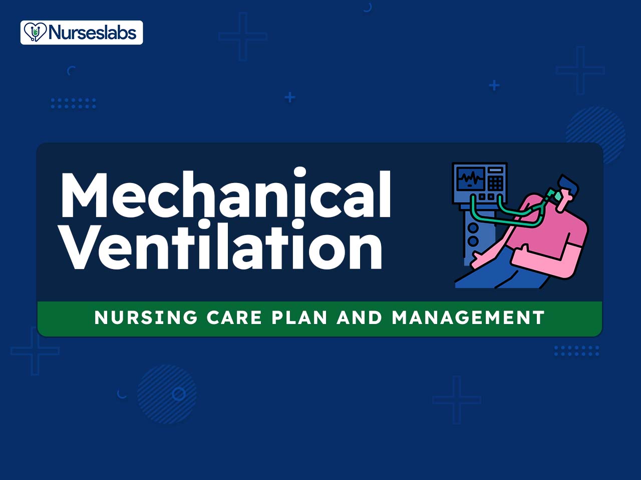
A mechanical ventilator is a positive- or negative-pressure breathing device that can maintain ventilation and oxygen delivery for a prolonged period. It is a machine that assists the client in breathing. Usually, the client is intubated before he is connected to the ventilator. An endotracheal tube or a tracheostomy tube is connected by oxygen tubing to the ventilator. If a client has evidence of respiratory failure or a compromised airway, endotracheal intubation and mechanical ventilation are indicated. This clinical evidence may be corroborated by a continuous decrease in oxygenation (PaO2), an increase in arterial carbon dioxide levels (PaCO2), and persistent acidosis (decreased pH).
Mechanical ventilators were traditionally classified according to the method by which they supported ventilation. The two general categories are negative-pressure and positive-pressure ventilators.
Positive-pressure Ventilators
Positive-pressure ventilators or PPVs inflate the lungs by exerting positive pressure on the airway, pushing air in, and forcing the alveoli to expand during inspiration. Endotracheal intubation or tracheostomy is usually necessary.
- Volume-cycled ventilators. These deliver a preset volume of air with each inspiration. Once this preset volume is delivered to the client, the ventilator cycles off, and exhalation occurs passively. The volume of air delivered by the ventilator is relatively constant.
- Pressure-cycled ventilators. When the pressure-cycled ventilator cycles on, it delivers a flow of air until it reaches a preset pressure, and then cycles off, and expiration occurs.
- High-frequency oscillatory support ventilators. These ventilators deliver very high respiratory rates (180 to 900 breaths/minute) that are accompanied by very low tidal volumes and high airway pressures. These small pulses of oxygen-enriched air move down the center of the airways, allowing alveolar air to exit the lungs along the margins of the airways.
- Noninvasive positive-pressure ventilation (NIPPV). NIPPV is a method of positive-pressure ventilation that can be given via facemasks that cover the nose and mouth, nasal masks, or other oral or nasal devices such as the nasal pillow. NIPPV eliminates the need for endotracheal intubation or tracheostomy and decreases the risk of nosocomial infections such as pneumonia. Pressure-controlled ventilation with pressure support, the most comfortable mode, eases the work of breathing and enhances gas exchange.
Negative-pressure Ventilators
Negative-pressure Ventilators or NPVs are applied to the thorax and abdomen to achieve lung inflation by distending the rib cage and abdomen. NPVs are not used as often as PPVs but many hospitals utilized this method for patients with acute respiratory failure until the Copenhagen polio epidemic in the 1950s.
- Iron-lung or Tank ventilators. These are large, sealed horizontal cylinders or “tanks” in which the patient lies, with their head protruding from a sealed opening at one end of the tank. The lowering and raising of air pressure in the cylinder cause the patient’s chest to rise and fall, stimulating inhalation and exhalation through the patient’s nose and mouth.
- Cuirass ventilator. A much smaller version of the iron lung, this ventilator is known as a chest shell, turtle shell, or tortoiseshell ventilator. This ventilator only encloses the patient’s torso and is sealed around the patient’s neck and waist, and is depressurized and repressurized by an external pump or portable ventilator.
- Exovent ventilator. This ventilator is a modern device similar to the cuirass ventilator. This NPV was developed in 2020, in response to the COVID-19 pandemic.
- Jacket ventilator. Also known as a poncho or raincoat ventilator. This NPV is a lighter version of the iron lung or the cuirass ventilator, made from an airtight material lined inside with a plastic or metal grid, a suction pump, and a back plate that goes to the patient’s hips, and depressurized and repressurized by a portable ventilator.
Ventilator mode refers to how breaths are delivered to the client. The most commonly used modes are controlled mechanical ventilation, continuous mandatory ventilation or assist-control (A/C), intermittent mandatory ventilation (IMV), synchronized intermittent mandatory ventilation (SIMV), pressure support ventilation, and airway pressure release ventilation.
Nursing Care Plans & Management
The major goals for patients with endotracheal intubation and/or tracheostomy receiving mechanical ventilation include improvement of gas exchange, maintenance of a patent airway, prevention of trauma, promoting optimal communication, minimizing anxiety, and absence of cardiac and pulmonary complications.
Nursing Problem Priorities
The following are the nursing priorities for patients receiving mechanical ventilation :
1. Ensuring a patent airway and proper placement of the endotracheal tube
2. Monitoring and managing the patient’s respiratory status, including assessing lung sounds, oxygen saturation levels, and end-tidal carbon dioxide (EtCO2) monitoring.
3. Monitoring and managing sedation levels and pain control for patient comfort and to promote synchrony with the ventilator.
4. Assessing for and managing any potential complications or emergencies, such as tube dislodgement, pneumothorax, or airway obstruction, and promptly initiating appropriate interventions.
5. Preventing complications associated with mechanical ventilation, such as ventilator-associated pneumonia and ventilator-induced lung injury
6. Collaborating with the healthcare team to optimize ventilator settings, weaning protocols, and extubation readiness assessments, while providing ongoing patient and family education and support.
Nursing Assessment
Assess for the following subjective and objective data:
- Adventitious breath sounds
- Apnea
- Apprehension
- Arterial ph less than 7.35
- Decreased tidal volume
- Decreased oxygen saturation (Sao2 <90%)
- Decreased Pao2 level (>50 to 60 mm Hg)
- Diminished lung sounds
- Dyspnea
- Forced vital capacity less than 10 mL/kg
- Increased Paco2 level (50 to 60 mm Hg or higher)
- Increased or decreased respiratory rate
- Inability to maintain airway (emesis, depressed gag, depressed cough).
- Restlessness
- Abnormal breath sounds
- Excessive secretions
- Increased peak airway pressure
- Ineffective cough
Assess for factors related during mechanical ventilation:
- Acute respiratory failure
- Noncompliant lung tissue
- Respiratory muscle weakness or paralysis
- Altered O2/CO2 ratio
- Decreased energy and fatigue
- Endotracheal intubation
- Stasis of secretions
Nursing Diagnosis
Following a thorough assessment, a nursing diagnosis is formulated to specifically address the challenges associated with this condition based on the nurse’s clinical judgement and understanding of the patient’s unique health condition. While nursing diagnoses serve as a framework for organizing care, their usefulness may vary in different clinical situations. In real-life clinical settings, it is important to note that the use of specific nursing diagnostic labels may not be as prominent or commonly utilized as other components of the care plan. It is ultimately the nurse’s clinical expertise and judgment that shape the care plan to meet the unique needs of each patient, prioritizing their health concerns and priorities.
Nursing Goals
The primary goals for the patient include ensuring optimal gas exchange, maintaining a clear airway, preventing trauma and infection, promoting mobility, adapting to nonverbal communication methods, developing effective coping strategies, and preventing complications. Goals and expected outcomes may include:
- The client will maintain spontaneous gas exchange resulting in reduced dyspnea, normal oxygen saturation, and normal arterial blood gases (ABGs) within client parameters.
- The client will demonstrate an absence of complications from mechanical ventilation.
- The client will participate in efforts to wean within the individual ability as appropriate.
- The caregiver will demonstrate behaviors necessary to maintain the client’s respiratory function.
- The client will maintain clear, open airways, as evidenced by normal breath sounds after suctioning.
- The caregiver will identify potential complications and initiate appropriate actions.
- The client will demonstrate reduced anxiety as evidenced by a calm manner and cooperative behavior.
Nursing Interventions and Actions
Nursing care for mechanically ventilated patients requires both technical expertise and effective communication skills. While interventions may vary depending on the patient’s condition and setting, pulmonary auscultation and interpretation of arterial blood gas measurements are essential. Nurses play a crucial role in detecting changes in assessment findings and identifying potential complications. Astute nursing assessment and a strong nurse-patient relationship are vital in providing optimal care for mechanically ventilated patients. Therapeutic interventions and nursing actions for patients with mechanical ventilation and endotracheal intubation may include:
1. Managing Mechanical Ventilation
In general, maintenance of spontaneous breathing in clients diagnosed with acute respiratory failure (ARF) under ventilatory support such as endotracheal intubation and/or tracheostomy has many positive effects such as improving oxygenation, preventing the mass loss and atrophy of the peripheral muscles, protecting against the diaphragm dysfunction, reducing the need for pharmacological sedation, and curbing the incidence of delirium. In clients diagnosed with acute respiratory distress syndrome (ARDS), the inspiratory effort might be affected by different stimuli not always subjected to ventilatory support. Different degrees of lung inflammation could influence the respiratory drive irrespective of gas exchange impairment. In clients diagnosed with COVID-19, the direct invasion of respiratory centers due to the virus may cause an alteration of the respiratory drive, thus affecting inspiratory effort (Tonelli et al., 2021). The following are assessment and nursing interventions for managing mechanical ventilation:
Prior intubation assessment:
1. Investigate the etiology of respiratory failure.
Understanding the underlying cause of the client’s particular ventilatory problem is essential to the care of the client, for example, decisions about future capabilities and ventilation needs and the most appropriate type of ventilatory support. The client should have a correctable underlying problem that can be resolved with the support of mechanical ventilation because there is no mode of mechanical ventilation that can cure a disease process (Jackson et al., 2020).
2. Observe changes in the level of consciousness.
Early signs of hypoxia include disorientation, irritability, and restlessness, while lethargy, stupor, and somnolence are considered as late signs. Mechanical ventilation is indicated for both hypercapnic respiratory failure and hypoxemic respiratory failure (Amitai & Mosenifar, 2020).
3. Assess the client’s respiratory rate, depth, and pattern, including the use of accessory muscles.
Changes in the respiratory rate and rhythm are early signs of possible respiratory distress. As moving air in and out of the lungs becomes more difficult, the breathing pattern changes to include the use of accessory muscles to increase chest excursions. Mechanical ventilation is indicated when the client’s spontaneous ventilation is inadequate to sustain life. It is indicated as a measure to control ventilation in critically ill clients and as prophylaxis for the impending collapse of other physiologic functions such as respiratory or mechanical insufficiency and ineffective gas exchange (Jackson et al., 2020).
4. Assess the client’s heart rate and blood pressure.
Tachycardia may result from hypoxia. Increased blood pressure happens in the initial phases then followed by lowered blood pressure as the condition progresses. Sufficiently severe hypoxia can result in tachycardia to provide sufficient oxygen to the tissues (Bhutta et al., 2022).
5. Auscultate the lung for normal or adventitious breath sounds.
Adventitious breath sounds such as wheezes and crackles are an indication of respiratory difficulties. Quick assessment allows for early detection of deterioration or improvement. Bilateral basilar crackles may indicate pulmonary edema or volume overload, other signs of that includes jugular vein distention and lower limb edema. Wheezing and rhonchi can be found in obstructive lung disease. Absent unilateral air entry can be caused by either massive pleural effusion or pneumothorax. Clear lung fields in the presence of hypoxia should raise suspicion of pulmonary embolism, especially if the client is tachycardic and has evidence of DVT (Bhutta et al., 2022).
6. Assess the skin color and examine the lips and nailbeds for cyanosis.
Bluish discoloration of the skin (cyanosis) indicates an excessive concentration of deoxygenated blood and that breathing pattern is ineffective to maintain adequate tissue oxygenation. Cyanosis might indicate severe hypoxia (Bhutta et al., 2022).
7. Monitor oxygen saturation using pulse oximetry.
Pulse oximetry is useful in detecting early changes in oxygen. Oxygen saturation levels should be between 92% and 98% for an adult without any respiratory difficulties. Resting arterial oxygen saturation (SaO2) less than or equal to 95% or exercise desaturation greater than or equal to 5% is considered abnormal. However, clinical correlation is always necessary as the exact cut-off below which tissue hypoxia ensues has not been defined (Bhutta et al., 2022).
8. Monitor arterial blood gases (ABGs) as indicated.
Increasing PaCo2 and decreasing PaO2 indicate respiratory failure. If the client’s condition begins to fail, the respiratory rate and depth decrease, and PaCo2 begins to rise. PaCO2 is an indirect measure of the exchange of CO2 with the air via the alveoli, its level is related to minute ventilation (Bhutta et al., 2022).
After intubation assessment:
9. Assess for correct endotracheal (ET) tube placement through observation of a symmetrical rise of both chest sides, auscultation of bilateral breath sounds, and X-ray confirmation.
Correct ET tube placement is important for effective mechanical ventilation. Changes in chest symmetry may indicate improper placement of the ET tube or the development of barotrauma. Frequent crackles or rhonchi that do not clear with coughing or suctioning may indicate developing complications, such as atelectasis, pneumonia, acute bronchospasm, and pulmonary edema.
10. Assess for the client’s comfort and the ability to cooperate while on mechanical ventilation.
Client discomfort may be secondary to incorrect ventilator settings that result in insufficient oxygenation. Once intubated and breathing on the mechanical ventilator, the client should be breathing easily and not “fighting or bucking” the ventilator.
11. Assess the ventilator settings and alarm system every hour.
The assessment ensures that settings are accurate and alarms are functional. Controls and settings are adjusted according to the client’s primary disease and results of diagnostic testing to maintain parameters within appropriate limits. Do not turn off alarms, even for suctioning. Ventilators have a series of visual and audible alarms, such as oxygen, low volume or apnea, high pressure, and inspiratory/expiratory (I:E) ratio. Turning off or failing to reset alarms places the client at risk for unobserved ventilator failure or respiratory distress or arrest.
12. Count the client’s respirations for 1 full minute and compare with desired respirations and ventilator set rate.
Respirations may vary depending on the problem requiring ventilatory assistance; for example, the client may be totally ventilator dependent or be able to take breaths on their own between ventilator-delivered breaths. Rapid client respirations can produce respiratory alkalosis and prevent the desired volume from being delivered by the ventilator. Slow client respirations and hypoventilation increases PaCO2 levels and may cause acidosis.
13. Maintain the client’s airway. Use the oral or nasal airway as needed.
An artificial airway is used to prevent the tongue from occluding the oropharynx. Different-sized oral appliances are available and measured from the lip to the angle of the jaw. These are useful for clients with spontaneous respirations who need help to keep their airways open (Avva et al., 2022).
14. Maintain the client in a High-Fowler’s position as tolerated. Frequently check the position.
This position promotes oxygenation via maximum chest expansion and is implemented during events of respiratory distress. Do not let the client slide down; this causes the abdomen to compress the diaphragm, which could cause respiratory change.
Preparation for endotracheal intubation:
15. Notify the respiratory therapist to bring a mechanical ventilator.
Mechanical ventilators are classified according to the method by which they support ventilation. The two types are negative-pressure and positive-pressure ventilators (used most frequently). Volume-cycled ventilation is the most common form of ventilator cycling used in adult medicine because it provides a consistent breath-to-breath tidal volume (Jackson et al., 2020).
16. Prepare the following equipment: ET tubes of different sizes; blades, laryngoscope, and stylet; syringe, benzoin, and waterproof tape or other securing materials; and local anesthetic agent (e.g., Xylocaine spray or jelly, benzocaine spray, cocaine, lidocaine, and cotton-tipped applicators.
Endotracheal tubes come in various sizes and shapes. Adult sizes range from 7 to 9 mm. Selection is based on the client’s size. Blades and scopes facilitate the opening of the upper airway and visualization of the vocal cords for the placement of oral ET tubes. A stylet makes the ET tube firmer and gives additional support to direction during intubation. A syringe is used to inflate the balloon (cuff) after the ET tube is in position. Tape and benzoin are used to secure the ET tube. These anesthetic agents suppress the gag reflex and promote general comfort.
17. Administer sedation as ordered.
Sedation facilitates comfort and ease of intubation. Pretreatment agents may be used to mitigate the physiologic response to laryngoscopy and induction and paralysis, which may be undesirable in certain clinical situations. Pretreatment medications are typically administered two to three minutes prior to induction and paralysis. These medications can be remembered by using the mnemonic LOAD (Lidocaine, Opioid analgesic, Atropine, Defasciculating agents) (Lafferty & Soo, 2020).
Assisting with intubation
18. Place the client in a supine position, hyperextending the neck unless contraindicated, and align the client’s oropharynx, posterior oropharynx, and trachea.
This position is necessary to promote the visualization of landmarks for accurate tube insertion. In the neutral position, the oral, pharyngeal, and laryngeal axes are not aligned to permit adequate visualization of the glottic opening. Place the client in the sniffing position for adequate visualization; flex the neck and extend the head. Studies have shown that simple head extension alone was as effective as the sniffing position in facilitating endotracheal intubation (Lafferty & Soo, 2020).
19. Apply cricoid pressure (Sellick maneuver) as directed by the healthcare provider.
Use of cricoid pressure to prevent passive regurgitation during rapid sequence intubation. It may also prevent passive regurgitation of gastric and oesophageal contents. This maneuver may lack supporting literature and may impede the laryngeal view, it is still initiated to prevent the regurgitation of gastric contents. Initiate this maneuver upon observing the beginning of unconsciousness. Maintain pressure throughout the intubation sequence until the position of the ET tube is verified (Lafferty & Soo, 2020).
20. Preoxygenate the client as indicated.
Administer 100% oxygen via a nonrebreather mask for three minutes for nitrogen washout. This is done without positive pressure ventilation using a tight seal. Assist ventilation with a bag-valve-mask (BVM) system only if needed to obtain an oxygen saturation equal to 90%. The client must be adequately pre-oxygenated to prevent desaturation during the apnea after the paralytic agent has been administered.
21. Assist with the verification of correct ET tube placement. Use an end-tidal carbon dioxide detector as indicated.
Correct placement is needed for effective mechanical ventilation to prevent complications associated with malpositioning such as vomiting, hypoxia, gastric distention, and lung trauma. The current criterion standard is end-tidal carbon dioxide detection, using either a calorimetric capnometer that changes color from purple to yellow with CO2 exposure or a quantitative capnometer that measures CO2 levels and can display a waveform. The yellow color change should occur rapidly within one to two breaths (Lafferty & Soo, 2020). Other capnography devices that provide numerical measurements of end-tidal carbon dioxide (normal value is 35 to 45 mm Hg) and Capnograms may also be used.
22. Continue with manual Ambu bag ventilation until the ET tube is stabilized. Assist in securing the ET tube once tube placement is confirmed.
Stabilization is necessary before initiating mechanical ventilation. Blow-by high-flow oxygen via a nonrebreather mask is usually used, but for clients who are noted to desaturate beyond 90%, breaths delivered via 100% oxygen BVM may be required. A client who is hypoxemic during attempts at intubation should undergo positive pressure ventilation with a BVM to raise PaO2 levels (Lafferty & Soo, 2020).
23. Document the ET tube position, noting the centimeter reference marking on the ET tube.
Documentation provides a reference for determining possible tube displacement, usually 21 cm for women and 23 cm at the lips for men. Traditionally, an endotracheal tube size 7.0 is used for women, while an 8.0 is used for men. Variations in size depend on the client’s height and whether they will require bronchoscopy (Alvarado & Panakos, 2022).
24. Institute mechanical ventilation with prescribed settings.
Modes for ventilating (assist/control, synchronized intermittent mandatory ventilation), tidal volume, rate per minute, a fraction of oxygen in inspired gas (FIO2), pressure support, positive end-expiratory pressure, and the like must be preset and carefully evaluated for response. In most circumstances, the initial mode of ventilation should be AC mode. The client can affect the frequency and timing of the breaths. If the client makes an inspiratory effort, the ventilator senses a decrease in the circuit pressure and delivers the preset tidal volume. This way, the client can dictate a comfortable respiratory pattern (Jackson et al., 2020).
25. Anticipate the need for nasogastric and/or oral gastric suction.
Abdominal distention may indicate gastric intubation and can also occur after cardiopulmonary resuscitation when the air is inadvertently blown or bagged into the esophagus, as well as the trachea. Suction prevents abdominal distention. Oral gastric suctioning may also reduce the risk of sinusitis.
26. Administer muscle-paralyzing agents, sedatives, and opioid analgesics as ordered.
See Pharmacologic Management
27. Examine the cuff volume by checking whether the client can talk or make sounds around the tube or whether exhaled volumes are significantly less than the volumes delivered. To correct this, slowly inflate the cuff with air until no leak is detected. Notify the respiratory therapist to check cuff pressure.
Cuff pressure should be maintained at 20 to 30 mm Hg. Maintenance of low-pressure cuffs prevents many tracheal complications formerly associated with ET tubes. Notify the healthcare provider if the leak persists. The ET tube cuff may be defective, requiring the HCP to change the tube.
28. Position the client by elevating the head of the bed if possible.
Elevating the client’s head and helping the client get out of bed while still on the ventilator is both physically- helps decrease the risk of aspiration– and psychologically beneficial. All clients on mechanical ventilation should have the head of the bed elevated to at least 30 degrees. According to a Cochrane review on ventilator-associated pneumonia, “ a semi-recumbent position reduced clinically suspected VAP by 25.7% when compared to a supine position” (Hickey & Giwa, 2022).
29. Inflate the endotracheal tube cuff properly. Check cuff inflation every 4 to 8 hours.
The cuff must be properly inflated to ensure adequate ventilation and delivery of desired tidal volume and to decrease the risk of aspiration. Endotracheal cuff pressures should be monitored initially after intubation and every 4 hours thereafter, with goal pressures of 20 to 30 cm water, as an increased risk of pneumonia has been associated with cuff pressures less than 20 cm water (Amitai & Mosenifar, 2020).
30. Note inspired humidity and temperature; use a heat moisture exchanger (HME), as indicated.
The usual warming and humidifying function of the nasopharynx are bypassed with intubation. Dehydration can dry up normal pulmonary fluids, cause secretions to thicken, and increase the risk of infection. The temperature should be maintained at about body temperature to reduce the risk of damage to cilia and hyperthermia reactions. The introduction of a heated wire circuit to the traditional system significantly reduces the problem of “rainout” or condensation in the tubing.
2. Promoting Patent Airway Clearance
In a client under mechanical ventilation such as endotracheal intubation and/or tracheostomy, the primary mechanisms of secretion clearance, like mucociliary transport and cough, are impaired. The presence of the artificial airway, poor humidification of inspired gases, and relative immobility are the major causes of pulmonary accretion retention in this population (Volpe et al., 2020). The following are the assessment and nursing interventions to promote effective airway clearance:
1. Assess airway patency.
Obstruction may be caused by the accumulation of secretions, mucous plugs, hemorrhage, bronchospasm, and problems with the position of the tracheostomy or ET tube. Accumulation of secretions, if extensive, starts a self-sustaining cycle of ventilation/perfusion mismatch, impairment of gas exchange, increased work of breathing, and subsequent augmented risk of mechanical ventilation dependence (Volpe et al., 2020).
2. Observe the color, odor, quantity, and consistency of sputum.
Thick, tenacious secretions increase airway resistance and the work of breathing. A sign of infection is discolored odoriferous sputum. In addition, retained secretions are a sequestered growth medium for bacteria, which increases the risk of pneumonia and keeps feeding this cycle (Volpe et al., 2020).
3. Auscultate the lungs for the presence of normal or adventitious breath sounds.
Diminished lung sounds or the presence of adventitious sounds may indicate an obstructed airway and the need for suctioning. This assessment will also determine the presence of rhonchi that do not clear with coughing, labored breathing, tachypnea, mental status changes, restlessness, cyanosis, and fever, which are all signs of respiratory system compromise.
4. Monitor oxygen saturation prior to and after suctioning using pulse oximetry.
This assessment provides an evaluation of the effectiveness of therapy. Pulse oximetry is a non-invasive measure of arterial oxygen saturation. Values of 92% or less are consistent with hypoxia and probably signal the need for oxygen supplementation or workup to determine the cause of desaturation.
5. Assess arterial blood gases (ABGs).
Signs of respiratory compromise include decreasing PaO2 and increasing PaCO2. An ABG measurement is frequently obtained 10 to 15 minutes after the institution of mechanical ventilation. The measured arterial PaO2 should verify the transcutaneous pulse oximetry readings and direct the reduction of FiO2 to a value of less than 0.5. The measured arterial PaCO2 can suggest adjustments of minute ventilation but should be interpreted in light of the client’s overall acid-base status (Amitai & Mosenifar, 2020).
6. Monitor for peak airway pressures and airway resistance.
Increases in these parameters signal the accumulation of secretions or fluid and the potential for ineffective ventilation. It should be recognized that peak inspiratory and plateau pressures will be increased by extrapulmonary pressure, from stiff chest walls or a distended abdomen, and do not reflect the true risk of barotrauma (Amitai & Mosenifar, 2020).
7. Monitor ET tube placement.
The ET tube may slip into the right main-stem bronchus, obstructing airflow to the left lung and putting the client at risk for a tension pneumothorax. Note the lip line marking and compare it with desired placement.
8. Note excessive coughing, increased dyspnea, high-pressure alarm on the ventilator, and visible secretions in the endotracheal or tracheostomy tube.
The intubated client often has an ineffective cough reflex, or the client may have a neuromuscular or neurosensory impairment, altering the ability to cough. The client will usually depend on suctioning to remove secretions.
9. Explain the suctioning procedure to the client; give reassurance throughout the procedure.
Suctioning can be frightening to the client. Reinforce the need to maintain a patent airway. Provide sedation and pain relief as indicated. Secretion clearance in clients with artificial airways is mainly performed through endotracheal suctioning. However, this procedure is only able to aspirate secretions located in the large airways up to the third-generation bronchi (Volpe et al., 2020).
10. Encourage deep breathing and coughing exercises. Promote early ambulation when possible
These actions expand alveoli and mobilize secretions. The effects of anesthesia and immobility may collapse alveoli and place the client at risk for nosocomial pneumonia and atelectasis. However, turning, coughing, and deep breathing is less effective than ambulation. Ambulation makes these techniques unnecessary for the vast majority of clients.
11. Turn the client in every two hours.
Turning mobilizes secretions and helps prevent ventilator-associated pneumonia. This promotes the drainage of secretions and ventilation to all lung segments, thereby reducing atelectasis.
12. Institute airway suctioning as indicated based on the presence of adventitious breath sounds and/or increased ventilatory pressure.
The frequency of suctioning should be based on the client’s clinical status, not on a preset routine such as every two hours. Over suctioning can cause hypoxia and injury to bronchial and lung tissue. It has been recommended to apply shallow suctioning instead of deep suctioning, which limits the insertion of the suction catheter to the tip of the artificial airway and further restrains the level of action (Volpe et al., 2020).
13. Use closed in-line suction.
This technique decreases the infection rate, may reduce hypoxia, and is often less expensive. Sterile technique is a priority. It also reduces the infection rate for healthcare workers and helps maintain oxygen saturation and PEEP when used.
14. Hyperoxygenated as ordered.
Hyperoxygenation before, during, and after endotracheal suctioning decreases hypoxia and cardiac dysrhythmias related to the suctioning procedure. Preoxygenation with 100% oxygen should be initiated prior to suctioning. This is in preparation for the hypoxia that is precipitated by suctioning, both from mechanical interruption and cessation of oxygen flow briefly (Sinha et al., 2022).
15. Instruct client in coughing techniques, if possible, during suctioning, such as splinting, the timing of breathing, and “step-cough”, as indicated.
Coughing techniques enhance the effectiveness of cough effort and secretion clearing. If the client has a weak cough, try the “step-cough” technique. A few weak coughs in a row may stimulate a larger, productive cough at the end of the cycle to clear the bronchial tree of secretions.
16. Administer intravenous therapy and aerosol bronchodilators as indicated.
Maintaining hydration increases ciliary action to remove secretions and reduces the viscosity of secretions. It is easier to mobilize thinner secretions with coughing. This also promotes ventilation and the removal of secretions by the relaxation of smooth muscles or bronchospasm.
17. Administer humidified oxygen as prescribed.
This intervention supplements oxygen and prevents further drying of respiratory passageways and secretions via added humidity.
18. Consult a respiratory therapist for chest physiotherapy as indicated.
Chest physiotherapy includes the techniques of postural drainage and chest percussion to loosen and mobilize secretions. This promotes ventilation of all lung segments and aids in the drainage of secretions.
3. Reducing Anxiety and Fear
Dependence on a ventilator is frightening to both the client and the family and disrupts even the most stable families. Clients may become withdrawn or depressed, especially after prolonged ventilation. Verbalization of feelings, provision of information, and provision of diversions and stress reduction techniques all contribute to lessening the client’s and the family’s anxiety. The following are the assessment and nursing interventions to reduce anxiety:
1. Assess the client’s understanding of the need for mechanical ventilation and the threat presented by the situation.
An accurate appraisal can facilitate the development of appropriate treatment strategies. This will also define the scope of individual problem separate from physiological causes and influences the choice of interventions.
2. Assess the client for signs of anxiety.
Being on a mechanical ventilator can be a drastic change that will produce a high level of anxiety. Anxiety can affect the respiratory rate and pattern, resulting in rapid, shallow breathing and leading to arterial blood gas abnormalities, and the client “fighting or bucking” the ventilator. High to severe dyspnea is associated with anxiety in clients receiving noninvasive mechanical ventilation in the ICU. Dyspnea can lead to prolonged mechanical ventilation (Roberts, 2020).
3. Observe the client’s physical responses.
The client may become restless, show repetitive movements, and display changes in vital signs. These signs are useful in evaluating the extent or degree of concerns, especially when compared with “verbal” comments.
4. Assess previous coping strengths of the client and family members and their current areas of ability and control.
Looking back on previous successful coping mechanisms may focus the client’s attention on their own capabilities, increasing the sense of control. Qualitative studies on coping families of ICU survivors have shown factors such as humor, spirituality, social support, communication, and hope as important to coping which in turn respondents relate to life quality (Nandig et al., 2016).
5. Encourage clients and family members to acknowledge and express fears. Acknowledge their expressions of concern.
Verbalization of fears provides an opportunity for dealing with concerns, clarifies reality, and reduces anxiety to a more manageable level. Acknowledging the client’s and family’s concerns validates the reality of the situation without minimizing the emotional impact. This also provides an opportunity for the client and family members to accept and begin to deal with what has happened, reducing anxiety.
6. Reduce distracting stimuli. Inform the client of alarms on the ventilatory system, and reassure the client about the close proximity of health care personnel to respond to the alarms.
Decreasing stimuli provides a quiet environment that enhances rest. Anxiety may escalate with excessive noise, conversation, and equipment around the client. Mechanical ventilation alarms provide both audible alarms and non-audible visual alerts designed to ensure safe mechanical ventilatory support. Beyond alarms from other devices, mechanical ventilators themselves have a plethora of alarm features and alarm sounds, some of which vary in priority, and all of these alarms can contribute to alarm fatigue and anxiety (Scott et al., 2019).
7. Educate the client and family about safety precautions when managing mechanical ventilators, such as backup power and oxygen supplies and emergency equipment for suctioning.
Discussing these precautions provides reassurance to help allay unnecessary anxiety, reduce concerns of the unknown, and preplan for response in emergency situations. The current standard of care is an approach that emphasizes interactivity and empowerment of the client and family members through family engagement (Urner et al., 2018).
8. Display a confident, calm manner and understanding attitude. Be available to the client for support, as well as for explanations of the client’s care and progress.
The presence of a trusted person may be helpful during periods of anxiety. An ongoing relationship establishes a basis for comfort in communicating anxious feelings. Explaining procedures every time they are performed helps to reduce anxiety and familiarizes the client with ventilator procedures.
9. Provide relaxation techniques.
Using anxiety-reduction techniques enhances the client’s sense of personal mastery and confidence. Results from a randomized control trial indicated the benefits of music therapy. Clients were randomized to standard-of-care, noise-canceling headphones, and self-initiated music therapy. All of the clients in the study were mechanically ventilated. Clients in the music therapy group exhibited decreased anxiety and need for sedation based on the self-reported evaluation (Roberts, 2020).
10. Encourage sedentary diversional activities.
These activities enhance the client’s quality of life and help pass time. Provide diversions, such as watching television, playing music or taking a walk (if appropriate), creating handicrafts, and writing. Although handicapped by dependence on ventilators, activities that are normal or desired by the individual should be encouraged to enhance the quality of life.
11. Encourage visiting family and friends and promote optimism.
The presence of significant others reinforces feelings of security for the client. Healthcare professionals may understandably focus on the need to be ‘clinically realistic’, but novel strategies to support family optimism and convey needed clinical information might help to decrease psychological stress (Nandig et al., 2016).
12. Promote spiritual care as appropriate.
Spiritual care is one of the alternative therapies that can be beneficial in decreasing anxiety. Reiki therapy is a natural healing technique that uses the laying of the hands to channel positive energy. While this practice has ancient ties to Buddhist practices, it has made its way into modern medicine with a number of experimental and quasi-experimental studies published since the 1980s (Roberts, 2020).
13. Reinforce education about cognitive behavioral therapy (CBT).
CBT has also been suggested as a method of easing anxiety in mechanically ventilated clients. A psychological treatment for common mental health disorders, CBt targets unhelpful thoughts or behaviors. A case study published in 2019 described two clients who received CBT while attempting to wean from mechanical ventilation. Despite their medical setback, at the end of the trial, each client was successfully weaned from the ventilator (Roberts, 2020).
14. Refer to the psychiatric liaison clinical nurse specialist, psychiatrist, or hospital chaplain, as appropriate.
Specialty expertise may provide a wider range of treatment options and may be needed to achieve successful outcomes. A family engagement concept in the critical care setting consists of a multidisciplinary framework, which involves providers, nurses, social workers, and spiritual care. Multiple opportunities are available at different levels to support families during the client’s critical illness and admission to the ICU (Urner et al., 2018).
4. Administering Medications and Pharmacological Support
Medications used during mechanical ventilation therapy aim to optimize respiratory function and support patient comfort. These may include sedatives and analgesics to ensure patient comfort, neuromuscular blocking agents to facilitate ventilator synchrony and reduce oxygen consumption, and bronchodilators or mucolytic agents to improve airway clearance and lung function. Here are the following medications used during mechanical ventilation:
1. Induction agents
Induction agents provide a rapid loss of consciousness that facilitates ease of intubation and avoids psychic harm to the client.
- 1.1. Etomidate
It has a rapid onset, short duration, is cerebroprotective, and is not associated with a significant drop in blood pressure. It is hemodynamically neutral compared with other agents, such as sodium thiopental. - 1.2. Ketamine
It produces a “dissociative” state, has analgesic properties, is a bronchodilator, and may decrease rather than increase intracranial pressure (Lafferty & Soo, 2020).
2. Paralyzing agents
These agents provide neuromuscular blockade and are administered immediately after the induction agent. The neuromuscular blockade does not provide sedation, analgesia, or amnesia; thus, administering a potent induction agent is important.
- 2.1. Succinylcholine
It has a rapid onset (45 to 60 seconds) and the shortest duration of action (8 to 10 minutes). - 2.2. Rocuronium
It has a slightly longer onset of action (60 to 75 seconds) and a longer duration of action (30 to 60 minutes) (Lafferty & Soo, 2020).
3. Opioids
It’s important to closely monitor the administration of opioids during mechanical ventilation to ensure patient safety. Careful assessment of vital signs, respiratory status, and the level of sedation is crucial to avoid complications such as respiratory depression or over-sedation.
- 3.1. Morphine
Morphine is a potent opioid analgesic that acts on the central nervous system to reduce pain perception, helping to alleviate pain and improve patient comfort during mechanical ventilation. - 3.2. Fentanyl
Fentanyl is a potent opioid analgesic commonly used in mechanical ventilation to provide pain relief and sedation. It helps to reduce anxiety and patient agitation, allowing for better synchronization between the patient and the ventilator.
4. Diuretics
Diuretics may be useful to maintain fluid balance if fluid retention is a problem. Loop diuretics have been approved by the FDA for treating edema and volume overload. Vasopressors and inotropes are medications used to create vasoconstriction or increase cardiac contractility, which improves cardiac output (Cardoso, 2022).
- 4.1. Furosemide (Lasix)
Furosemide is a loop diuretic that works by inhibiting the reabsorption of sodium and chloride in the kidney, leading to increased urine output. It can be used to reduce fluid overload in patients who have developed volume overload during mechanical ventilation. - 4.2. Hydrochlorothiazide (HCTZ)
Hydrochlorothiazide is a thiazide diuretic that acts by increasing sodium and water excretion. It may be used in situations where a milder diuretic effect is needed.
5. Vasopressors and inotropes
Vasopressors and inotropes are medications used to create vasoconstriction or increase cardiac contractility, which improves cardiac output (Cardoso, 2022).
- 5.1. Norepinephrine
Norepinephrine is a potent vasopressor that acts on alpha-adrenergic receptors, causing vasoconstriction and increasing blood pressure. It is commonly used to maintain blood pressure and perfusion in patients with septic shock or other forms of distributive shock. - 5.2. Epinephrine
Epinephrine is a potent vasopressor and inotrope that acts on both alpha- and beta-adrenergic receptors. It increases heart rate, myocardial contractility, and systemic vascular resistance, thereby improving blood pressure and cardiac output. Epinephrine is commonly used in cases of severe hypotension or cardiac arrest. - 5.3. Dopamine
Dopamine can act as both a vasopressor and an inotrope, depending on the dosage. At lower doses, it primarily acts on dopamine receptors and helps improve renal blood flow and urine output. At higher doses, it stimulates alpha-adrenergic receptors, causing vasoconstriction and increasing blood pressure. - 5.4. Dobutamine
Dobutamine is an inotrope that primarily acts on beta-1 adrenergic receptors, increasing myocardial contractility and cardiac output. It is commonly used to improve cardiac function in patients with heart failure or cardiogenic shock.
6. Broad-spectrum antibiotics
The use of broad-spectrum antibiotics such as includes ceftriaxone, cefepime, or piperacillin-tazobactam during mechanical ventilation is aimed at providing coverage against a wide range of of gram-positive and gram-negative bacteria that could cause respiratory infections.
7. Vancomycin and Linezolid
These antibiotics have activity against MRSA and are effective in treating or preventing MRSA-related respiratory infections in patients on mechanical ventilation.
8. Antifungals agents
- 8.1. Fluconazole
Fluconazole is an antifungal medication that may be used during mechanical ventilation to treat or prevent fungal respiratory infections, such as Candida or Aspergillus species. - 8.2. Voriconazole
Voriconazole is an antifungal medication that is usually used during mechanical ventilation to treat or prevent invasive fungal infections, particularly those caused by Aspergillus species.
5. Preventing Respiratory Injury Risk
Complications can occur in clients under endotracheal intubation and/or tracheostomy or at any stage of mechanical ventilation and are sometimes life-threatening. With a ventilator-induced lung injury, the alveolar epithelium is at risk for both barotrauma and volutrauma. Barotrauma refers to the rupture of the alveolus with the subsequent entry of air into the pleural space and/or the tracking of air along the vascular bundle to the mediastinum. Volutrauma refers to the local overdistention of normal alveoli (Hickey & Giwa, 2022). The following are the assessment and nursing interventions to prevent respiratory injury during mechanical ventilation:
1. Review the ventilator settings every hour, especially the tidal volume and plateau pressures. Notify the respiratory unit of any discrepancy in the ventilator settings immediately:
Frequent assessment guarantees that the client is receiving the correct mode, rate, tidal volume, FiO2, positive end-respiratory pressure (PEEP), and pressure support. Important attention to detail can prevent problems. As the tidal volume increases, so does the pressure required to force that volume into the lung. Persistent breath-to-breath peak pressures greater than 45 cm in water are a risk factor for barotrauma. Some researchers also suggested that plateau pressures should be monitored as a means to prevent barotrauma when the plateau pressure is maintained at less than 30 cm of water (Hickey & Giwa, 2022).
2. Assess respiratory rate and rhythm including the work of breathing.
It is important to maintain the client in synchrony with the ventilator and not permit “bucking” it. Adjustments may be required in flow, tidal volume, respiratory rate, and dead space of the ventilator, or the client may need sedation to synchronize respiration and reduce the work of breathing and energy expenditures.
3. Assess arterial blood gas results and monitor oxygen saturation.
These objective data guide the ventilator settings and appropriate interventions. ABGs allow assessment of acid-base status, oxygenation, and ventilation, and, therefore, the consequences of barotrauma (Soo & Mosenifar, 2022).
4. Assess for the signs of barotrauma: the client with crepitus, subcutaneous emphysema, altered chest excursion, asymmetrical chest, abnormal ABGs, a shift in trachea, restlessness, evidence of pneumothorax on chest x-ray studies.
Barotrauma is damage to the lungs from positive pressure as seen in clients with an acute respiratory disease when high pressures are needed to ventilate stiff lungs or when PEEP is used. Frequent assessments are needed because barotrauma can occur at any time and the client will not show signs of dyspnea, shortness of breath, or tachypnea if heavily sedated to maintain ventilation (Soo & Mosenifar, 2022).
5. Auscultate breath sounds.
Decreased breath sounds on the side of the pneumothorax is an initial finding. After tension develops, accumulating air displaces the mediastinum and associated structures away from the pneumothorax (contralaterally). These findings may be detected by placing a finger in the space between the trachea and neck strap muscles just above the sternal notch. The space should be equivalent, and deviation decreases the amount of space palpable (Soo & Mosenifar, 2022).
6. Monitor chest x-ray reports daily and obtain a stat portable chest x-ray film if barotrauma is suspected.
Vigilant monitoring helps to reduce complications. The portable chest radiograph often provides the first indication of barotrauma, especially in an otherwise asymptomatic client. Initial findings can be subtle because clients often have other pulmonary opacities that may obscure the appearance of extra-alveolar air (Soo & Mosenifar, 2022).
7. Monitor plateau pressures with the respiratory therapist.
Monitoring for barotrauma can involve measuring plateau pressure, which is the pressure after delivery of the tidal volume but before the client is allowed to exhale. The ventilator is programmed so that after delivery of the tidal volume the client is not allowed to exhale for a half second. Therefore pressure must be maintained to prevent exhalation. Elevation of plateau pressures increases both the risk and incidence of barotrauma when the client is on mechanical ventilation. There has been less occurrence of barotrauma since guidelines have recommended lower standard tidal volumes (Hickey & Giwa, 2022).
8. Make sure that the ventilator alarms are on.
The alarm alerts the caregiver in cases of ventilation problems. A quick response to alarms ensures the correction of problems and the maintenance of adequate ventilation. The Joint Commission has included alarm safety as a National Patient Safety Goal. Although organizations have heightened their level of concern surrounding mechanical ventilation alarm management, standards and guidelines are needed to achieve safe and effective practices regarding mechanical ventilation alarms (Scott et al., 2019).
9. Listen for alarms. Know the range in which the ventilator will set off the alarm and how to troubleshoot.
The ventilator is a life-sustaining treatment that requires a prompt response to alarms. The apnea alarm is indicative of disconnection or absence of spontaneous respirations. The low exhale alarm indicates that the client is not returning the delivered TV (through disconnection or leak). The low-pressure alarm indicates a possible disconnection or mechanical ventilator malfunction. The high peak pressure alarm indicates bronchospasm, retained secretions, obstruction of the ET tube, atelectasis, acute respiratory distress syndrome (ARDS), or pneumothorax, among others.
10. Suction the client only when necessary.
The use of suction in these clients is somewhat controversial. Some clients are treated with suction to facilitate the evacuation of the pleural space air. However, continuous suction may also promote the persistence of the bronchopleural fistula as the pressure gradients continue to favor flow from the airways to the pleural space (Soo & Mosenifar, 2022).
11. Lower the ventilator tidal volume settings, as indicated.
Clinicians have recognized the hazards of alveolar overdistention in all clients, and lower tidal volumes (in the range of 8 to 10 mL/kg) have generally been adopted for all clients. Low tidal volume was also associated with improvements in ventilator-free days and in the incidence of nonpulmonary organ failure.
12. Provide early nutritional support, as appropriate.
The respiratory muscles become weak or atrophied after just a few days of mechanical ventilation, especially if nutrition is inadequate. Adequate protein intake is important in increasing respiratory muscle strength. Protein intake should be approximately 25% of total daily kilojoules. Total carbohydrate intake should not exceed 25%.
13. Ensure proper sedation and pain management.
The need for and the dose of IV sedation should be assessed on a daily basis. Sedation is essential for the client’s comfort, but also to minimize adverse effects that may occur with client agitation and client-ventilator dyssynchrony. The neuromuscular blockade, such as succinylcholine or rocuronium could virtually eliminate client-ventilator dyssynchrony and its adverse effects while maximizing efficient airflow and improving oxygenation (Soo & Mosenifar, 2022).
14. Assist in performing tube thoracostomy, emergency needle thoracostomy, or large-bore thoracostomy.
Effective management of barotrauma requires prompt evacuation of pleural air and placement of a device to permit the excess air to egress. The urgency and type of tube thoracostomy device depend on the client’s clinical status. Emergency needle thoracostomy is indicated for clients with a tension pneumothorax that requires immediate decompression (Soo & Mosenifar, 2022).
15. Observe for air leaks in the water-seal chamber.
Bedside examination of the water-seal chamber for air determines whether an air leak has been sealed or resolved. Closure of the air leak is obvious when the air leak is large because the air would appear with every inspiratory cycle in a mechanically ventilated client (Soo & Mosenifar, 2022).
16. Clamp the tubing to determine the origin of the air leak, as indicated.
When an air leak persists, it is important to determine that the air leak is from the pleural space rather than a break in the tubing apparatus to the pleural drainage. Clamping the tube at the site of exit from the chest wall can help in this determination. Air leaks that continue even with the tube clamped indicate a leak somewhere in the system (Soo & Mosenifar, 2022).
6. Optimizing Cardiac Function
The heart, great vessels, and pulmonary vasculature lie within the chest cavity and are subject to the increased intrathoracic pressures associated with mechanical ventilation. The result is a decrease in cardiac output due to decreased venous return to the right heart, right ventricular dysfunction, and altered left ventricular distensibility (Amitai & Mosenifar, 2020). The following are the assessment and nursing interventions to optimize cardiac function during mechanical ventilation:
1. Assess the client’s level of consciousness, blood pressure, heart rate, and hemodynamic parameters if in place (central venous pressure, pulmonary artery diastolic pressure (PADP), and pulmonary capillary wedge pressure, cardiac output).
Mechanical ventilation can produce a decreased venous return to the heart, resulting in decreased BP, compensatory increased heart rate, and decreased cardiac output. This may happen abruptly with ventilator changes: rate, tidal volume, or positive-pressure ventilation. The level of consciousness will decrease if cardiac output is severely compromised. Therefore close monitoring during ventilator changes is imperative.
2. Assess the capillary refill, skin temperature, and peripheral pulses.
Pulses are weak with reduced stroke volume and cardiac output. Capillary refill is slow with reduced cardiac output. Cold, pale, clammy skin is secondary to compensatory sympathetic nervous system stimulation and is associated with low cardiac output and oxygen desaturation.
3. Monitor for dysrhythmias.
Cardiac dysrhythmias may result from a low perfusion state, acidosis, or hypoxia. The decrease in cardiac output from a reduction of right ventricular preload is more pronounced in the hypovolemic client and in those with a low ejection fraction (Amitai & Mosenifar, 2020).
4. Auscultate heart sounds.
Decreased cardiac output results in diminished, weak, or thready pulses. Irregularities suggest dysrhythmias, which may require further evaluation and monitoring.
5. Monitor fluid balance and urine output.
Optimal hydration status is needed to maintain effective circulating blood volume and counteract the ventilatory effects on cardiac output. With positive pressure ventilation, pressure from the diaphragm decreases blood flow to the kidneys and could result in a drop in urine output. The brain is very sensitive to a decrease in blood flow and may respond by releasing antidiuretic hormone (ADH) (to increase water and sodium retention), further reducing urine output. After the initial decrease in venous return to the heart, volume receptors in the right atrium signal a decrease in volume, which triggers an increase in the release of ADH from the posterior pituitary and the retention of water by the kidneys (de Haro et al., 2019).
6. Assess the client’s response to activity and promote rest appropriately.
Overexertion increases oxygen consumption and demand and can compromise myocardial function. The chronic presentation is usually less dramatic, however, with dyspnea on exertion as the most common complaint (Bhutta et al., 2022).
7. Monitor liver function test results.
Hepatic function is adversely affected by decreased cardiac output, increased hepatic vascular resistance, and elevated bile duct pressure (Amitai & Mosenifar, 2020).
8. Maintain an optimal fluid balance.
Volume therapy may be required to maintain adequate filling pressures and optimize cardiac output. However, if PADP and/or pulmonary capillary wedge pressure rises and cardiac output remains low, a fluid restriction may be necessary. Positive-pressure ventilation is responsible for an overall decline in renal function with decreased urine volume and sodium excretion (Amitai & Mosenifar, 2020).
9. Provide small, easily digested meals and instruct to limit caffeine intake, as appropriate.
Large meals may increase myocardial workload and cause vagal stimulation, resulting in bradycardia or ectopic beats. Caffeine is a direct cardiac stimulant that can increase heart rate but may not be a problem for everyone, such as for some clients with regular daily caffeine intake.
10. Measure cardiac output parameters and other functional parameters as appropriate.
Cardiac index, preload, afterload, contractility, and cardiac work can be measured noninvasively with the thoracic electrical bioimpedance (TEB) technique. This is useful in evaluating responses to therapeutic interventions and identifying the need for more aggressive or emergency care.
11. Notify the healthcare provider immediately of signs of a decrease in cardiac output and anticipate possible ventilator setting changes.
Vigilant monitoring reduces the risk of complications. Hypotension and decreased cardiac output may be related to the positive-pressure ventilator itself or the use of positive end-expiratory pressure (PEEP) mode. An exaggerated respiratory variation on the arterial pressure waveform is a clue that positive-pressure ventilation is significantly affecting venous return and cardiac output (Amitai & Mosenifar, 2020).
12. Assist in inserting a Swan-Ganz catheter in the ICU settings, and perform PEEP studies.
PEEP studies may be performed for clients with a Swan-Ganz catheter. This is performed by adjusting PEEP, monitoring oxygenation by peripheral oxygen saturation or arterial oxygen measurement via blood gas sampling, and measuring the associated cardiac output. The process is repeated at various PEEP settings, and the results are recorded (Amitai & Mosenifar, 2020)
13. Administer medications as ordered (diuretics, inotropic agents).
See Pharmacologic Management
7. Facilitating Weaning Process
Respiratory weaning, the process of withdrawing the client from dependence on the ventilator, takes place in three stages: gradual removal from the ventilator, then from the tube, and finally from oxygen therapy. Weaning from mechanical ventilation is performed at the earliest possible time, consistent with the client’s safety, and involves collaboration between the healthcare provider, physiotherapist, and nurse. The following are the assessment and nursing interventions to facilitate weaning process:
1. Assess the client’s vital signs.
If the client has stable vital signs and showing signs of improvement or reversal of the disease or condition that caused the need for mechanical ventilation, weaning indices should be assessed. The heart has to work harder to meet the increased energy needs associated with weaning. The healthcare provider may defer weaning if tachycardia, pulmonary crackles, or hypertension are present. An increase of 1℉ (0.6℃) in body temperature raises metabolic rate and oxygen demands by 7%.
2. Assess nutritional status and muscle strength.
Weaning is hard work. The client must not only be able to withstand the stress of weaning but also have the stamina to breathe spontaneously for extended periods. Success in weaning the long-term ventilator-dependent client requires early and aggressive but judicious nutritional support.
2. Determine the client’s psychological readiness for weaning.
Weaning provokes anxiety for the client regarding concerns about the ability to breathe on their own and the long-term need for ventilation. Clients are often frightened by having to breathe on their own again and need reassurance that they are improving and are well enough to handle spontaneous breathing.
3. Note restlessness; changes in BP, heart rate, and respiratory rate; use of accessory muscles; discoordinated breathing with ventilator; inability to cooperate; and skin color changes.
These are indicators that the client may require slower weaning and an opportunity to stabilize, or may need to stop the program. During T-piece trials, the nurse monitors the client closely for signs of hypoxia. Signs of exhaustion and hypoxia correlated with deterioration in the blood gas measurements indicate the need for ventilatory support.
4. Monitor cardiopulmonary response to activity.
Excessive oxygen consumption and demand increase the possibility of failure. If the rapid, shallow breathing index is less than 105, the client is likely to be weaned from mechanical ventilation. Clinical judgment and experience still play a large role in the provider’s decision to withdraw mechanical ventilatory support (Jackson et al., 2020).
5. Monitor laboratory tests as indicated.
Laboratory studies such as CBC, serum albumin and prealbumin, transferrin, total iron-binding capacity, and electrolytes verify that nutrition is adequate to meet energy requirements for weaning.
6. Review chest radiograph results and ABGs.
Chest x-rays should show clear lungs or marked improvement in pulmonary congestion or infiltrates. ABGs should demonstrate satisfactory oxygenation on an FiO2 of 40% or less.
7. Clarify weaning techniques for clients and family members.
This assists the client and family members in preparing for the weaning process, helps limit the fear of the unknown, promotes cooperation, and enhances the likelihood of a successful outcome. The three general approaches to weaning are synchronized intermittent mandatory ventilation (SIMV), pressure-support ventilation (PSV), and a spontaneous breathing trial (SBT). The preferred method of weaning is the SBT (Jackson et al., 2020).
8. Schedule undisturbed rest and sleep periods.
This maximizes the client’s energy for the weaning process and limits fatigue and oxygen consumption. Reestablishing independent spontaneous ventilation can be physically exhausting, therefore, it is crucial that the client has enough energy reserve to succeed. Weaning is accomplished by decreasing the number of mandatory breaths, and gradually increasing the workload of the respiratory muscles (Jackson et al., 2020).
9. Provide encouragement and recognition of the client’s efforts.
Positive feedback provides reassurance and support for the continuation of the weaning process. Responsiveness to the client’s wishes and experiences is an important part of the decision-making process. It is necessary to create a trusting relationship with the client and reflect on their physical and mental needs. When preparing a care plan, the nurse has to reflect over and take account of the way in which the client experiences the weaning, while at the same time considering their physical needs (Tingsvik et al., 2014).
10. Collaborate with a dietitian and nutritional support team for adjustments in the client’s diet.
The respiratory muscles become weak or atrophied after just a few days of mechanical ventilation, especially if nutrition is inadequate. Compensation for inadequate nutrition must be undertaken with care; excessive intake can increase the production of carbon dioxide and the demand for oxygen and lead to prolonged ventilator dependence and difficulty in weaning.
11. Terminate the weaning process if adverse reactions occur, as ordered by the healthcare provider.
Adverse reactions to weaning include a heart rate increase of 20 beats/minute, systolic blood pressure increase of 20 mm Hg, a decrease in oxygen saturation to less than 90%, respiratory rate of less than 8 or greater than 20 breaths/minute, ventricular arrhythmias, fatigue, panic, cyanosis, erratic or labored breathing, and paradoxical chest movement. If any of these signs are observed, the weaning process may be terminated with permission from the healthcare provider to avoid respiratory failure and delay in extubation.
8. Promoting Communication & Alternative Communication Methods
Mechanical ventilation greatly affects the production of voice, leaving clients unable to communicate their needs with nurses and their family. Over 50% of clients report communication challenges during mechanical ventilation as moderately to extremely stressful. Inability to communicate causes great distress to clients who are treated with mechanical ventilation which leads to various psychological stress like anxiety, depression, anger and fear, panic, sleep disorders, and decreased self-esteem (Thapa et al., 2019). The following are assessment and nursing interventions in promoting communication among patients receiving mechanical ventilation:
1. Assess the client’s ability to communicate by alternative means.
Reasons for long-term ventilatory support are various; the client may be alert and adept at writing, or may be lethargic, comatose, or paralyze. The method of communicating with the client is highly individualized.
2. Evaluate the need for or appropriateness of talking tracheostomy tube.
A client with adequate cognitive and muscular skills may have the ability to manipulate a talking tracheostomy tube. The use of a talking or fenestrated tube tracheostomy tube allows the client to talk while on the ventilator.
3. Assess the presence of communication barriers in invasive mechanically ventilated (IMV) clients.
Caring within the ICU setting can be viewed as different from caring in other nursing environments, especially when it concerns caring for IMV clients who are awake. Caring for awake IMV clients may increase uncertainty among ICU nurses, as it is less predictable than caring for sedated clients. Also, it is not always possible for nurses to calm and reassure restless and agitated IMV clients (Hussein, 2018).
4. Establish a means of communication.
Eye contact assures the client of interest in communicating. If the client is able to move their head, blink their eyes, or is comfortable with simple gestures, a great deal can be done with yes or no questions. Pointing to letter boards or writing is often tiring to the client, who can become frustrated with the effort needed to attempt conversations. The use of picture boards that express a concept or routine needs may simplify communication. Family members and other caregivers may be able to assist and interpret needs.
5. Educate the client and family members about available systems of communication.
The American Speech-Language-Hearing Association defines augmentative and alternative communication (AAC) as the means of communication when oral speech cannot be achieved. AAC systems are classified as either aided or unaided. Aided alternative communication systems are either electronic or non-electronic and are used to transmit or receive messages. Unaided AAC systems are those in which the physical functioning of the body is used as a means to communicate, such as gesturing, pointing, or other body language movements (Hussein, 2018).
6. Plan the form of communication before placing an IV line.
An IV line positioned in the hand or wrist may limit the client’s ability to write questions. For example, if the client is right-handed, the IV line may be placed in the left arm if possible so that the right hand is free.
7. Place a call light or bell within the client’s reach, ensuring that the client is physically capable of using it. Answer the call light or bell immediately.
Ventilator-dependent clients may be better able to relax, feel safe and not abandoned, and breathe with the ventilator knowing that the nurse is vigilant and needs will be met. The nurse should also inform the client that the nurse is available immediately as much as possible should assistance is required.
8. Inform staff on duty at the nurses’ station that the client is unable to speak.
This will alert all the staff members to respond to the client at the bedside instead of over the intercom. The client’s encounter with the staff should be characterized by positive experiences, and nurses play a vital role in this process.
9. Encourage family members to talk to the client.
The family members may feel self-conscious in one-sided conversation, but the knowledge that they are assisting the client to regain or maintain contact with reality and enabling the client to feel part of the family unit may reduce feelings of awkwardness. Encourage them to include information about the family at home and daily happenings to let the client catch up with their daily activities.
10. Consult a speech therapist to assist the client in choosing an appropriate communication method.
A speech therapist can assist in determining the most appropriate method of communication. A speech-language pathologist is an expert in communication disorders. They can conduct training for nurses on matching communication strategies and resources to meet the individual client’s needs and preferences (Hussein, 2018).
9. Initiating Measures for Infection Control & Management
Clients with endotracheal intubation or a tracheostomy are at increased risk of immunocompromise and infection. The oral cavity is a primary source of contamination of the lungs in the intubated and compromised client. Ventilator-associated pneumonia (VAP) is a new infection of the lung parenchyma that develops within 48 hours after intubation. It is a life-threatening complication with mortality rates of 33 to 50%. The risk of VAP is highest immediately after intubation (Jackson et al., 2020). The following are assessment and nursing interventions to provide measures for infection control and management:
1. Identify risk factors for the occurrence of infection.
Intubation interferes with the normal defense mechanism that keeps microorganisms out of the lungs. ET tubes, especially cuffed ones, interfere with the mucociliary transport system that helps clear airway secretions. Secretions that accumulate below and above the ET tube cuff are an ideal growth medium for pathogens.
2. Assess the characteristics of the client’s sputum.
Yellow or green purulent odorous sputum is indicative of infection; thick, tenacious sputum suggests dehydration. VAP is suspected when a new or changing pulmonary infiltrate is seen in conjunction with fever, leukocytosis, and purulent tracheobronchial secretions (Jackson et al., 2020).
3. Auscultate breath sounds.
The presence of rhonchi and wheezes suggests retained secretions requiring expectoration or suctioning. Assessing and reporting early indicators of infection will enable prompt intervention and treatment.
4. Assess for the signs of pulmonary infection including increased temperature, purulent secretions, elevated white blood cell count, positive bacterial cultures, and evidence of pulmonary infection on chest X-ray studies.
VAPs occur in up to 28% of clients on ventilators. Mortality rates of 40% to 50% have been reported for these clients. Most ventilator-associated infections are caused by bacterial pathogens, with gram-negative bacilli being common. The diagnostic triad for VAP consists of pulmonary infection (fever, purulent secretions, leukocytosis), bacteriologic evidence of pulmonary infection, and radiologic suggestion of pulmonary infection. When the combination of radiologic infiltrates and 2 clinical criteria are observed, the sensitivity of diagnosing VAP is 69% and the specificity is 75% (Amanullah & Mosenifar, 2015).
5. Obtain sputum culture as indicated.
A sputum culture may be needed to identify pathogens and appropriate antibiotics. Samples of respiratory secretions may be obtained from the proximal and/or distal airways by using bronchoscopy or non bronchoscopic techniques (Amanullah & Mosenifar, 2015).
6. Encourage the client, family members, and other staff members to engage in proper hand hygiene.
An artificial airway bypasses the normal protective mechanisms of the upper airways. Promote proper hand hygiene by hand washing or alcohol-based hand rubs, and wear the appropriate PPE when handling respiratory secretions and contaminated equipment. These factors may be the simplest but are the most important keys to the prevention of hospital-acquired infection.
7. Encourage deep breathing and coughing exercises.
Deep breathing and effective coughing maximize lung expansion and mobilization of secretions to prevent or reduce atelectasis and accumulation of sticky, thick secretions.
8. Provide oral hygiene every two hours, including the use of a dental oral antibiotic rinse. Instruct in proper secretion disposal.
Oral hygiene reduces oral bacterial flora, which could be aspirated. Encourage the client to brush their teeth two to three times per day and rinse with a chlorhexidine-based mouthwash. Swabs and toothbrushes with built-in suction catheter capability may facilitate oral care. Instruct the client and family members in secretion disposal such as disposing of tissues and soiled tracheostomy dressings to reduce the transmission of fluid-borne organisms.
9. Limit visitors and avoid contact with persons with respiratory infections.
The client is already compromised and is at increased risk for exposure to infections. VAP has been implicated as the most common infectious complication occurring in ICU clients and results in prolonged ICU and hospital lengths of stay, prolonged duration of mechanical ventilation, and overall increased cost of care (Amitai & Mosenifar, 2020).
10. Keep the head of the bed elevated to 30 to 45 degrees or perform subglottic suctioning unless it is medically contraindicated.
Elevation promotes better lung expansion. It also reduces gastric reflux and aspiration. Placing the client in the semi-recumbent position is associated with approximately a 3-fold reduction in the risk of hospital-acquired pneumonia, especially during enteral feeding (Amanullah & Mosenifar, 2015).
11. Use a continuous subglottic suction endotracheal (ET) tube for intubation that is expected to be longer than 24 hours.
This intervention prevents the accumulation of secretions that can be aspirated. This system will further reduce contamination of the lower airway by removing stagnant oropharyngeal secretions above the cuff that might otherwise be aspirated.
12. Use sterile suctioning procedures and reduce the number of times the ventilator tubes are open.
This technique decreases the introduction of microorganisms into the airway. The CDC recommends changing the tubing no more often than every 48 hours. Research indicates that less frequent tubing changes every five to seven days may be acceptable (Amanullah & Mosenifar, 2015).
13. Promote the use of humidifiers or heat moisture exchangers.
Passive humidifiers or heat moisture exchangers are preferred to reduce colonization of the ventilator circuit. Ventilatory-circuit condensation should be prevented from entering the endotracheal tubes and any inline nebulizer (Amanullah & Mosenifar, 2015).
14. Attempt a spontaneous awakening trial as indicated by the HCP per facility protocol.
These measures promote early extubation and minimize sedation. A spontaneous awakening trial involves reducing sedation until the client is awake and can follow simple commands or becomes agitated. During this “awake” time, a weaning trial may test the client’s ability to breathe spontaneously. Decreasing sedation reduces the amount of time spent on mechanical ventilation and, therefore, the risk of VAP.
15. Administer antibiotics or antifungals as prescribed.
For clients with early-onset VAP and no risk factors for multidrug-resistant (MDR) pathogens, currently recommended initial empiric antibiotics include ceftriaxone, fluoroquinolones, ampicillin-sulbactam, and ertapenem. Outcomes after VAP improve with the early administration of appropriate antibiotic regimens and with adequate dosing of antibiotics (Amanullah & Mosenifar, 2015). See More on Pharmacological Management
10. Promoting Optimal Nutrition Balance
Nutritional support for ventilated clients with endotracheal tubes and/or tracheostomy is challenging because they are often under anesthesia and unable to protect their airways. These clients are at high risk of regurgitation and aspiration. Anesthetic-related ileus can also become an important concern (Clare & Hopper, 2005). The gastric mucosa does not have autoregulatory capability. Thus mucosal ischemia and secondary bleeding may result from increased gastric venous pressure (Amitai & Mosenifar, 2020). The following are the assessment and nursing interventions for promoting optimal nutrition among patients receiving mechanical ventilation:
1. Weigh the client regularly.
Significant and recent weight loss (7 to 10% body weight) and poor nutritional intake provide clues regarding catabolism, muscle glycogen stores, and ventilatory drive sensitivity. According to a study, clients with a high risk for malnutrition may require a longer duration, about 50.34 days for mechanical ventilation (Koontalay et al., 2021).
2. Evaluate the client’s ability to eat.
The client with a tracheostomy tube may be able to eat, but a client with an ET tube must be tube fed or parenterally nourished. Nutrition status is the best predictor of mechanical ventilator duration for critical clients (Koontalay et al., 2021).
3. Observe for generalized muscle wasting and loss of subcutaneous fat.
These symptoms are indicative of depletion of muscle energy and can reduce respiratory muscle function. Malnutrition prevalence is associated with chronic diseases, particularly in 40 to 60% of clients resulting from changes in systematic functions, such as loss of body mass, respiratory function impairment, diaphragm muscle weakness, fat or protein depletion, and prolonged admission time (Koontalay et al., 2021).
4. Auscultate for bowel sounds and measure abdominal girth. Observe for diarrhea and constipation.
A functioning GI system is essential for the proper utilization of enteral feedings. Mechanically ventilated clients are at risk of developing abdominal distention due to trapped air or ileus and gastric bleeding caused by stress ulcers.
5. Monitor gastric residual volumes.
If the client is administered with enteral feedings, gastric residual volumes should be monitored to avoid gastric distention and an increased risk of regurgitation.
6. Offer food that the client desires and document when oral intake resumes.
The client’s appetite is usually poor and intake of essential nutrients may be reduced. Offering favorite foods can enhance oral intake.
7. Provide small, frequent meals of soft and easily digested foods as appropriate.
This prevents excessive fatigue, enhances intake, and reduces the risk of gastric distress. The updated ASPEN guidelines emphasize adequate protein delivery to critically ill adults. Protein is the most important nutrient to support wound healing and immune function and to maintain lean body mass (VanBlarcom & McCoy, 2018).
8. Encourage increased oral intake of at least 2500 mL per day or as tolerated within cardiac limits.
This prevents dehydration that can be exacerbated by increased insensible losses (ventilator or intubation) and reduces the risk of constipation.
9. Provide a diet that can meet the client’s respiratory needs as indicated.
A high intake of carbohydrates, protein, and calories may be desired or needed during ventilation to improve respiratory muscle function. Carbohydrates may be reduced and fat somewhat increased just before weaning attempts to prevent excessive carbon dioxide production and reduced respiratory drive. The American Society of Parenteral and Enteral Nutrition (ASPEN) recommended that 75.6% of clients receive adequate protein and 61.2% meet energy requirements within the first seven days of admission. It can improve their respiratory function, and structure, increase weaning ability, and reduce mechanical ventilation duration (Koontalay et al., 2021).
10. Administer early enteral feedings as needed.
These methods provide adequate nutrients to meet individual needs when oral intake is insufficient or inappropriate. ASPEN recommended that ICU clients should start enteral nutrition support within 24 to 48 hours of admission or after resuscitation in order to maintain systemic immune functions and structure major organs (Koontalay et al., 2021).
11. Provide parenteral feedings if enteral feedings are not tolerated.
When clients are severely malnourished at the time of admission to the ICU and nutritional feedings are delayed for a week or longer, the mortality rate is significantly higher. Beginning parenteral feeding in these clients is not a benign intervention. However, acute care clients who receive parenteral feeding are at a higher risk for hyperglycemia, infections, electrolyte and mineral disruptions, and volume overload (VanBlarcom & McCoy, 2018).
11. Providing Patient Education & Health Teachings
Preparing the client and family for home care involves educating them about the maintenance of mechanical ventilators such as suctioning, tracheostomy care, signs of pulmonary infection, cuff inflation and deflation, and assessment of vital signs. Education often begins in the hospital and continues at home.
1. Assess the client’s and caregiver’s perception and understanding of mechanical ventilation.
This information provides an important starting point in education. Participants in a study reported key knowledge aspects were the need to know about the modifiable contributors to their family member’s experiencing mechanical ventilation, what the proposed medical treatments entailed, what is going on, and the details of the client’s clinical progress (Dale et al., 2020).
2. Assess the client’s readiness and ability to learn.
Educational interventions must be designed to meet the learning limitations, motivation, and needs of the client. Clients in acute care may not be able to take in much information because of fatigue, pain, sensory overload, hypoxemia, and the like. The caregiver may feel inadequate, afraid of machinery, and have reservations about the ability to learn or deal with the overall situation.
3. Encourage the client or significant others to express feelings and ask questions.
Questions facilitate open communication between the client and healthcare professionals and allow the verification of understanding and the opportunity to correct misconceptions. Participants in a study reported that detached and distant HCP behavior was contrary to their need for ongoing proactive emotional support. These attitudes may aggravate uncertainty in terms of understanding what is going on (Dale et al., 2020).
4. Schedule teaching sessions during quiet, non stressful times. Arrange information according to the learners’ pace.
Ensure that all participants are well rested to enhance the learner’s ability to focus on and absorb the information provided. Place all data in a logical sequence, progressing from simple to more complex material to allow the learner to build on information learned from previous sessions. This way, the information provided is less threatening and overwhelming.
5. Provide material in multiple formats, such as books and pamphlets, audiovisuals, hands-on demonstrations, and take-home instruction sheets.
These materials use multiple senses to stimulate learning and retention of information. They also provide resources for review following discharge. In South Korea, the main teaching methods include oral presentations and printed handouts, and limited demonstrations of how to provide care for clients using ventilators are performed during rental agent visits; thus, caregivers in South Korea do not receive video-based education (Kim et al., 2021).
6. Encourage the client and caregiver to evaluate the impact of ventilatory dependence on their lifestyle.
Quality of life must be resolved by the ventilator-dependent client and caregivers who need to understand that home ventilatory support is a 24-hour job that affects everyone. Participants in a study reported that they were given few anticipatory guidance from HCPs regarding the potential for debilitating health changes or prompts to engage in activities essential to safeguarding mental, emotional, and physical well-being. These unanticipated and long-term health changes contributed to a reduced capacity to perform family caregiving and decision-making roles (Dale et al., 2020).
7. Explain the importance of frequent assessment of vital signs, auscultation of breath sounds, and ventilation checks.
This information also helps reduce anxiety by providing a basis for actions. Tracheal suctioning is performed when adventitious breath sounds are detected or whenever secretions are present. Unnecessary suctioning can initiate bronchospasm and cause mechanical trauma to the tracheal mucosa.
6. Explain to the client the reason for the inability to talk while intubated. Explain alternative efforts for communicating.
The endotracheal tube passes through the vocal cords and attempts to talk can cause more trauma to the cords. However, clients must understand how to use supplementary methods for communication such as paper, pen, and pictures. Not being able to communicate verbally or using assistive equipment is one of the worst experiences for these clients and leads to anger and hopelessness among them. Communications about pain can also become unsuccessful, resulting in client frustration, fear, and anger (Hosseini et al., 2018).
8. Explain that the client will not be able to eat or drink while intubated but assure him or her that alternative measures (IV fluids, gastric feedings, or hyperalimentation) will be taken to provide nourishment.
The risk for aspiration is high if the client eats or drinks while intubated. Clients may eat and drink after a swallow evaluation in long-term care settings. Other measures such as early placement of a nasogastric tube and oral care with a soft toothbrush and chlorhexidine rinses may also be considered (Amitai & Mosenifar, 2020).
9. Explain that alarms may periodically sound off, which may be normal, and that the staff will be in close proximity.
Explaining expected events can help reduce anxiety. A 2014 report by the Association for the Advancement of Medical Instrumentation identified challenges regarding mechanical ventilation alarms. Some alarm systems do not properly distinguish life-threatening alarms from nuisance alarms. This overexposure to non actionable alarms results in decreased alertness and lower confidence in the accuracy and urgency of audible alarms (Scott et al., 2019).
10. Explain the need for suctioning as needed.
This information can help reduce anxiety associated with the procedure. If the client is pending discharge to the home setting with a tracheostomy tube, the nurse should ensure that suction and other appropriate equipment are in place in the home prior to discharge. When a tracheostomy or endotracheal tube is in place, it is usually necessary to suction the client’s secretions because of the decreased effectiveness of the cough mechanism.
11. Explain the weaning process and explain that extubation demonstrates adequate respiratory function and a decrease in pulmonary secretions.
This information aids the client in maintaining some control. Respiratory weaning, the process of withdrawing the client from dependence on the ventilator, takes place in three stages: gradual removal from the ventilator, then from the tube, and finally from oxygen therapy. The client needs to know what is expected of them during the procedure.
12. If long-term ventilation is anticipated, discuss or plan for long-term ventilator care management and use appropriate referrals: long-term ventilator facilitates versus home care management.
Continuity of care is facilitated through the use of specialized resources. The community nurse helps identify and contact community resources that may assist in the home management of the client with mechanical ventilation. Community resources may include food and meal services, domestic help, carer support, occupational therapy activities, and access to client support groups. The technical aspects of the ventilator are managed by vendor follow-up.
13. Recommend that caregivers learn cardiopulmonary resuscitation (CPR).
This provides a sense of security about the ability to handle emergency situations that might arise until help can be obtained. The nurse may also educate the family about CPR, including mouth-to-tracheostomy tube (instead of mouth-to-mouth) breathing.
14. Ensure that all safety concerns have been addressed and all needed equipment is in place.
Predischarge preparations can ease the transfer process. Planning for potential problems increases the sense of security for the client and caregiver. The nurse may also explain how to handle a power failure, which usually involves converting the ventilator from an electrical power source to a battery power source. Conversion is automatic in most home ventilators and lasts approximately one hour. Instruct the family on using a manual self-inflation bag should it be necessary.
Recommended Resources
Recommended nursing diagnosis and nursing care plan books and resources.
Disclosure: Included below are affiliate links from Amazon at no additional cost from you. We may earn a small commission from your purchase. For more information, check out our privacy policy.
Ackley and Ladwig’s Nursing Diagnosis Handbook: An Evidence-Based Guide to Planning Care
We love this book because of its evidence-based approach to nursing interventions. This care plan handbook uses an easy, three-step system to guide you through client assessment, nursing diagnosis, and care planning. Includes step-by-step instructions showing how to implement care and evaluate outcomes, and help you build skills in diagnostic reasoning and critical thinking.

Nursing Care Plans – Nursing Diagnosis & Intervention (10th Edition)
Includes over two hundred care plans that reflect the most recent evidence-based guidelines. New to this edition are ICNP diagnoses, care plans on LGBTQ health issues, and on electrolytes and acid-base balance.

Nurse’s Pocket Guide: Diagnoses, Prioritized Interventions, and Rationales
Quick-reference tool includes all you need to identify the correct diagnoses for efficient patient care planning. The sixteenth edition includes the most recent nursing diagnoses and interventions and an alphabetized listing of nursing diagnoses covering more than 400 disorders.

Nursing Diagnosis Manual: Planning, Individualizing, and Documenting Client Care
Identify interventions to plan, individualize, and document care for more than 800 diseases and disorders. Only in the Nursing Diagnosis Manual will you find for each diagnosis subjectively and objectively – sample clinical applications, prioritized action/interventions with rationales – a documentation section, and much more!

All-in-One Nursing Care Planning Resource – E-Book: Medical-Surgical, Pediatric, Maternity, and Psychiatric-Mental Health
Includes over 100 care plans for medical-surgical, maternity/OB, pediatrics, and psychiatric and mental health. Interprofessional “patient problems” focus familiarizes you with how to speak to patients.

See Also
Other recommended site resources for this nursing care plan:
- Nursing Care Plans (NCP): Ultimate Guide and Database MUST READ!
Over 150+ nursing care plans for different diseases and conditions. Includes our easy-to-follow guide on how to create nursing care plans from scratch. - Nursing Diagnosis Guide and List: All You Need to Know to Master Diagnosing
Our comprehensive guide on how to create and write diagnostic labels. Includes detailed nursing care plan guides for common nursing diagnostic labels.
Other nursing care plans related to respiratory system disorders:
- Asthma
- Aspiration Risk & Aspiration Pneumonia
- Airway Clearance Therapy & Coughing
- Bronchiolitis
- Bronchopulmonary Dysplasia (BPD)
- Chronic Obstructive Pulmonary Disease (COPD)
- Cystic Fibrosis
- Hemothorax and Pneumothorax
- Influenza (Flu)
- Ineffective Breathing Pattern (Dyspnea)
- Impairment of Gas Exchange
- Lung Cancer
- Mechanical Ventilation
- Near-Drowning
- Pleural Effusion
- Pneumonia
- Pulmonary Embolism
- Pulmonary Tuberculosis
- Tracheostomy
References
To further your research and reading about mechanical ventilation, check out these sources:
- Alvarado, A. C., & Panakos, P. (2022, July 13). Endotracheal Tube Intubation Techniques – StatPearls – NCBI Bookshelf. NCBI. Retrieved December 21, 2022.
- Amanullah, S., & Mosenifar, Z. (2015, December 31). Ventilator-Associated Pneumonia: Overview of Nosocomial Pneumonias, Epidemiology of VAP, Clinical Presentation of VAP. Medscape Reference. Retrieved December 26, 2022.
- Amitai, A., & Mosenifar, Z. (2020, April 7). Ventilator Management: Introduction to Ventilator Management, Modes of Mechanical Ventilation, Methods of Ventilatory Support. Medscape Reference. Retrieved December 21, 2022.
- Avva, U., Lata, J. M., & Kiel, J. (2022, May 1). Airway Management – StatPearls – NCBI Bookshelf. NCBI. Retrieved December 21, 2022.
- Bhutta, B. S., Alghoula, F., & Berim, I. (2022, August 9). Hypoxia – StatPearls – NCBI Bookshelf. NCBI. Retrieved December 21, 2022.
- Cardoso, S. (2022, August 18). Inotropes And Vasopressors – StatPearls – NCBI Bookshelf. NCBI. Retrieved December 23, 2022.
- Cheever, K. H., & Hinkle, J. L. (2018). Brunner & Suddarth’s Textbook of Medical-surgical Nursing. Wolters Kluwer.
- Clare, M., & Hopper, K. (2005, April). Mechanical Ventilation: Ventilator Settings, Patient Management, and Nursing Care. Compendium.
- Dale, C. M., Carbone, S., Istanboulian, L., Fraser, I., Cameron, J. I., Herridge, M. S., & Rose, L. (2020, June). Support needs and health-related quality of life of family caregivers of patients requiring prolonged mechanical ventilation and admission to a specialised weaning centre: A qualitative longitudinal interview study. Intensive and Critical Care Nursing, 58.
- De Haro, C., Ochagavia, A., Lopez-Aguilar, J., Fernandez-Gonzalo, S., Navarra-Ventura, G., Magrans, R., Montanya, J., Blanch, L., & Asynchronies in the Intensive Care Unit (ASYNICU) Group. (2019). Patient-ventilator asynchronies during mechanical ventilation: current knowledge and research priorities. Intensive Care Medicine Experimental, 7(43).
- Farrell, M. (2017). Smeltzer And Bare’s Textbook Of Medical-Surgical Nursing (First ed.). Lippincott Williams & Wilkins.
- Hickey, S. M., & Giwa, A. O. (2022). Mechanical Ventilation – StatPearls – NCBI Bookshelf. NCBI. Retrieved December 21, 2022.
- Hosseini, S.-R., Valizad-Hasanloei, M.-A., & Feizi, A. (2018, September-October). The Effect of Using Communication Boards on Ease of Communication and Anxiety in Mechanically Ventilated Conscious Patients Admitted to Intensive Care Units. Iranian Journal of Nursing and Midwifery Research, 23(5), 358-362.
- Hussein, K. (2018, July 24). Communication with invasive mechanically ventilated patients and the use of alternative devices: integrative review. NCBI. Retrieved December 24, 2022.
- Jackson, C. D., Mosenifar, Z., & Poe, G. (2020, September 15). Mechanical Ventilation: Background, Classifications of Positive-Pressure Ventilators, Indications for Mechanical Ventilation. Medscape Reference. Retrieved December 21, 2022.
- Kim, H. S., Lee, C. E., & Yang, Y. S. (2021, July). Factors associated with caring behaviors of family caregivers for patients receiving home mechanical ventilation with tracheostomy: A cross-sectional study. PLoS One, 16(7).
- Koontalay, A., Suksatan, W., Sadang, J. M., & Prabsangob, K. (2021, June 10). Optimal Nutritional Factors Influencing the Duration of Mechanical Ventilation Among Adult Patients with Critical Illnesses in an Intensive Care Unit. NCBI. Retrieved December 26, 2022.
- Lafferty, K. A., & Soo, G. W. (2020, April 7). Rapid Sequence Intubation: Background, Indications, Contraindications. Medscape Reference. Retrieved December 21, 2022.
- Murr, A. C., Moorhouse, M. F., & Doenges, M. E. (2016). Nurse’s Pocket Guide: Diagnoses, Prioritized Interventions, and Rationales. F.A. Davis Company.
- Nandig, N., Huff, N. G., Cox, C. E., & Ford, D. (2016, September). Coping as a Multi-Faceted Construct: Associations with Psychological Outcomes among Family Members of Mechanical Ventilation Survivors. Critical Care Medicine, 44(9), 1710-1717.
- Roberts, K. (2020, October 1). Assessment and Treatment of Anxiety During Mechanical Ventilation. AARC Times Digital.
- Scott, J. B., De Vaux, L., Dills, C., & Strickland, S. L. (2019, October). Mechanical Ventilation Alarms and Alarm Fatigue. Respiratory Care, 64(10), 1308-1313.
- Sinha, V., Semien, G., & Fitzgerald, B. M. (2022, September 25). Surgical Airway Suctioning – StatPearls – NCBI Bookshelf. NCBI. Retrieved December 21, 2022.
- Soo, G. W., & Mosenifar, Z. (2022, February 11). Barotrauma and Mechanical Ventilation: Practice Essentials, Pathophysiology, Etiology. Medscape Reference. Retrieved December 23, 2022.
- Swearingen, P. L. (2018). All-in-one Nursing Care Planning Resource: Medical-surgical, Pediatric, Maternity, and Psychiatric-mental Health (J. Wright & P. L. Swearingen, Eds.). Elsevier.
- Thapa, D., Dahal, A., & Singh, R. (2019). Communication Difficulties and Psychological Stress in Patients Receiving Mechanical Ventilation. Birat Journal of Health Sciences, 4(2).
- Tingsvik, C., Johansson, K., & Martensson, J. (2014). Weaning from mechanical ventilation: factors that influence intensive care nurses’ decision-making. British Association of Critical Care Nurses.
- Tonelli, R., Marchioni, A., Tabbi, L., Fantini, R., Busani, S., Castaniere, I., Andrisani, D., Gozzi, F., Bruzzi, G., Manicardi, L., Demurtas, J., Andreani, A., Cappiello, G. F., Samarelli, A. V., & Clini, E. (2021, March). Spontaneous Breathing and Evolving Phenotypes of Lung Damage in Patients with COVID-19: Review of Current Evidence and Forecast of a New Scenario. Journal of Clinical Medicine, 10(5).
- Treas, L. S., & Wilkinson, J. M. (2014). Basic Nursing: Concepts, Skills, & Reasoning. F.A. Davis Company.
- Urner, M., Ferreyro, B. L., Doufle, G., & Mehta, S. (2018, December). Supportive Care of Patients on Mechanical Ventilation. Respiratory Care, 63(12), 1567-1574.
- VanBlarcom, A., & McCoy, M. A. (2018, June). New Nutrition Guidelines: Promoting Enteral Nutrition via a Nutrition Bundle. Critical Care Nurse, 38(3).
- Volpe, M. S., Guimaraes, F. S., & Morais, C. C. (2020, August). Airway Clearance Techniques for Mechanically Ventilated Patients: Insights for Optimization. Respiratory Care, 65(8), 1174-1188.


Leave a Comment