Disseminated intravascular coagulation (DIC) is a life-threatening condition that requires immediate medical attention. As a nurse, it is crucial to understand the nursing care plans and nursing diagnosis for DIC to provide the best care for patients. This guide provides a comprehensive overview of DIC nursing care plans and nursing diagnosis, including common symptoms, nursing management, nursing interventions, and treatment options.
What is Disseminated Intravascular Coagulation?
Disseminated intravascular coagulation (DIC) is a coagulation disorder that prompts overstimulation of the normal clotting cascade and results in simultaneous thrombosis and hemorrhage. The formation of micro clots affects tissue perfusion in the major organs, causing hypoxia, ischemia, and tissue damage. Coagulation occurs in two different pathways: intrinsic and extrinsic. These pathways are responsible for the formation of fibrin clots and blood clotting, which maintains homeostasis. In the intrinsic pathway, endothelial cell damage commonly occurs because of sepsis or infection. The extrinsic pathway is initiated by tissue injuries such as from malignancy, trauma, or obstetrical complications. DIC may present as an acute or chronic condition.
Essential medical management of DIC is primarily aimed at treating the underlying cause, managing complications from both primary and secondary causes, supporting organ function, stopping abnormal coagulation, and controlling bleeding. Morbidity and mortality depend on the underlying cause and severity of coagulopathy.
Nursing Care Plans & Management
Nursing care for patients with DIC includes monitoring vital signs and bleeding, administering fluids and blood products, practicing bleeding precautions, wound care, providing respiratory support, offering psychosocial support, and maintaining effective communication.
Nursing Problem Priorities
The following are the nursing priorities for patients with disseminated intravascular coagulation:
- Minimizing bleeding risk
- Respiratory support
- Patient education
Nursing Assessment
Assess for the following subjective and objective data:
- Abnormal arterial blood gases (ABGs)
- Abnormal breathing (rate, depth, and rhythm)
- Confusion
- Dyspnea
- Hypercapnia
- Hypoxemia
- Hypoxia
- Irritability
- Restlessness
- Somnolence
- Abnormal blood profile
- Capillary refill >3 seconds
- Changes in the level of consciousness
- Chest pain
- Cyanosis
- Hematuria
- Oliguria
Assess for factors related to the cause of disseminated intravascular coagulation:
- The altered oxygen-carrying capacity of the blood
- Blood circulation disruption
- Microthrombi
- Abnormal blood profile (depleted coagulation factors)
- Drug therapy (adverse effects of heparin)
- Complexity of treatment
- Emotional state affecting learning
- New condition and/or treatment
- Unfamiliar environment
Nursing Diagnosis
Following a thorough assessment, a nursing diagnosis is formulated to specifically address the challenges associated with disseminated intravascular coagulation based on the nurse’s clinical judgement and understanding of the patient’s unique health condition. While nursing diagnoses serve as a framework for organizing care, their usefulness may vary in different clinical situations. In real-life clinical settings, it is important to note that the use of specific nursing diagnostic labels may not be as prominent or commonly utilized as other components of the care plan. It is ultimately the nurse’s clinical expertise and judgment that shape the care plan to meet the unique needs of each patient, prioritizing their health concerns and priorities.
Nursing Goals
Goals and expected outcomes may include:
- The client will maintain optimal gas exchange, as evidenced by ABGs within the client’s usual range; oxygen saturation of 90% or greater; alert, responsive mentation or no further reduction in the level of consciousness; and relaxed breathing and baseline HR for the client.
- The client will maintain optimal peripheral tissue perfusion in the affected extremity, as evidenced by strong palpable pulses, reduction in and/or absence of pain, warm, and dry extremities, and adequate capillary refill.
- The client will experience reduced episodes of bleeding and hematomas.
- The client will experience reduced side effects of medication therapy.
- The client will maintain therapeutic levels of coagulation laboratory profiles (prothrombin, partial prothrombin time, fibrinogen, fibrin split products, and bleeding time).
- The client and/or significant others verbalize a basic understanding of the disease condition, risk factors, and treatment regimen.
Nursing Interventions and Actions
Therapeutic interventions and nursing actions for patients with disseminated intravascular coagulation may include:
1. Promoting Effective Gas Exchange
DIC is a complex condition characterized by abnormal clotting and bleeding throughout the body, which can lead to tissue damage and organ dysfunction, including the lungs. Nurses play a vital role in ensuring adequate oxygenation and carbon dioxide elimination by closely monitoring respiratory parameters, such as oxygen saturation and arterial blood gases. Interventions to promote effective gas exchange may include supplemental oxygen therapy, positioning techniques to optimize ventilation, and maintaining proper fluid balance. Prompt identification and management of underlying causes, such as treating infections or addressing coagulation abnormalities, are also essential in improving gas exchange and minimizing respiratory complications. By prioritizing effective gas exchange, healthcare professionals aim to enhance the patient’s respiratory function, support organ perfusion, and contribute to their overall recovery from DIC.
1. Assess for changes in the level of consciousness.
Early signs of cerebral hypoxia are restlessness and irritability; later signs are confusion and somnolence.
2. Assess the respiratory depth, rate, and rhythm.
The client will adapt breathing patterns over time to facilitate gas exchange. Rapid, shallow respirations may result from hypoxia or from acidosis with the shock state. The development of hypoventilation indicates that immediate ventilator support is needed.
3. Assess the client’s breath sounds. Assess cough for signs of bloody sputum.
Changes in breath sounds may reveal the cause of impaired gas exchange. Hemoptysis is an indication of bleeding in the respiratory tract.
4. Assess for tachycardia, shortness of breath, and use of accessory muscles.
These signify an increased work of breathing. With initial hypoxia, HR increases. The use of accessory muscles increases chest excursion to facilitate effective breathing.
5. Monitor oxygen saturation and assess arterial blood gases (ABGs).
Pulse oximetry is a useful tool to detect early changes in oxygen saturation. Oxygen saturation should be kept at 90% or greater. Increasing PaCo2 and decreasing PaO2 are signs of hypoxemia and respiratory acidosis.
6. Provide reassurance and allay anxiety by staying with the client during the acute episodes of respiratory distress.
Anxiety increases dyspnea, the work of breathing, and the respiratory rate.
7. Change the client’s positioning every 2 hours, and perform chest physiotherapy.
These maneuvers facilitate the movement and drainage of secretions.
8. Position the client in a high-Fowler’s position as indicated.
An upright position allows for adequate diaphragmatic and lung excursion and promotes optimal lung expansion.
9. Assist with coughing or suction as indicated.
Productive coughing is the most effective way to remove moist secretions. If the client is unable to perform independently, suctioning may be needed to promote airway patency and reduce the work of breathing.
10. Maintain an oxygen administration device as ordered.
The appropriate amount of oxygen must be delivered continuously so that the client maintains an oxygen saturation of 90% or greater.
11. Anticipate the need for intubation and mechanical ventilation.
Early intubation and mechanical ventilation are recommended to prevent full decompensation of the client. Mechanical ventilation provides supportive care to maintain adequate oxygenation and ventilation for the client.
2. Enhancing Tissue Perfusion
The formation of small blood clots in the blood vessels can lead to the blockage of oxygen and nutrient supply to organs and tissues, resulting in organ dysfunction and potentially life-threatening complications. Additionally, the consumption of platelets and coagulation factors during clotting can lead to bleeding, which further contributes to ineffective tissue perfusion. Healthcare providers especially nurses play a vital role in closely monitoring hemodynamic parameters, such as blood pressure, heart rate, and urine output, to assess tissue perfusion. Interventions aimed at enhancing tissue perfusion may include fluid resuscitation to maintain intravascular volume, administration of vasopressor medications to support blood pressure, and targeted treatments to address the underlying causes of DIC.
1. Assess for contributing factors:
When the proteins used in the normal clotting process become overly active, it can cause DIC. Infection, severe trauma, inflammation, surgery, Obstetrical complications (such as abruptio placenta, and intrauterine fetal death), and cancer are all known to contribute to DIC.
2. Assess for the signs and symptoms of DIC.
Bleeding, from mucous membranes, venipunctures sites, and areas from the gastrointestinal and urinary tracts.
3. Assess for chest pain and shortness of breath.
Blood clots may form in the blood vessels of the lungs and heart, therefore, blocking the blood flow that can cause these symptoms.
4. Assess the amount and color of urine.
Hematuria and oliguria occur (urine output less than 30 ml/hour) due to decreased perfusion to the kidneys as a result of tissue injury and clotted capillary beds.
5. Assess the client’s level of consciousness.
A decreased level of consciousness can be precipitated by hemorrhagic changes or insufficient oxygenation of the brain.
6. Assess arterial blood gases (ABGs).
Arterial blood gases may reveal a compensatory respiratory alkalosis in an attempt to decrease hydrogen ion concentration from hypoxia striking at the tissue level.
7. Monitor platelet count.
Thrombocytopenia (a common cause of abnormal bleeding, can occur as a result of insufficient platelet production by the bone marrow, or due to increased platelet destruction).
8. Monitor PT (prothrombin time) and PTT (partial thromboplastin time).
These laboratory values are typically prolonged as coagulation factors are consumed.
9. Monitor D-dimer levels.
This is a test that detects a protein that results from clot breakdown; it is often markedly elevated with DIC.
10. Position the client in a semi-Fowler’s to high-Fowler’s as tolerated.
Upright positioning promotes improved alveolar gas exchange.
11. Provide oxygen therapy as necessary.
This saturates circulating hemoglobin and augments the efficiency of blood that is reaching the ischemic tissues.
12. Administer parenteral fluids as prescribed.
The maintenance of an adequate blood volume is vital for maintaining cardiac output and systemic perfusion.
13. Administer heparin as prescribed.
Treatment with anticoagulant is used primarily to prevent the formation of new clots by decreasing the normal activity of the clotting mechanism.
3. Preventing Bleeding Risk and Injury
Clients with disseminated intravascular coagulation (DIC) are at risk for bleeding due to the depletion of coagulation factors, which occurs as a result of widespread activation of the clotting system. As the body attempts to form clots, the available supply of clotting factors can become depleted, leading to impaired clotting ability and increased bleeding risk. Additionally, the use of heparin, a medication commonly used to prevent blood clots, can increase the risk of bleeding in clients with DIC, particularly if their clotting factors are already depleted. Important nursing interventions include closely monitoring coagulation parameters, such as platelet count, prothrombin time, and fibrinogen levels, and administering appropriate blood products, such as platelets or clotting factors, when necessary. Careful management of invasive procedures, including frequent assessment of catheter sites and careful administration of medications, can help minimize the risk of bleeding complications.
1. Assess for the underlying cause of DIC.
DIC is not a primary disease but occurs in response to a precipitating factor such as an infection or tumor. Successful treatment of DIC includes the management of the underlying disorder.
2. Assess the client’s heart rate and blood pressure. Observe for signs of orthostatic hypotension.
Tachycardia and hypotension are signs of decreased cardiac output. Orthostasis (a drop of more than 15 mm Hg when changing from a supine to a sitting position) indicates reduced circulating fluids.
3. Observe for signs of internal bleeding, such as pain or changes in the level of consciousness. Institute a neurological checklist.
Changes in the level of consciousness may occur with decreased fluid volume or with decreasing hemoglobin.
4. Observe for signs of external bleeding from the gastrointestinal (GI) and genitourinary (GU) tracts.
One of the diagnostic hallmarks of acute DIC can be manifested as bleeding simultaneously from at least three unrelated sites associated with shock, respiratory failure, or renal failure. For example, the client may have increased skin bruising, hemoptysis, and hematuria.
5. Note any hemoptysis or blood obtained during suctioning.
These are the common manifestation of acute DIC.
6. Examine the skin surface for signs of bleeding. Note petechiae; purpura; hematomas; oozing of blood from IV sites, drains, and wounds; and bleeding from the mucous membranes. Prolonged oozing of blood from injection sites or venipuncture sites could be the first indication of DIC.
7. Monitor hemoglobin and hematocrit levels.
Decreased hemoglobin and hematocrit levels are associated with bleeding from DIC.
8. Monitor serial coagulation profiles.
Initially, accelerated clotting is noted. As the clotting then stimulates the fibrinolytic system, clotting factors become depleted and large quantities of proteins are produced as part of the fibrin degradation process. Common laboratory values in DIC are PT greater than 15 seconds, PTT greater than 60 to 90 seconds, hypofibrinogenemia, thrombocytopenia, elevated fibrin split products (FSPs), elevated d-dimers, and prolonged bleeding time. All put the client at risk for increased bleeding. Specific deficiencies guide treatment therapy.
9. If heparin therapy is initiated, observe for an increase in bleeding from IV sites, GI/GU tracts, respiratory tract, or wounds; new purpura, petechiae, or hematoma.
Heparin is used for milder cases when clotting is more of a problem than bleeding. It aborts the clotting process by blocking thrombin production.
10. Institute precautionary measures such as avoiding intramuscular injections, avoiding unnecessary venipunctures; drawing all laboratory specimens through an existing line: arterial line or venous heparin lock line, and applying pressure to any oozing sites.
Intramuscular injections can cause bleeding or hematoma formation, which is the accumulation of blood in the tissue. In DIC patients, this can lead to further clotting and organ damage. Therefore, must be avoided. Instead use alternative routes of medication administration, such as oral or subcutaneous routes, can be used in DIC patients. These routes are less invasive and put a lower risk of bleeding or hematoma formation.
11. Provide gentle oral care, using saline and water rinses instead of toothbrushes.
Maintaining good oral care is important in DIC patients to prevent oral infections, but some patients may not be able to tolerate toothbrushing due to the risk of bleeding. In such cases, saline and water rinses can be used instead of toothbrushing.
12. Use gentle chest physiotherapies, such as turning, repositioning, coughing, deep breathing, percussion, and vibration.
Nursing interventions should be planned and implemented to eliminate potential sources of bleeding and to control the amount of potential bleeding and tissue injury.
13. Administer heparin therapy as prescribed. The dose may be titrated based on laboratory values and the clinical situation. If bleeding is increased, notify the physician of the possible need to decrease the IV drip.
Heparin is used for milder cases when clotting is more of a problem than bleeding. Heparin augments antithrombin III activity that interrupts the clotting cycle and conversion of fibrinogen to fibrin. It also blocks the intrinsic and extrinsic pathways by inhibiting factor X, which slows clot formation. As the clinical situation improves, the need for heparin decreases. The challenge lies in differentiating the blood loss as an untoward effect of heparin therapy from a worsening DIC.
14. Administer parenteral fluids as prescribed. Anticipate the need for an IV fluid challenge with the immediate infusion of fluids for clients with hypotension.
The maintenance of an adequate blood volume is vital for maintaining cardiac output and systemic perfusion.
15. Administer blood products as prescribed: red blood cells (RBCs), fresh frozen plasma (FFP), cryoprecipitate, and platelets.
Blood and plasma transfusions replace blood clotting factors. RBCs increase oxygen-carrying capacity; FFP replaces clotting factors and inhibitors; platelet and cryoprecipitate provide proteins for coagulation.
16. Administer additional medications or investigational drugs as ordered.
See Pharmacologic Management
4. Initiating Health Teachings and Patient Education
Clients with disseminated intravascular coagulation (DIC) may experience deficient knowledge related to the complexity of treatment because managing DIC often involves multiple interventions and medications that can be difficult to understand and adhere to. Nurses play a vital role in empowering patients with knowledge about their condition, treatment options, and self-care measures. Patient education may include information about the causes and risk factors of DIC, signs and symptoms of bleeding or clotting, the importance of medication adherence, and strategies to minimize complications. By initiating health teachings and providing comprehensive patient education, healthcare professionals aim to enhance patient understanding, promote self-management, and facilitate active participation in the management and prevention of DIC-related complications.
1. Assess the client’s knowledge of DIC.
DIC usually occurs acutely, so the client and family have no prior knowledge of it. An assessment provides a baseline for teaching.
2. Carefully explain the underlying cause that precipitated DIC.
Clients are better able to ask questions when they have basic information about what to expect.
3. Explain the purpose of drug and transfusion therapy.
The controversial nature of treatment may be difficult for the client or significant others to understand in the acute setting. In addition, the frequent use of blood components may cause fear regarding the transmission of infectious diseases such as hepatitis or human immunodeficiency virus (HIV).
4. Instruct the client or significant others to notify the nurse of new bleeding from wounds or IV sites.
This notification can aid in achieving early intervention at bleeding sites. However, any new episodes of bleeding may have a traumatic impact on the client and family.
5. Assessing and Monitoring for Potential Complications
Nurses closely monitor vital signs, including blood pressure, heart rate, and respiratory rate, to identify early signs of deteriorating organ function or bleeding. Regular laboratory assessments, such as complete blood counts, coagulation profiles, and renal and hepatic function tests, help evaluate the progression of DIC and guide treatment decisions. Imaging studies, such as ultrasound or computed tomography scans, may be conducted to assess organ damage or identify clot formations. Close observation for signs of bleeding, such as petechiae, purpura, or hematuria, is essential. By diligently assessing and monitoring for potential complications, healthcare professionals can promptly detect and manage any changes, optimizing patient care and minimizing the risks associated with DIC.
1. Monitor patient’s vital signs regularly.
Regularly assessing vital signs such as blood pressure, heart rate, respiratory rate, and temperature helps identify any abnormalities that may indicate the presence of complications. Prompt recognition allows for early intervention and prevents further deterioration.
2. Monitor patient’s cardiac rhythm.
Continuous monitoring of the patient’s cardiac rhythm and electrocardiogram (ECG) helps identify any arrhythmias or changes in cardiac function. This allows for early detection and intervention in case of cardiac complications.
3. Assess patient’s neurological status.
Regular assessment of neurological status, including level of consciousness, motor strength, and sensory function, helps identify any changes that may indicate compromised perfusion to the brain. Prompt evaluation and intervention can prevent further neurological complications.
4. Assess patient’s respiratory status.
Monitoring respiratory rate, oxygen saturation, and lung sounds helps identify any signs of respiratory distress or compromised gas exchange. Timely intervention can be initiated to maintain adequate oxygenation.
5. Assess bleeding or clotting tendencies.
Close observation for signs of bleeding, such as petechiae, purpura, or hematuria, as well as signs of clot formation, helps detect potential complications related to disseminated intravascular coagulation (DIC). Early recognition allows for appropriate management and prevention of further complications.
6. Monitor laboratory studies.
Regular laboratory tests, including complete blood counts, coagulation profiles, and renal and hepatic function tests, help assess organ function and detect any abnormalities or complications that may arise. Monitoring these parameters provides valuable information for treatment planning and intervention.
7. Monitor patient’s fluid balance.
Regular monitoring of fluid intake and output, along with assessment of hydration status, helps ensure adequate tissue perfusion and prevent complications related to hypovolemia or fluid overload.
8. Assess the patient’s medication regimen and monitor for potential side effects or interactions.
Some medications used in the management of DIC, such as anticoagulants or blood products, carry inherent risks and require close monitoring.
9. Collaborate with interdisciplinary team.
Collaborating with other healthcare professionals, such as physicians, pharmacists, and laboratory staff, ensures comprehensive assessment and monitoring. Sharing information and collaborating on care plans enhances patient safety and improves outcomes.
10. Provide information about the disease to patient and family members.
Providing education to patients and their families about potential complications, signs and symptoms to watch for, and the importance of reporting any changes promptly is essential. Educating patients on self-monitoring and encouraging compliance with follow-up appointments promotes their active involvement in their care.
6. Administering Medications and Pharmacologic Support
DIC is a complex condition characterized by abnormal clotting and bleeding, and pharmacotherapy plays a significant role in its management. Healthcare providers administer medications to address underlying causes, manage complications, and restore hemostatic balance. Careful administration of medications, close monitoring for potential side effects or interactions, and regular assessment of therapeutic response are vital components of providing pharmacologic support in patients with DIC.
1. Heparin
Heparin is an anticoagulant medication used to prevent the formation and progression of blood clots in DIC. It works by inhibiting certain clotting factors and preventing the formation of fibrin clots.
2. Antithrombin III concentration
This is a cofactor of heparin used for more severe cases. The anti-inflammatory properties may be of benefit when sepsis is the causative factor. Antithrombin III is a natural protein in the body that plays a crucial role in regulating blood clotting. It inhibits certain clotting factors, such as thrombin and factor Xa, thereby preventing excessive clot formation.
3. Epsilon-aminocaproic acid (Amicar)
This antifibrinolytic agent is reserved for when other measures have failed. It works by inhibiting the breakdown of blood clots, specifically by blocking the activity of plasmin, an enzyme responsible for clot dissolution. Amicar is frequently utilized in situations where there is a risk of excessive bleeding, such as during surgeries, dental procedures, or in patients with certain bleeding disorders. Its use can lead to organ failure from large vessel thrombosis, and thus its use is controversial.
4. Hirudin
This is a thrombin inhibitor and neutralizer; Hirudin is commonly used in medical practice as an anticoagulant medication. It is particularly useful in situations where the use of traditional anticoagulants, such as heparin, may be contraindicated or ineffective. Limited clinical experience exists with this drug.
5. Recombinant human-activated protein C
This inhibits factors Va and VIIIa of the coagulation cascade.
6. Platelet transfusions
Platelet transfusions are used to increase the platelet count and improve clotting in patients with severe thrombocytopenia in DIC. Platelets are essential for proper blood clot formation.
7. Fresh frozen plasma (FFP)
FFP is a blood product that contains various clotting factors. It is used to replenish these factors in DIC patients with coagulation abnormalities and to support proper clotting.
8. Cryoprecipitate
Cryoprecipitate is another blood product used in DIC that contains concentrated fibrinogen, von Willebrand factor, and other clotting factors. It is administered to manage severe bleeding or correct coagulation abnormalities.
7. Monitoring Laboratory and Diagnostic Procedures
Regular monitoring of these laboratory and diagnostic procedures allows healthcare providers to assess the progression or resolution of DIC, guide treatment decisions, and evaluate the patient’s response to therapy. These procedures provide valuable information about the patient’s coagulation status, organ function, and overall response to treatment.
1. Complete Blood Count (CBC)
The CBC helps assess the patient’s platelet count, red blood cell count, and white blood cell count. It provides essential information about the severity of thrombocytopenia (low platelet count) and anemia, which are common in DIC.
2. Coagulation Profiles
Coagulation profiles, such as prothrombin time (PT), activated partial thromboplastin time (aPTT), and international normalized ratio (INR), help evaluate the patient’s clotting ability. Abnormal results indicate coagulation abnormalities associated with DIC.
3. Fibrinogen Level
Fibrinogen is a key clotting factor. Monitoring fibrinogen levels helps assess the patient’s clotting capacity and indicates the progression or resolution of DIC.
4. D-dimer Assay
D-dimer is a degradation product of fibrin clots. Elevated D-dimer levels suggest ongoing fibrinolysis and clot breakdown, which are commonly observed in DIC. Serial measurements of D-dimer can be used to monitor response to treatment.
5. Liver and Renal Function Tests
DIC can affect liver and kidney function. Monitoring liver enzymes (such as ALT, AST) and renal function (such as serum creatinine) helps assess organ involvement and guide management decisions.
6. Imaging Studies
Imaging modalities such as ultrasound, computed tomography (CT) scans, or magnetic resonance imaging (MRI) may be used to evaluate organ damage or identify thrombus formation in DIC.
7. Arterial Blood Gas Analysis
Arterial blood gas analysis helps assess the patient’s acid-base balance and oxygenation status, providing important information about respiratory function and tissue perfusion.
Recommended Resources
Recommended nursing diagnosis and nursing care plan books and resources.
Disclosure: Included below are affiliate links from Amazon at no additional cost from you. We may earn a small commission from your purchase. For more information, check out our privacy policy.
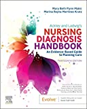
Ackley and Ladwig’s Nursing Diagnosis Handbook: An Evidence-Based Guide to Planning Care
We love this book because of its evidence-based approach to nursing interventions. This care plan handbook uses an easy, three-step system to guide you through client assessment, nursing diagnosis, and care planning. Includes step-by-step instructions showing how to implement care and evaluate outcomes, and help you build skills in diagnostic reasoning and critical thinking.

Nursing Care Plans – Nursing Diagnosis & Intervention (10th Edition)
Includes over two hundred care plans that reflect the most recent evidence-based guidelines. New to this edition are ICNP diagnoses, care plans on LGBTQ health issues, and on electrolytes and acid-base balance.

Nurse’s Pocket Guide: Diagnoses, Prioritized Interventions, and Rationales
Quick-reference tool includes all you need to identify the correct diagnoses for efficient patient care planning. The sixteenth edition includes the most recent nursing diagnoses and interventions and an alphabetized listing of nursing diagnoses covering more than 400 disorders.

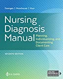
Nursing Diagnosis Manual: Planning, Individualizing, and Documenting Client Care
Identify interventions to plan, individualize, and document care for more than 800 diseases and disorders. Only in the Nursing Diagnosis Manual will you find for each diagnosis subjectively and objectively – sample clinical applications, prioritized action/interventions with rationales – a documentation section, and much more!

All-in-One Nursing Care Planning Resource – E-Book: Medical-Surgical, Pediatric, Maternity, and Psychiatric-Mental Health
Includes over 100 care plans for medical-surgical, maternity/OB, pediatrics, and psychiatric and mental health. Interprofessional “patient problems” focus familiarizes you with how to speak to patients.

See also
Other recommended site resources for this nursing care plan:
- Nursing Care Plans (NCP): Ultimate Guide and Database MUST READ!
Over 150+ nursing care plans for different diseases and conditions. Includes our easy-to-follow guide on how to create nursing care plans from scratch. - Nursing Diagnosis Guide and List: All You Need to Know to Master Diagnosing
Our comprehensive guide on how to create and write diagnostic labels. Includes detailed nursing care plan guides for common nursing diagnostic labels.
Other care plans for hematologic and lymphatic system disorders:


Leave a Comment