Utilize this comprehensive nursing care plan and management guide to provide effective care for patients experiencing hypovolemic shock. Gain valuable insights on nursing assessment, interventions, goals, and nursing diagnosis specifically tailored for hypovolemic shock in this guide.
Table of Contents
- What is hypovolemic shock?
- Nursing Care Plans & Management
- Recommended Resources
- See also
What is hypovolemic shock?
Hypovolemic shock, characterized by decreased intravascular volume, is a medical condition resulting from blood loss, leading to reduced cardiac output and inadequate tissue perfusion. It can be caused by external fluid losses, such as traumatic blood loss, or internal fluid shifts, like severe dehydration or edema. Symptoms vary based on the severity of fluid or blood loss, but all symptoms of shock require immediate medical treatment. Hypovolemic shock occurs when there is a reduction in intravascular volume by 15% to 30%, representing a loss of approximately 750 to 1,500 mL of blood in a 70-kg person. This reduction in intravascular volume leads to decreased venous return, decreased ventricular filling, decreased stroke volume, decreased cardiac output, and ultimately, inadequate tissue perfusion.
The primary objectives in treating hypovolemic shock are to restore intravascular volume, redistribute fluid, and address the underlying cause of fluid loss. These goals are typically addressed concurrently to reverse the progression of inadequate tissue perfusion.
Nursing Care Plans & Management
Nursing care management for patients with hypovolemic shock involves rapid assessment to determine the cause and severity of hypovolemia, administering intravenous fluids for volume resuscitation, closely monitoring vital signs and perfusion parameters, using invasive hemodynamic monitoring when needed, providing oxygen therapy and respiratory support, addressing the underlying cause of hypovolemia, and collaborating with the healthcare team for timely interventions and adjustments.
Nursing Problem Priorities
The following are the nursing priorities for patients with hypovolemic shock:
- Monitoring vital signs and perfusion parameters. Continuous assessment and monitoring of vital signs and perfusion parameters (skin color, temperature, capillary refill time, and urine output) are essential in determining the patient’s hemodynamic status and response to therapeutic management.
- Fluid resuscitation. This is to restore blood volume, enhance cardiac output, and improve tissue perfusion. Close monitoring of the patient’s fluid balance, including input and output, is crucial in preventing fluid overload or under-resuscitation.
- Oxygen administration and providing airway support. Maintaining adequate oxygenation and tissue perfusion by assessing oxygen saturation, providing oxygen therapy, and assisting with airway management as needed.
- Achieving hemodynamic stability. Collaborating with the healthcare team to monitor invasive hemodynamic parameters (such as central venous pressure, and arterial blood pressure) if indicated, to assist with fluid resuscitation and optimize cardiac output.
- Emotional support and patient/family education. Providing emotional support, clear and concise communication, and education to the patient and family members about the condition, treatment, and possible complications can help decrease anxiety and promote understanding and involvement in the care.
Nursing Assessment
Assess for the following subjective and objective data:
- Abnormal arterial blood gases (ABGs) indicating hypoxemia and acidosis.
- Prolonged capillary refill time (>3 seconds).
- Cardiac dysrhythmias.
- Altered level of consciousness.
- Cold and clammy skin.
- Decreased skin turgor.
- Dizziness.
- Dry mucous membranes.
- Increased thirst.
- Narrowing of pulse pressure.
- Orthostatic hypotension.
- Tachycardia.
- Variable urine output ranging from normal (>30 ml/hr) to as low as 20 ml/hr.
Assess for factors related to the cause of hypovolemic shock:
- Alterations in heart rate and rhythm (tachycardia).
- Decreased ventricular filling (preload) and diminished venous return.
- Fluid volume loss of 30% or more (severe blood loss).
- Late uncompensated hypovolemic shock.
- Active fluid volume loss (abnormal bleeding, diarrhea, diuresis, or abnormal drainage).
- Internal fluid shifts.
- Inadequate fluid intake and/or severe dehydration.
- Regulatory mechanism failure.
- Trauma.
- Decreased urinary output (less than 30 ml per hour).
- Decreased peripheral pulses and decreased pulse pressure.
- Decreased blood pressure.
- Decreased stroke volume and decreased preload.
Nursing Diagnosis
After conducting a thorough assessment of a patient experiencing hypovolemic shock, the next step of the nursing process is to formulate an appropriate nursing diagnosis. This diagnosis reflects the patient’s current health condition and needs and guides the development of a tailored care plan.
Nursing Goals
Goals and expected outcomes may include:
- The client will maintain adequate cardiac output, as evidenced by strong peripheral pulses, systolic BP within 20 mm Hg of baseline, HR 60 to 100 beats per minute with a regular rhythm, urinary output of 30 ml/hr or greater, warm and dry skin, and normal level of consciousness.
- The client will be normovolemic as evidenced by HR 60 to 100 beats per minute, systolic BP greater than or equal to 90 mm Hg, absence of orthostasis, urinary output greater than 30ml/hr, and normal skin turgor.
- The client will experience a decrease in anxiety levels.
Nursing Interventions and Actions
Preventing shock is a key aspect of nursing care. Close monitoring of at-risk patients and prompt fluid replacement can help prevent hypovolemic shock. Nursing care also involves supporting the treatment of the underlying cause and restoring intravascular volume. Safe administration of fluids and medications, documentation of their effects, and the use of volumetric IV pumps for vasopressor medications are important nursing responsibilities. Monitoring for complications and side effects and promptly reporting them are crucial in providing comprehensive care. Therapeutic interventions and nursing actions for patients with hypovolemic shock may include:
- 1. Managing Decrease in Cardiac Output
- 2. Improving Deficiencies in Fluid Volume
- 3. Improving Cardiac Tissue Perfusion
- 4. Monitoring and Preventing Complications
- 5. Reducing Anxiety and Providing Emotional Support
1. Managing Decrease in Cardiac Output
In hypovolemic shock, there is a significant loss of blood volume, resulting in reduced venous return to the heart. This leads to decreased preload, impairing the ability of the heart to fill adequately, which in turn decreases cardiac output. Furthermore, the body initiates compensatory mechanisms such as increased heart rate and systemic vascular resistance but these can only partially offset the decrease in cardiac output. The following are assessment and nursing interventions for managing decrease in cardiac output:
1. Administer fluid and blood and fluid replacement therapy as prescribed.
Safe administration of blood transfusions is a critical nursing responsibility. Prompt acquisition of blood specimens, baseline blood tests, and blood typing are important in emergency situations. Close monitoring of patients receiving blood products is essential to detect adverse effects. Complications related to fluid replacement, such as cardiovascular overload and transfusion-associated circulatory overload, must be carefully monitored. Older adults, patients with preexisting cardiac disease, and those receiving multiple blood products are at increased risk. Transfusion-related acute lung injury is a potential complication characterized by pulmonary edema and respiratory distress. Monitoring parameters include hemodynamic pressure, vital signs, blood gases, lactate levels, hemoglobin and hematocrit levels, bladder pressure, and fluid intake and output. Temperature should also be closely monitored to prevent hypothermia. Physical assessment focuses on jugular vein distention and jugular venous pressure, which increases with fluid overload. Close monitoring of cardiac and respiratory status is crucial, with prompt reporting of any changes to the primary provider.
2. Assess the client’s HR and BP, including peripheral pulses. Use direct intra-arterial monitoring as ordered.
Sinus tachycardia and increased arterial BP are seen in the early stages to maintain an adequate cardiac output. Hypotension happens as the condition deteriorates. Vasoconstriction may lead to unreliable blood pressure. Pulse pressure (systolic minus diastolic) decreases in shock. Older clients have reduced response to catecholamines; thus their response to decreased cardiac output may be blunted, with less increase in HR.
3. Assess the client’s ECG for dysrhythmias.
Cardiac dysrhythmias may occur from the low perfusion state, acidosis, or hypoxia, as well as from side effects of cardiac medications used to treat this condition.
4. Assess capillary refill time.
Capillary refill is slow and sometimes absent.
5. Assess the respiratory rate, rhythm, and auscultate breath sounds.
Characteristics of a shock include rapid, shallow respirations and adventitious breath sounds such as crackles and wheezes.
6. Monitor oxygen saturation and arterial blood gases.
Pulse oximetry is used in measuring oxygen saturation. The normal oxygen saturation should be maintained at 90% or higher. As shock progresses, aerobic metabolism stops and lactic acidosis occurs, resulting in an increased level of carbon dioxide and decreasing pH.
7. Monitor the client’s central venous pressure (CVP), pulmonary artery diastolic pressure (PADP), pulmonary capillary wedge pressure, and cardiac output/cardiac index.
CVP provides information on filling pressures of the right side of the heart; pulmonary artery diastolic pressure and pulmonary capillary wedge pressure reflect left-sided fluid volumes. Cardiac output provides an objective number to guide therapy.
8. Assess for any changes in the level of consciousness.
Restlessness and anxiety are early signs of cerebral hypoxia while confusion and loss of consciousness occur in the later stages. Older clients are especially susceptible to reduced perfusion to vital organs.
9. Assess urine output.
The renal system compensates for low BP by retaining water. Oliguria is a classic sign of inadequate renal perfusion from reduced cardiac output.
10. Assess skin color, temperature, and moisture.
Cool, pale, clammy skin is secondary to a compensatory increase in sympathetic nervous system stimulation and low cardiac output and desaturation.
11. Provide electrolyte replacement as prescribed.
Electrolyte imbalance may cause dysrhythmias or other pathological states.
12. If possible, use a fluid warmer or rapid fluid infuser.
Fluid warmers keep core temperature. Infusing cold blood is associated with myocardial dysrhythmias and paradoxical hypotension. Macropore filtering IV devices should also be used to remove small clothes and debris.
2. Improving Deficiencies in Fluid Volume
Identifying and addressing the underlying cause of hypovolemia, such as controlling bleeding or correcting dehydration, is important in managing the patient’s condition effectively. To improve fluid volume deficiency in patients with hypovolemic shock, immediate administration of intravenous fluids such as crystalloids (e.g., normal saline, lactated Ringer’s solution) and colloids (e.g., albumin) is essential. These fluids help replenish blood volume, increase preload, and restore cardiac output. The following are assessment and nursing interventions for enhancing fluid volume deficit:
1. Monitor BP for orthostatic changes (changes seen when changing from a supine to a standing position).
A common manifestation of fluid loss is postural hypotension. The incidence increase with age. Note the following orthostatic hypotension significances:
- Greater than 10 mm Hg: circulating blood volume decreases by 20%.
- Greater than 20 to 30 mm Hg drop: circulating blood volume is decreased by 40%.
2. Assess the client’s HR, BP, and pulse pressure. Use direct intra-arterial monitoring as ordered.
Sinus tachycardia and increased arterial BP are seen in the early stages to maintain an adequate cardiac output. Hypotension happens as the condition deteriorates. Vasoconstriction may lead to unreliable blood pressure. Pulse pressure (systolic minus diastolic) decreases in shock. Older clients have reduced response to catecholamines; thus their response to decreased cardiac output may be blunted, with less increase in HR.
3. Assess for changes in the level of consciousness.
Confusion, restlessness, headache, and a change in the level of consciousness may indicate an impending hypovolemic shock.
4. Monitor for possible sources of fluid loss.
Sources of fluid loss may include diarrhea, vomiting, wound drainage, severe blood loss, profuse diaphoresis, high fever, polyuria, burns, and trauma.
5. Assess the client’s skin turgor and mucous membranes for signs of dehydration.
Decreased skin turgor is a late sign of dehydration. It occurs because of the loss of interstitial fluid.
6. Monitor the client’s intake and output.
Accurate measurement is important in detecting negative fluid balance and guiding therapy. Concentrated urine denotes a fluid deficit.
7. Monitor coagulation studies, including INR, prothrombin time, partial thromboplastin time, fibrinogen, fibrin split products, and platelet count as ordered.
Specific deficiencies guide treatment therapy.
8. Obtain a spun hematocrit, and reevaluate every 30 minutes to 4 hours, depending on the client’s ability.
Hematocrit decreases as fluids are administered because of dilution. As a rule of thumb, hematocrit decreases by 1% per liter of normal saline solution or lactated Ringer’s used. Any other hematocrit decrease must be evaluated as an indication of continued blood loss.
9. Place patient in a modified Trendelenburg position.
Proper positioning of the patient can aid in fluid redistribution in addition to fluid administration. The modified Trendelenburg position, also known as passive leg raising, is recommended for patients with hypovolemic shock. Elevating the legs facilitates venous blood return and serves as a dynamic assessment of fluid responsiveness. The nurse monitors vital signs, specifically blood pressure and pulse pressure, for improvement. It is important to note that a full Trendelenburg position is not recommended as it may impede breathing and does not increase blood pressure or cardiac output.
10. If hypovolemia is a result of severe diarrhea or vomiting, administer antidiarrheal or antiemetic medications as prescribed, in addition to IV fluids.
Treatment is guided by the cause of the problem.
11. Encourage oral fluid intake if able.
The oral route supports maintaining fluid balance.
12. Prepare to administer a bolus of 1 to 2 L of IV fluids as ordered. Use crystalloid solutions for adequate fluid and electrolyte balance.
The client’s response to treatment relies on the extent of the blood loss. If blood loss is mild (15%), the expected response is a rapid return to normal BP. If the IV fluids are slowed, the client remains normotensive. If the client has lost 20% to 40% of circulating blood volume or has continued uncontrolled bleeding, a fluid bolus may produce normotension, but if fluids are slowed after the bolus, BP will deteriorate. Extreme caution is indicated in fluid replacement in older clients. Aggressive therapy may precipitate left ventricular dysfunction and pulmonary edema.
13. Initiate IV therapy. Start two shorter, large-bore peripheral IV lines.
Maintaining adequate circulating blood volume is a priority in treating hypovolemic shock. The amount of fluid infused is more important than the specific type of fluid used. Large-bore IV catheters should be used to maximize the volume that can be infused. Rapid access for fluid administration is achieved through the insertion of two or more large-gauge IV lines. In challenging cases, an intraosseous catheter may be considered. Multiple IV lines allow for simultaneous administration of fluids, medications, and blood components. The choice of fluids aims to restore intravascular volume and prevent intracellular fluid shifts. For further details on commonly used fluids in shock treatment, refer to Table 14-3.
14. Administer blood products (e.g., packed red blood cells, fresh frozen plasma, platelets) as prescribed. Transfuse the client with whole blood-packed red blood cells.
Preparing fully crossmatched blood may take up to 1 hour in some laboratories. Consider using uncross-matched or type-specific blood until cross-matched blood is available. If type-specific blood is not available, type O blood may be used for exsanguinating clients. If available, Rh-negative blood is preferred, especially for women of childbearing age. Autotransfusion may be used when there is massive bleeding in the thoracic cavity.
15. Monitor the client’s central venous pressure (CVP), pulmonary artery diastolic pressure (PADP), pulmonary capillary wedge pressure, and cardiac output/cardiac index.
CVP provides information on filling pressures of the right side of the heart; pulmonary artery diastolic pressure and pulmonary capillary wedge pressure reflect left-sided fluid volumes. The cardiac output provides an objective number to guide therapy.
3. Improving Cardiac Tissue Perfusion
Enhancing cardiac tissue perfusion is vital in hypovolemic shock for multiple reasons. It prevents myocardial ischemia, supports organ function, and maintains hemodynamic stability. The following are interventions to improve cardiac tissue perfusion:
1. Assess for signs of decreased tissue perfusion.
Particular clusters of signs and symptoms occur with differing causes. Evaluation provides a baseline for future comparison.
2. Assess for rapid changes or continued shifts in mental status.
Restlessness and anxiety are early signs of cerebral hypoxia while confusion and loss of consciousness occur in the later stages.
3. Assess capillary refill.
Capillary refill is slow and sometimes absent.
4. Observe for pallor, cyanosis, mottling, and cool or clammy skin. Assess the quality of every pulse.
The nonexistence of peripheral pulses must be reported or managed immediately. Systemic vasoconstriction resulting from reduced cardiac output may be manifested by diminished skin perfusion and loss of pulses. Therefore, assessment is required for constant comparisons
5. Record BP readings for orthostatic changes (a drop of 20 mm Hg systolic BP or 10 mm Hg diastolic BP with position changes).
Stable BP is needed to keep sufficient tissue perfusion. Medication effects such as altered autonomic control, decompensated heart failure, reduced fluid volume, and vasodilation are among many factors potentially jeopardizing optimal BP.
6. Use pulse oximetry to monitor oxygen saturation and pulse rate.
Pulse oximetry is a useful tool to detect changes in oxygen saturation.
7. Review laboratory data (ABGs, BUN, creatinine, electrolytes, international normalized ratio, and prothrombin time or partial thromboplastin time) if anticoagulants are utilized for treatment.
Blood clotting studies are being used to conclude or make sure that clotting factors stay within therapeutic levels. Gauges of organ perfusion or function. Irregularities in coagulation may occur as an effect of therapeutic measures.
8. Assist with position changes.
Gently repositioning the patient from a supine to a sitting/standing position can reduce the risk of orthostatic BP changes. Older patients are more susceptible to such drops of pressure with position changes.
9. Provide oxygen therapy if indicated.
Oxygen is administered to increase the amount of oxygen carried by available hemoglobin in the blood.
10. Administer IV fluids as ordered.
Sufficient fluid intake maintains adequate filling pressures and optimizes cardiac output needed for tissue perfusion.
4. Monitoring and Preventing Complications
Monitoring and preventing complications during hypovolemic shock is important for timely intervention and optimizing patient outcomes, as it allows for early recognition of worsening conditions, appropriate resuscitation, and prevention of organ damage or failure. The following are nursing assessments and interventions to prevent complications for patients experiencing hypovolemic shock:
1. If the only visible injury is an obvious head injury, look for other causes of hypovolemia (e.g., long-bone fractures, internal bleeding, external bleeding).
Hypovolemic shock following trauma usually results from hemorrhage.
2. If trauma has occurred, evaluate and document the extent of the client’s injuries; use a primary survey (or another consistent survey method) or ABCs: airway with cervical spine control, breathing, and circulation.
A primary survey helps identify potentially life-threatening injuries. This serves as a quick primary assessment.
3. Perform a secondary survey after all life-threatening injuries are ruled out or treated.
A secondary survey uses a methodical head-to-toe inspection.
4. For post-surgical clients, monitor blood loss (mark skin area, weigh dressing to determine fluid loss, monitor chest tube drainage).
It is important to observe an expanding hematoma or swelling or increased drainage to identify bleeding or coagulopathy.
5. Control the external source of bleeding by applying direct pressure to the bleeding site.
External bleeding is controlled with firm, direct pressure on the bleeding site, using a thick dry dressing material. Prompt, effective treatment is needed to preserve vital organ function and life.
6. For trauma victims with internal bleeding (e.g., pelvic fracture), military antishock trousers (MAST) or pneumatic antishock garments (PASGs) may be used.
These devices are useful for tamponade bleeding. Hypovolemia from long-bone fractures (e.g., femur or pelvic fractures) may be uncontrolled by splinting with air splints. Hare traction splints or MAST and/or PASG trousers may be used to reduce tissue and vessel damage from the manipulation of unstable fractures.
7. If hypovolemia is a result of severe burns, calculate the fluid replacement according to the extent of the burn and the client’s body weight.
Formulas such as the Parkland formula, which follows, guide fluid replacement therapy:
% BSA (body surface area) burned x weight in kg x 4 ml lactated Ringer’s = Total fluid to be infused over 24 hours: half given intravenously over 8 hours and a half given over the next 16 hours.
8. If the client’s condition progressively deteriorates, initiate cardiopulmonary resuscitation or other lifesaving measures according to Advanced Cardiac Life Support guidelines, as indicated.
Shock unresponsive to fluid replacement can worsen into cardiogenic shock. Depending on etiological factors, vasopressors, inotropic agents, antidysrhythmics, or other medications can be used.
9. If bleeding is secondary to surgery, anticipate or prepare for a return to surgery.
Surgery may be the only option to fix the problem.
5. Reducing Anxiety and Providing Emotional Support
Anxiety in hypovolemic shock may arise from physiological stress responses and awareness of the critical condition. Nursing assessments and interventions can effectively alleviate anxiety in these patients. The following are nursing assessments and interventions to help reduce anxiety in patients experiencing hypovolemic shock:
1. Assess the previous coping mechanisms used.
Anxiety and ways of decreasing perceived anxiety are highly individualized. Interventions are most effective when they are consistent with the client’s established coping pattern. However, in the acute care setting these techniques may no longer be feasible.
2. Assess the client’s level of anxiety.
Shock can result in an acute life-threatening situation that will produce high levels of anxiety in the client as well as in significant others.
3. Acknowledge awareness of the client’s anxiety.
Acknowledgment of the client’s feelings validates the client’s feelings and communicates acceptance of those feelings.
4. Encourage the client to verbalize his or her feelings.
Talking about anxiety-producing situations and anxious feelings can help the client perceive the situation in a less threatening manner.
5. Reduce unnecessary external stimuli by maintaining a quiet environment. If medical equipment is a source of anxiety, consider providing sedation to the client.
Anxiety may escalate with excessive conversation, noise, and equipment around the client.
6. Explain all procedures as appropriate, keeping explanations basic.
The information helps reduce anxiety. Anxious clients were unable to understand anything more than simple, clear, brief instructions.
7. Maintain a confident, assured manner while interacting with the client. Assure the client and significant others of close, continuous monitoring that will ensure prompt intervention.
The staff’s anxiety may be easily perceived by the client. The client’s feeling of stability increases in a calm and non-threatening atmosphere. The presence of a trusted person may help the client feel less threatened.
Evaluation
Expected outcomes for the patient with hypovolemic shock include:
- Maintained fluid volume at a functional level.
- Reported understanding of the causative factors of fluid volume deficit.
- Maintained normal blood pressure, temperature, and pulse.
- Maintained elastic skin turgor, most tongue and mucous membranes, and orientation to person, place, and time.
Discharge and Home Care Guidelines
The discharge and home care guidelines for patients recovering from hypovolemic shock may vary depending on the severity of the condition and the specific needs of the individual. However, here are some general considerations:
- Follow-up appointments: The importance of complying with the follow-up appointments with the healthcare provider to evaluate the progress of recovery and to adjust treatment if needed.
- Rest and recovery: Ensure an adequate period of rest and gradually increase activity levels as advised by the healthcare provider.
- Hydration and nutrition: Ensure adequate hydration by increasing fluid intake as indicated by the HCP. Follow any dietary recommendations to promote healing and restore lost nutrients.
- Wound care: Adhere to instructions on proper wound care, such as keeping the area clean, changing dressings as directed, and observing for signs of infection if there are any wounds or surgical incisions.
- Patient and family education: Recognizing the warning signs and symptoms that may indicate a worsening condition or potential complications.
- Emotional support: Hypovolemic shock can be a traumatic experience. Seeking emotional support from friends, family, or support groups to help cope with any anxiety, or emotional distress that may arise.
Documentation Guidelines
The focus of documentation includes:
- Degree of deficit and current sources of fluid intake.
- I&O, fluid balance, changes in weight, presence of edema, urine specific gravity, and vital signs.
- Results of diagnostic studies.
- Functional level and specifics of limitations.
- Needed resources and adaptive devices.
- Availability and use of community resources.
- Plan of care.
- Teaching plan.
- Client’s responses to interventions, teachings, and actions performed
- Attainment or progress towards desired outcomes.
- Modifications to plan of care.
Recommended Resources
Recommended nursing diagnosis and nursing care plan books and resources.
Disclosure: Included below are affiliate links from Amazon at no additional cost from you. We may earn a small commission from your purchase. For more information, check out our privacy policy.
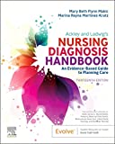
Ackley and Ladwig’s Nursing Diagnosis Handbook: An Evidence-Based Guide to Planning Care
We love this book because of its evidence-based approach to nursing interventions. This care plan handbook uses an easy, three-step system to guide you through client assessment, nursing diagnosis, and care planning. Includes step-by-step instructions showing how to implement care and evaluate outcomes, and help you build skills in diagnostic reasoning and critical thinking.

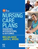
Nursing Care Plans – Nursing Diagnosis & Intervention (10th Edition)
Includes over two hundred care plans that reflect the most recent evidence-based guidelines. New to this edition are ICNP diagnoses, care plans on LGBTQ health issues, and on electrolytes and acid-base balance.

Nurse’s Pocket Guide: Diagnoses, Prioritized Interventions, and Rationales
Quick-reference tool includes all you need to identify the correct diagnoses for efficient patient care planning. The sixteenth edition includes the most recent nursing diagnoses and interventions and an alphabetized listing of nursing diagnoses covering more than 400 disorders.

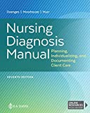
Nursing Diagnosis Manual: Planning, Individualizing, and Documenting Client Care
Identify interventions to plan, individualize, and document care for more than 800 diseases and disorders. Only in the Nursing Diagnosis Manual will you find for each diagnosis subjectively and objectively – sample clinical applications, prioritized action/interventions with rationales – a documentation section, and much more!

All-in-One Nursing Care Planning Resource – E-Book: Medical-Surgical, Pediatric, Maternity, and Psychiatric-Mental Health
Includes over 100 care plans for medical-surgical, maternity/OB, pediatrics, and psychiatric and mental health. Interprofessional “patient problems” focus familiarizes you with how to speak to patients.

See also
Other recommended site resources for this nursing care plan:
- Nursing Care Plans (NCP): Ultimate Guide and Database MUST READ!
Over 150+ nursing care plans for different diseases and conditions. Includes our easy-to-follow guide on how to create nursing care plans from scratch. - Nursing Diagnosis Guide and List: All You Need to Know to Master Diagnosing
Our comprehensive guide on how to create and write diagnostic labels. Includes detailed nursing care plan guides for common nursing diagnostic labels.
Other care plans for hematologic and lymphatic system disorders:
