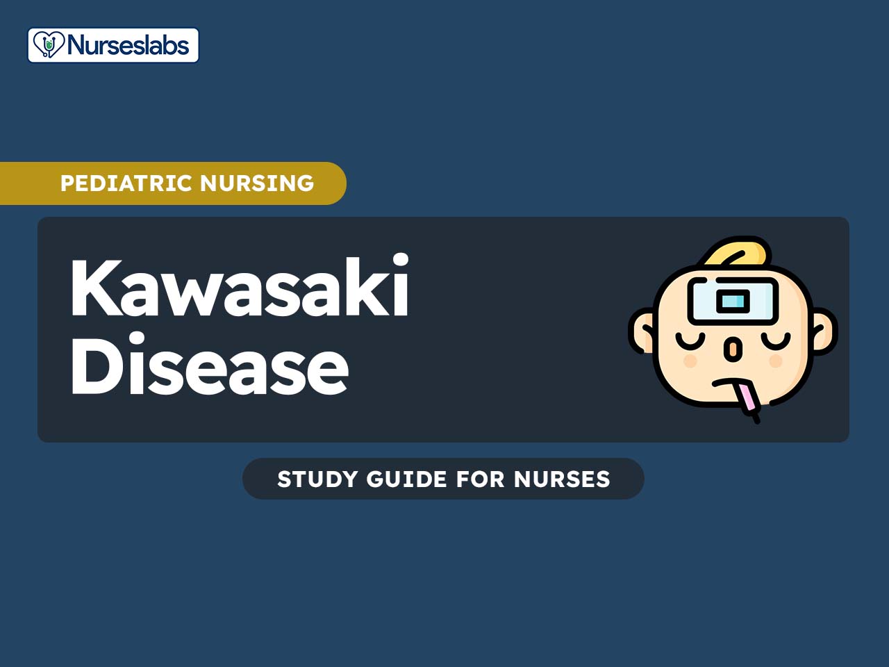
Kawasaki disease is a rare but serious inflammatory condition that primarily affects children under the age of five and is characterized by prolonged high fever, redness of the eyes, changes in the lips and mouth, rash, swollen lymph nodes, and inflammation of the blood vessels. If left untreated, Kawasaki disease can lead to potentially life-threatening complications, such as coronary artery aneurysms.
What is Kawasaki Disease?
Some cardiovascular system disorders occur as a result of a congenital heart disorder or a disease such as Kawasaki disease.
- Kawasaki disease or mucocutaneous lymph node syndrome is an acute, febrile disease that is most often seen in boys younger than 5 years.
- Kawasaki disease (KD) is an acute febrile vasculitic syndrome of early childhood that, although it has a good prognosis with treatment, can lead to death from coronary artery aneurysm (CAA) in a very small percentage of patients.
- The disorder has also been called mucocutaneous lymph node syndrome and infantile periarteritis nodosa.
Pathophysiology
Despite the prominent mucocutaneous clinical findings that define the illness, Kawasaki disease is best regarded as a generalized vasculitis that involves small- to medium-sized arteries.
- After infection, an alteration in the immune system occurs; most cases occur in the late winter or early spring.
- In the earliest stages of the disease, the endothelial cells and the vascular media become edematous, but the internal elastic lamina remains intact.
- Then, approximately 7-9 days after the onset of fever, an influx of neutrophils occurs, which is quickly followed by a proliferation of CD8+ (cytotoxic) lymphocytes and immunoglobulin A–producing plasma cells.
- The inflammatory cells secrete various cytokines (ie, tumor necrosis factor, vascular endothelial growth factor, monocyte chemotactic and activating factor), interleukins, and matrix metalloproteinases that target the endothelial cells and result in a cascade of events that eventuates in fragmentation of the internal elastic lamina and vascular damage.
- Over the next few weeks to months, the active inflammatory cells are replaced by fibroblasts and monocytes, and fibrous connective tissue begins to form within the vessel wall.
- The intima proliferates and thickens; the vessel wall eventually becomes narrowed or occluded owing to stenosis or a thrombus.
- Most of the pathology of the disease is induced by medium vessel arterial vasculitis.
- Initially, neutrophils are present in great numbers, but the infiltrate rapidly switches to mononuclear cells, T lymphocytes, and immunoglobulin A (IgA)–producing plasma cells. Inflammation involves all 3 layers of vessels.
- The period during of the greatest vascular damage is when a concomitant progressive increase in the serum platelet count occurs, and this is the point of the illness when the risk of death is most significant.
Statistics and Incidences
Epidemics of Kawasaki disease primarily occur in the late winter and spring, at 2- to 3-year intervals.
- Approximately 3,000 children with Kawasaki disease are hospitalized annually in the United States.
- The approximate annual race-specific incidence per 100,000 children younger than 5 years is 32.5 cases for Americans of Asian and Pacific Island descent, 16.9 cases for non-Hispanic African Americans, 11.1 cases for Hispanics, and 9.1 cases for whites.
- Outside the United States, the disease is most frequently observed in Japan, Taiwan, and Korea.
- The prevalence of Kawasaki disease increased from 1967 to the mid-1980s and has leveled out at 5000-6000 cases per year.
- The highest incidence of Kawasaki disease has been reported in Japan, where the frequency of the disease is 10 to 20 times higher than in Western countries.
- Approximately 5000-6000 cases are reported each year in Japan; the incidence in 2000 was 134.2 cases per 100,000 children younger than 5 years.
- The annual incidence reported in white populations outside the United States is similar to that reported in the US population, with 11.3-14.7 cases per 100,000 children younger than 5 years in Canada and 3.6 cases per 100,000 children younger than 5 years in Australia.
- From 1999-2000, the incidence in the United Kingdom was 8.1 cases per 100,000 children.
- Ontario has the highest rate of Kawasaki disease outside of Asia, with a yearly incidence of 26.2 cases per 100,000 population younger than 5 years.
- Although Kawasaki disease has been reported in children of all ethnic origins, it occurs most commonly in Asian children, especially those of Japanese descent.
- Kawasaki disease is slightly more common in males than in females; the male-to-female ratio ranges from 1.3-1.83:1 depending on the country from which the statistics are reported.
- Approximately 85-90% of Kawasaki disease cases occur in children younger than 5 years; 90-95% of cases occur in children younger than 10 years.
Causes
The etiology is unknown, but the disease appears to be caused by an infectious agent.
- Genetic factors. Siblings of affected children have a 10-20 times higher probability of developing Kawasaki disease than the general population, and children in Japan whose parents had Kawasaki disease seem to have a more severe form of the disease and to be more susceptible to recurrence.
- Infection. Features of Kawasaki disease that are consistent with an infectious etiology include the occurrence of epidemics primarily in late winter and spring with 3-year intervals and the wavelike geographic spread of those epidemics; the self-limited nature of the disease; and the characteristic fever, adenopathy, and eye signs.
Clinical Manifestations
The clinical presentation of Kawasaki disease varies over time, with the clinical course conventionally divided into three stages: acute, subacute, and convalescent.

Acute Febrile Stage
The acute stage begins with an abrupt onset of fever and lasts approximately 7-14 days; the fever is typically high-spiking and remittent, with peak temperatures ranging from 102-104°F (39-40°C) or higher; in addition to fever, signs and symptoms of this phase may include the following:
-
Irritability
-
Nonexudative bilateral conjunctivitis (90%)
-
Anterior uveitis (70%)
-
Perianal erythema (70%)
-
Strawberry tongue and lip fissures
-
Hepatic, renal, and GI dysfunction
-
Myocarditis and pericarditis
-
Lymphadenopathy (75%), generally a single, enlarged, nonsuppurative cervical node measuring approximately 1.5 cm
Subacute Stage
The subacute stage begins when the fevers have abated, and it continues until week 4-6; the hallmarks of this stage are desquamation of the digits, thrombocytosis (the platelet count may exceed 1 million/μL), and the development of coronary aneurysms; the risk for sudden death is highest at this stage.
Convalescent Stage
The convalescent phase is marked by the complete resolution of clinical signs of the illness, usually within 3 months of presentation; this stage begins with the return to baseline of the acute phase reactants (eg, erythrocyte sedimentation rate, C-reactive protein) and other laboratory abnormalities; during this stage, most of the clinical findings resolve; however, deep transverse grooves across the nails (Beau lines) may become apparent 1-2 months after the onset of fever.
Physical Examination
Because no specific test can be performed for Kawasaki disease and no clinical feature is pathognomonic, the diagnosis of Kawasaki disease is based on the presence of a constellation of clinical findings.
-
Changes in the peripheral extremities. Initial reddening or edema of the palms and soles, followed by membranous desquamation of the finger and toe tips or transverse grooves across the fingernails and toenails (Beau lines).
-
Polymorphous rash (not vesicular). Usually generalized but may be limited to the groin or lower extremities.
-
Oropharyngeal changes. Erythema, fissuring, and crusting of the lips; strawberry tongue; diffuse mucosal injection of the oropharynx.
-
Conjunctivitis. Bilateral, nonexudative, painless bulbar conjunctival injection.
-
Lymphadenopathy. Acute nonpurulent cervical lymphadenopathy with lymph node diameter greater than 1.5 cm, usually unilateral.
Assessment and Diagnostic Findings
No specific laboratory test is used to diagnose Kawasaki disease; however, certain abnormalities coincide with various stages.
- Urine proteins. More recently, 2 urine proteins hold promise as biomarkers of Kawasaki disease: meprin A or filamin C; these 2 biomarkers were diagnostically superior to ESR or CRP; investigators identified more than 190 proteins that were present only in children with Kawasaki disease, including the proteins associated with endothelial and myocardial cell injury (filamin C) and immune regulators (meprin A).
- CBC. On complete blood counts (CBCs), mild-to-moderate normochromic anemia is observed in the acute stage; the white blood cell count (WBC) is moderate to high (50% of patients have a WBC greater than 15,000/µL), with a left shift, which is a predominant sign of immature and mature granulocytes.
- Platelet count. During the subacute stage, thrombocytosis is the outstanding marker; the platelet count begins to rise in the second week and continues to rise during the third week; platelet counts average 700,000/μL, but levels as high as 2 million have been observed.
- Cholesterol. Serum cholesterol, high-density lipoprotein, and apolipoprotein A levels are decreased; these values tend to persist beyond clinical resolution of the disease.
- Echocardiography. Echocardiography is the study of choice to evaluate for coronary artery aneurysms (CAAs), in both fully manifested and suspected incomplete cases of Kawasaki disease.
- Imaging studies. Magnetic resonance imaging (MRI), magnetic resonance angiography (MRA), and ultrafast computed tomography (CT) scanning are other noninvasive tests that can be used to evaluate coronary artery abnormalities.
- Electrocardiography. On electrocardiography (ECG), tachycardia, prolonged PR interval, ST-T wave changes, and decreased voltage of R waves may indicate myocarditis; Q waves or ST-T wave changes may indicate myocardial infarction.
- Cardiac enzymes. Cardiac enzyme levels (eg, creatine kinase [CK], creatine kinase myocardial band [CK-MB], cardiac troponin, lactate dehydrogenase [LD-1 >LD-2]) are elevated during a myocardial infarction.
Medical Management
The principal goal of treatment for Kawasaki disease is to prevent coronary artery disease and to relieve symptoms.
Pharmacologic Therapy
Full doses of intravenous immunoglobulin (IVIG) are the mainstay of treatment, along with other drugs.
- Intravenous immunoglobulin. IVIG relieves acute inflammation and has been shown to reduce the rate of coronary aneurysms from greater than 25% in untreated patients to 1-5% in treated patients; maximal benefits are seen when IVIG is given within the first 10 days of the illness.
- Aspirin. Aspirin has a synergistic effect with IVIG and has long been a standard part of therapy for Kawasaki disease; patients on prolonged aspirin therapy must be instructed that concomitant use of ibuprofen antagonizes the irreversible effect of platelet inhibition by aspirin and should be avoided during therapy; additionally, the risks of developing Reye syndrome during an active infection with influenza or varicella should be addressed.
- Other adjunctive agents. In addition to their use in treatment of IVIG-resistant Kawasaki disease, corticosteroids have been proposed as part of primary therapy; the roles of other adjunctive therapies, including pentoxifylline and abciximab, have not yet been definitively determined; pentoxifylline acts as an anti-inflammatory agent by inhibiting tumor necrosis factor-alpha and may reduce the incidence of aneurysms; abciximab is a platelet glycoprotein IIb/IIIa receptor inhibitor and has been used in conjunction with standard therapies in patients with Kawasaki disease and giant aneurysms.
- Anticoagulant therapy. Anticoagulants such as warfarin and low molecular weight heparin are used in patients with large aneurysms in whom the risk of thrombosis is high. The goal is to maintain an international normalized ratio (INR) of 2-2.5.
Nursing Management
The nursing management of a child with Kawasaki disease involves the following systematic approach:
Nursing Assessment
The child should be assessed in every phase of the disease:
- Acute febrile phase. The child appears severely ill and irritable; there is high, spiking fever for 5 or more days, bilateral conjuctival injection, oropharyngeal erythema, strawberry tongue, or red and dry lips, erythema and edema of hands and feet, periungual desquamation, erythematous generalized rash, and cervical lymphadenopathy greater than 0.6 inch (1.5 cm).
- Subacute phase. Acute symptoms of the acute stage subside; temperature returns to normal. The child remains irritable and anorectic.
- Convalescent phase. Check the child’s new set of diagnostic results to establish he disease’s status.
Nursing Diagnosis
Based on the assessment data, the major nursing diagnoses are:
- Chronic pain related to inflammation of the myocardium or pericardium.
- Risk for decreased cardiac output related to accumulation of fluid in the pericardial sac.
- Activity intolerance related to inflammation and degeneration of myocardial muscle cells.
- Impaired skin integrity related to inflammatory process, altered circulation, and edema formation.
- Impaired oral mucous membrane related to inflammatory process, dehydration, and mouth breathing.
Nursing Care Planning and Goals
Main Article: 6 Kawasaki Disease Nursing Care Plans
The goals for the patient include:
- The patient and family will express understanding of or demonstrate the following:
Kawasaki disease is an illness that causes swelling and inflammation of blood vessels. This swelling and inflammation may occur in vessels of the heart. This is why we monitor your child so closely. - The patient and family will understand the need for CRM and possibly oximetry.
- The patient and family will express understanding of medications used to treat inflammation.
- The patient and family will express understanding of or need for echocardiogram and possibly EKG during admission and as outpatient to follow up cardiac status.
- The patient and family will demonstrate the following:
Coping strategies (family and child), child life referral and follow of plan even at home as needed for compliance.
Nursing Interventions
Nursing interventions for the patient with Kawasaki disease are:
- Monitor pain. Monitor pain level and child’s response to analgesia.
- Cardiac monitoring and assessment. Take vital signs as directed by conditions; assess for signs of mycocarditis (tachycardia, gallop rhythm, chest pain); and monitor for heart failure.
- Monitor I&O. Closely monitor intake and output, and monitor hydration status by checking skin turgor, weight, urinary output, specific gravity, and presence of tears.
- Plan periods of rest and activities. Allow the child periods of uninterrupted rest; encourage the child to move about freely under supervision; provide soft toys and quiet play and encourage use of hands and fingers; and provide quiet, peaceful environment with diversional activities.
- Provide oral care. Offer cool liquids (ice chips and ice pops); progress to soft, bland foods; and give mouth care every 1 to 4 hours with special mouth swabs; use soft toothbrush only after healing has occurred.
Evaluation
Goals are met as evidenced by:
- Child’s symptoms are improving and overall the child’s condition has improved.
- No fever for at least 18 hours prior to discharge.
- Echocardiogram complete.
- Cardiologist has seen child
- Child’s physician has been contacted and discharge instructions and follow up plans
have been finalized. - Child has a confirmed appointment with physician within 48 hours of discharge.
- Cardiology follow up appointment(s) are scheduled.
Documentation Guidelines
Documentation in a child with Kawasaki disease involves the following:
- Duration of the problem and specific contributing factors.
- Perception of pain, effects on lifestyle, and expectations of therapeutic management.
- Baseline and subsequent findings and individual hemodynamic patterns, heart and breath sounds, ECG patterns, presence/strength of pulses, skin or tissue status, renal output, and mentation.
- Level of activity.
- Characteristics of lesions or condition, ulcer classification.
- Condition of oral mucous membranes, routine oral care habits and interferences.
- Plan of care.
- Teaching plan.
- Responses to interventions, teaching, and actions performed.
- Attainment or progress towards desired outcomes.
- Modifications to plan of care.


Leave a Comment