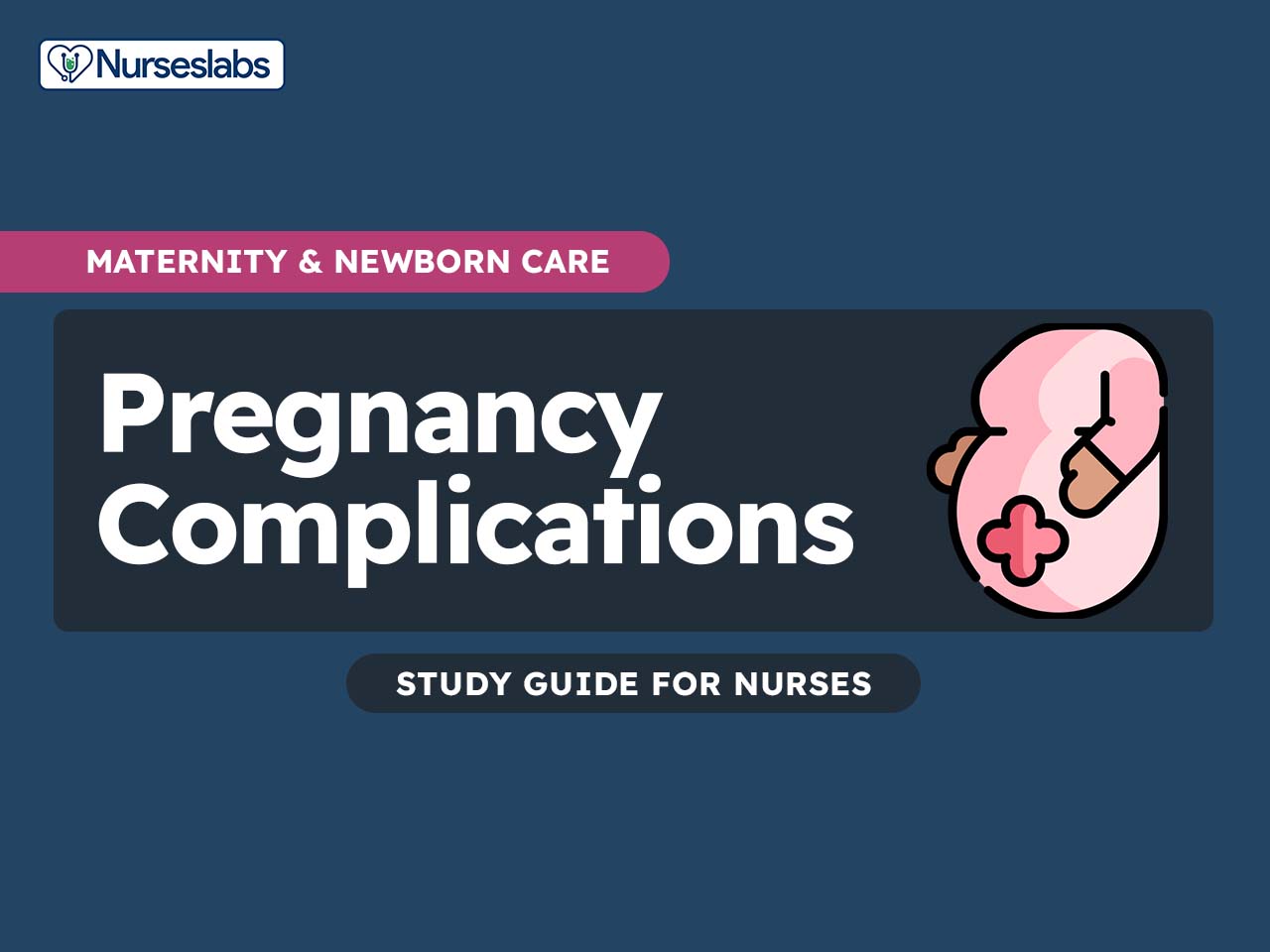
High-risk pregnancies and pregnancy complications present complex challenges for both expectant mothers and healthcare providers. These situations demand a specialized and compassionate nursing approach to ensure the best possible outcomes for maternal and fetal health. As frontline caregivers, nurses play a pivotal role in early detection, expert management, and the provision of emotional support to women navigating the uncertainties of high-risk pregnancies and pregnancy-related complications.
This article aims to serve as a comprehensive nursing guide, delving into the various aspects of high-risk pregnancies and pregnancy complications. By equipping nursing professionals with evidence-based practices, clinical insights, and a patient-centered focus, we seek to enhance the quality of care provided to women during these critical times.
High-Risk Pregnancy
How to Identify One
- More than one factor can contribute to the classification of a high-risk pregnancy.
- Women who already have a disorder before the pregnancy is termed to have a greater than normal risk.
- The factors that categorize the woman’s pregnancy as high risk were classified into minimal, moderate, or extensive.
- Psychological, social, and physical factors also break down the factors that categorize a high-risk pregnancy.
Psychological Factors
- History of drug dependence
- History of intimate partner abuse
- History of mental illness
- Loss of support person
- Poor acceptance of pregnancy
- Severely frightened by labor and birth experience
- Inability to participate because of anesthesia
- Illness in newborn
Social Factors
- Occupation involving handling of toxic materials
- Environmental contaminants
- Isolated
- Low economic level
- Poor access to transportation
- Poor housing
- Refusal or neglected prenatal care
- Disruptive family incident
- Conception less than one year after last pregnancy
- Lack of support person
- Inadequate home for infant care
- Lack of access to continued health care
Physical Factors
- Pelvic inadequacy or misshape
- Uterine incompetency, position or structure
- Secondary major illness
- Poor gynecologic or obstetric history
- Obesity
- Underweight
- PID
- Potential of blood incompatibility
- Younger than 18 years old and older than 35 years old
- Cigarette smoker
- Substance abuser
- Subject to trauma
- Bleeding disruption
- Gestational diabetes
- Nutritional deficiency
- Infection
- Hemorrhage
- Cephalopelvic disproportion
- Retained placenta
Pregnant women with special needs
A pregnant woman who has a special need needs more attention and care than an average woman warrants. Caregivers must understand that pregnancy for them is much more challenging, and they need every bit of guidance from us. With thorough assessment and an extra amount of care given, we could ensure that the pregnancy would be safe and less risky of them.
The Pregnant Adolescent
Teenage pregnancy is rampant nowadays. Since adolescent girls are not yet fully mature physically and emotionally, they need special care and education from their caregivers so they can have a safe and worry-free delivery.
- Adolescents have higher incidences of iron deficiency anemia, pregnancy-induced hypertension, premature labor, a low birth weight infant, disproportionate size between fetus and pelvis, and a high rate of intimate partner abuse.
- During the prenatal period, the adolescent can be more comfortable with primary nursing or case management approach because she is less exposed to a number of healthcare providers.
- Health history taking during the first prenatal visit is best done without the girl’s parents so a detailed history could be taken.
- The adolescent should be encouraged to continue with her prenatal visits, especially if she sought for prenatal care late in her pregnancy because she is protecting the pregnancy.
- If a parent accompanies their child, ask them separately about their concerns regarding their daughter, as they may be anxious too about hr health during pregnancy.
- The baby’s father may also accompany her during the pregnancy diagnosis, but he does not have a legal right to participate in the girl’s decisions regarding abortion and adoption because he is not married to her.
- Allow the father to offer support in the pregnancy so he will know more about himself and have education on how to prevent further pregnancies until he is more mature.
- Educate the adolescent about the common signs and symptoms associated with pregnancy and reassure her that they are part of a normal pregnancy so she would not attempt to treat the symptoms with drugs that could potentially harm the fetus.
- Some adolescents may have difficulty in telling their parents about the pregnancy, so involving them in role-playing or simulation might help them prepare to do this.
- Health teaching is compulsory for pregnant adolescents because they may not know the care measures that older women have gleaned through experience.
- Encourage the adolescent to eat a sufficient diet full of nutrients and minerals to accommodate the growth of the fetus and also her growing body.
- Also, teach her how to construct quick, healthy meals especially if she still attends school or how to buy nutritious cafeteria lunch at school.
- Remind the girl of the importance of taking her vitamins and supplements as adolescents may not be too compliant with medication taking.
- Assess the adolescent’s level of activity and participation in sports to determine what should be discontinued during pregnancy.
- Suggest alternative activities so they do not suffer from the loss of companionship, and also encourage them to plan their rest periods.
- The adolescent also needs substantial education on the physiologic changes during pregnancy and also with labor and delivery to help her gain more knowledge and treat the pregnancy as a growth experience.
- Encourage the adolescent to breastfeed like the average pregnant woman and assure them that their breast tissue would mature enough for them to be able to breastfeed.
The Pregnant Woman over the Age of 40
The dangers of getting pregnant over the age of 40 are mostly due to the complications and previously diagnosed conditions that these women may have. However, as long as prenatal care is done early in pregnancy, the serious complications in older pregnant women seem to decrease gradually.
- Women over the age of 40 may already have adequate information regarding the importance of early prenatal care and may already have an adequate insurance.
- Ask the woman about the present symptoms she is experiencing and how it fits into her lifestyle to ensure that she is not taking medicines to help relieve these symptoms.
- Assess the woman’s source of income and if stopping work because of the pregnancy could greatly reduce the family income to ascertain if the woman needs extra emotional support for feeling responsible for so many people.
- Assess the woman’s job, her recent diet, and exercise programs to determine if there should be any modifications for her pregnancy.
- Ask about personal habits such as cigarette smoking and alcohol consumption to determine if counseling is needed to halt or decrease these habits.
- A thorough physical examination should be performed on the first prenatal visit to identify any health problems especially those circulatory disturbances.
- Assess the woman’s breasts, the fetal heart sounds, and fetal movements during the prenatal visits because H-mole is also more common in women over 40.
- Obtain a urine specimen to check for the specific gravity, glucose, and protein to evaluate her renal function and the possibility of developing gestational or type 2 diabetes.
- Assess the woman’s number of meals especially those eaten outside her home or in restaurants to give her suggestions on how to adjust pregnancy nutrition so she can still obtain the same amount of nutrients when she eats at home or outside.
- Suggest attending prenatal classes where she could feel that she is one of the group, and makes sure that she plans to set aside time to do breathing exercises in preparation for her labor.
- Encourage the woman to undergo a triple screen testing (AFP, hCg, and unconjugated estriol levels) to detect any chromosomal defect or open spinal cord in the fetus.
Pregnant Woman with Physical and Mental Challenges
For so many years, women with disabilities were discouraged from getting pregnant because their condition may complicate the pregnancy. However, with the advent of technology, women who are physically and mentally challenged are given more ease during pregnancy and childbirth. Also with the guidance of their healthcare providers, they are now able to plan pregnancies and childbirth and their special concerns are now addressed in any healthcare facility across the country.
The Pregnant Woman who is Physically or Mentally Challenged
A physically or mentally challenged woman who wants to get pregnant is advised to engage in careful planning of her pregnancy so everything would be tailored to exactly what she needs.
- On the first prenatal visit, explore the nature of the woman’s disability to identify the alterations needed for a successful pregnancy.
- A woman who is house bound during pregnancy must be compliant in taking a vitamin D supplement because she might not be getting adequate sun exposure.
- Assess the woman’s ability to reach her emergency contacts for pregnancy-related emergencies.
- Assess the woman’s ability to come for a prenatal visit or if she can drive alone.
- Encourage a woman who uses a wheelchair to press with their hands against the armrests and lift their buttocks off the wheelchair seat for five seconds every hour to prevent pressure ulcers as the fetus is getting heavier.
- Encourage the woman to increase fluid intake to prevent urinary tract infections and to void frequently even though mobility is an effort.
- Physical examinations may need to be modified to accommodate the woman with disability.
- Women who are cognitively challenged may not know how they became pregnant, so if she was abused sexually, give ample time to talk to her regarding a pelvic examination.
- For a visually challenged woman, a trained guide dog may be brought during prenatal visits but since the guide dog’s main function is to offer directions, it still has protective instincts and might be threatened whenever people may pet it.
- Use demonstration aids when teaching a visually impaired woman, to allow her to feel or touch the aids.
- A cognitively challenged woman may need instructions limited to those few items related to safety.
- A visually challenged woman could not read pregnancy pamphlets, so either her support person would read the pamphlet to her or she can have a tape recorder with information recorded about pregnancy or any topic you want her to remember.
- Nutrition counseling needs to center on foods that can be prepared without cooking.
- For activity and exercise, if mobility Is an issue, let the woman understand that walking around her home is just like walking around the block as exercise.
- Encourage the woman to attend childbirth preparation classes if possible, especially those who are not working outside their home so they can practice breathing exercises.
- For a hearing impaired woman, show her printed words when teaching so she can see what your lip motions represents.
- If the woman has an interpreter for sign language, be certain that you are facing the woman while talking and not the interpreter.
- For a woman with spinal cord injury, instruct her to palpate her abdomen for uterine contractions so she can be aware that she is in labor.
- Cesarean and forceps birth may be necessary for women with muscle spasticity or spinal cord injury.
- A woman who cannot assume a lithotomy position could be positioned in a dorsal recumbent or Sims’ position during vaginal delivery.
The Pregnant Woman who is Substance Dependent
One of the major problems among pregnant women today is the use of illegal drugs. A person is substance dependent if she has withdrawal symptoms after discontinuing the use of illegal drugs. Assessment of the pregnant woman during the prenatal visit is necessary for the possibility of substance abuse.
- A woman with substance abuse problem may come in late for her prenatal care because she is afraid that her drug use would be discovered and reported to the authorities.
- She may also have difficulty in following instructions for proper nutrition as she may lack sufficient money to buy nutritious foods and drugs, as well as supplemental vitamins.
- Illicit drugs can cross the placenta, so the fetus of a substance dependent woman has a drug concentration of about 50% that of the mother.
- Drug abuse accounts for preterm birth and fetal abnormalities, and also the risk for hepatitis and HIV increases if the woman uses injected drugs.
- Cocaine is the most frequently abused drug during pregnancy and it causes extreme vasoconstriction that can compromise placental circulation leading to premature separation of the placenta and ultimately, preterm labor or fetal death.
- Infants of cocaine-dependent women may suffer from immediate effects of intracranial hemorrhage and withdrawal syndrome.
- Newborns born to women who use amphetamines show jitteriness, poor feeding at birth, and growth restriction.
- A woman who uses marijuana or hashish would not be able to breastfeed because of reduced milk production and the risk to the newborn from excretion of the drug in the milk.
- Narcotic agonists such as heroin would have effects on the pregnant woman such as fetal opiate dependence and severe withdrawal symptoms in the infant after birth.
- Women who are opiate dependent can enroll in a methadone maintenance program during pregnancy, but the infant would still not escape the withdrawal symptoms at birth, and methadone withdrawal has more severe reactions than heroin.
- However, the fetus would still be ensured of better nutrition, better prenatal care and less exposure to pathogens since methadone is given to her legally during the program.
- The woman may also be treated with buprenorphine if methadone programs are not available.
Musculoskeletal Disorders in a Pregnant Woman
The full function of the musculoskeletal system is needed by the pregnant woman all throughout her pregnancy. The following disorders would be a hindrance to her everyday tasks and should be therefore managed by the caregiver accordingly.
Scoliosis
- Scoliosis is the lateral curve of the spine.
- If left uncorrected, scoliosis would progress until it causes deformity, with interferences in respiration and heart functions due to chest compression.
- Pelvic distortion may interfere with birth, especially at the pelvic inlet, and administration of epidural or spinal anesthesia is difficult.
- Scoliosis is more noticeable in girls at the ages of 12 to 14 years old, so it is recommended that they wear a body brace to maintain an erect posture.
- The body brace cannot be worn during the last half of pregnancy, however, while have surgically implanted rods placed on both sides of the vertebrae.
- Surgically implanted rods do not interfere with pregnancy, and the back pain that the women with steel rods experience is also similar to the ones experienced by the average woman.
- Pelvic distortion and cephalopelvic disproportion could happen with scoliosis, so cesarean birth is necessary.
- Vaginal birth could be permitted with close monitoring of the labor process.
Cancer and Pregnancy
A malignant disease that grows within a person’s body could take the life out of the cells. This is a very fatal situation for a pregnant woman, therefore special care and attention should be given to her to save both the baby and the mother.
- The most common malignancies during pregnancy and the childbirth years are cervical, breast, ovarian, thyroid, leukemia, melanoma, and lymphomas.
- Women who opt to bear children at the age of 30 and above are more at risk for malignancies, especially breast cancer.
- If a malignancy is diagnosed in the first trimester, the couple would be asked to make decision between delaying the treatment to decrease teratogenic effects on the fetus, ending the pregnancy to continue the treatment, or continuing the pregnancy and the treatment with the knowledge that the fetus might end up with birth anomalies.
- On the second and third trimesters, the women can receive chemotherapy safely, but with radiation therapy the fetus could be put at risk if exposed directly.
- The cancer from the woman would not metastasize to the fetus because the placenta serves as a barrier and the fetus is capable of resisting foreign cells.
- If surgery to remove a tumor is done during pregnancy, there must be awareness that this might cause anoxia to the fetus during anesthesia.
- Cervical conization could also put the fetus at risk because the procedure might disrupt the pregnancy.
- The incidence of cervical cancer may go down in the future because of the HPV vaccine.
Mental Illness and Pregnancy
The mind is a complicated matter; illness springing from it could affect the entirety of the human being. Dealing with mental illness in pregnancy is a delicate thing yet could be achieved if every caregiver would take part in it.
- Schizophrenia most commonly occurs in young pregnant women; however, depression is seen as the most common mental illness among pregnant women.
- Childbirth and stress may reveal mental illness for the first time because normal levels of stress can still make coping difficult.
- A woman with an existing mental disease must have a psychiatric team and a prenatal group to make sure that the pregnancy would not exacerbate the disease and depression would not complicate the pregnancy.
- Psychotropic medications taken by the woman must be evaluated first because it might cause teratogenic effects to the fetus.
- Mental illness could also occur during the postpartum period.
Trauma in Pregnancy
During pregnancy, prioritizing safety is crucial. Trauma is an uncommon event for pregnant women due to the heightened precautions one takes. However, despite best efforts, accidents can still occur. Healthcare providers should possess the necessary skills to plan the care of pregnant women who experience trauma, as these situations may arise unexpectedly, albeit infrequently.
Assessment
Assessment must be done quickly yet thoroughly and both the physiological and physical status must be included.
- Assessment of an injured pregnant woman must be done concurrently with supportive reassurance to relieve the woman of her fear of fetal damage and also to remind her that she might be injured herself.
- Anxiety should be first reduced so the woman could cooperate more effectively during the interview.
- Assess the pregnancy history of the woman as well as her trauma history.
- Document the circumstances of trauma: what happened, the time of injury, signs and symptoms of the injury she is experiencing, and actions she had taken to counteract these.
- Evaluate whether the woman’s extent of injury is in proportion to the history.
- Assess the woman’s awareness of common safety measures.
- Body organs such as the lungs, heart, kidney, or brain must be analyzed first because injury to these body systems could place the fetus’ health in jeopardy.
Types of Trauma
The trauma that a pregnant woman might experience differs in every aspect, and the care given to each case must also be focused on the trauma that occurred.
Open Wounds
- To assess if an infection is occurring, serial measurements must be used because the WBC count is normally elevated during pregnancy.
- A bleeding laceration could be halted by placing pressure at the edges of the lacerations.
- For punctured wounds, tetanus immunization is given if the woman has not had tetanus immunization within 10 years, and tetanus toxoid for women who have had tetanus immunization within the past 10 years.
- To determine the depth and extent of the wound, a fistulogram can be done.
- Pregnant women bitten by animals or snakes can have rabies immune globulin and vaccine, and anti-venom serum for snake bites as these are not contraindicated during pregnancy.
- Caution a pregnant woman to avoid contact with unfamiliar dogs to prevent bites and to avoid feeding wild animals while camping.
Blunt Abdominal Trauma
- Blunt abdominal trauma usually occurs due to an automobile accident, when the woman’s abdomen strikes the steering wheel or dashboard, or when someone kicks or punches her abdomen.
- The injured underlying tissue becomes edematous and broken vessels form ecchymoses or hematomas while there is no visible break in the skin.
- To assess for abdominal bleeding, a diagnostic peritoneal lavage or ultrasound is done.
- Assessment should be done carefully because a traumatic blow could cause dislodgement of the placenta.
- To assess for vaginal bleeding or rupture of the amniotic membranes, a pelvic examination can be performed.
- Assure the woman that the fetus is unharmed by listening to its fetal heartbeat with the use of Doppler.
- If preterm labor occurred, a tocolytic can be administered, and fetal and uterine monitors should be attached too.
Gunshot Wounds
- Assessment of a gunshot wound includes inspection of the point of entry of the bullet and the point where it exited.
- If the uterus is punctured, there may be no entry point as the uterine walls are very thick.
- A gunshot wound is surgically cleaned and debrided and the woman is given a high-dose antibiotic that is safe for pregnant women such as Ampicillin.
- The incidence of fetal mortality is high if the bullet entered the uterus especially if the placenta is torn by the bullet.
- Stay with the woman as she recounts the history of the accident with law enforcement officers.
Poisoning
- A woman should contact the local poison control center and state that she is pregnant and what she accidentally swallowed, then follow the instructions of the personnel at the poison control center.
- Activated charcoal is the drug of choice to neutralize stomach poison.
- Investigate the circumstances of the poisoning afterward so you can educate the woman about safety with medications or food and discover possible suicidal intent.
Choking
- Dislodging the object that the woman choked on would be difficult because of a lack of space between the uterus and the end of the sternum and the average person could not reach from the rear around a woman’s enlarged abdomen.
- The rescuer might use successive chest thrusts instead late in pregnancy.
Orthopedic Injuries
- A woman late in her pregnancy has poor balance and can easily fall, so she automatically reaches out a hand to cushion her fall to prevent landing on her abdomen, which could cause serious wrist injury.
- Apply ice to the area to decrease the swelling as a first aid measure.
- To determine if the fracture is present, a radiograph can be performed.
- Assure the woman that a radiograph is safe as long as her abdomen is shielded during exposure.
- Assist the woman in identifying calcium-rich foods if she has a fracture so both she and the fetus can have adequate calcium for new bone growth.
- A woman who had a previous knee injury should be re-evaluated early in the pregnancy because a knee immobilizer might be needed for the last 3 months of pregnancy to prevent the joint from dislocating or the ligament from tearing again.
Burns
- Burns can cause thermal injury and inhalation of carbon monoxide from the fire which could lead to extreme fetal hypoxia.
- Prostaglandins are produced in response to severe trauma which could cause preterm labor.
- If more than 50% of the body’s surface area is burned, both the mother and the fetus could be in grave danger.
- Burn tissue heals quickly during pregnancy probably because of overall increased metabolism and an increase in the corticosteroid levels.
Respiratory Disorders in a Pregnant Woman
The respiratory system is one of the most important systems that should be assessed during pregnancy and also in the newborn. Acquiring a respiratory disorder in pregnant women could be fatal and detrimental to the health of both the fetus and the woman.
Acute Nasopharyngitis
- Estrogen stimulation causes nasal congestion, which makes nasopharyngitis more severe during pregnancy.
- Aspirin must be avoided because it can cause clotting interference in both the mother and the fetus and also prolonged pregnancy.
- Before taking any over-the-counter cough syrup, the woman must consult her healthcare provider.
- Antibiotic therapy is unnecessary unless it is to prevent a secondary infection.
Influenza
- Influenza spreads in epidemic form and is caused by viruses A, B, or C.
- Its symptoms are high fever, pains in the back and extremities, and a sore throat.
- Influenza can cause preterm labor but it is not linked to any congenital anomalies in the fetus.
- Antipyretics like Tylenol can be administered to control fever.
- Women can be safely immunized against influenza during pregnancy because the vaccine only contains killed virus.
Pneumonia
- Bacterial and viral pathogens such as S. pneumonia, H. influenza, and Mycoplasma pneumonia are responsible for the invasion of the lung tissue during pneumonia.
- An acute inflammatory response happens after the invasion in the lung alveoli.
- Bacteria or virus is trapped within the lung segments and lobes, filling the lung with fluid and blocking off the breathing space.
- Antibiotic therapy and oxygen administration are the treatment to be used.
- Ventilation support is only necessary in severe cases.
- Preterm labor may occur late in pregnancy because of oxygen deficit, so oxygen must be administered for the fetus.
Severe Acute Respiratory Syndrome
- SARS is characterized by persistent fever, muscle aches, chills, dry cough, malaise, headache, and dyspnea.
- Decreased lymphocyte and platelet counts are common laboratory findings.
- The pathogen for this illness is the coronavirus which has originated from southern China.
- The mode of transmission of SARS is close person to person contact via droplet transmission.
- Therapy includes intravenous antibiotic and respiratory support.
- SARS during pregnancy is associated with spontaneous miscarriage, intrauterine growth restriction, and preterm labor.
Asthma
- Asthma is marked by reversible airflow obstruction, airway hyperreactivity, and airway inflammation.
- Asthma is associated with perinatal complications.
- Symptoms would only be triggered by inhaled allergens, which release histamines and leukotrienes from an IgE/immunoglobulin interaction.
- Bronchial constriction occurs and thick bronchial secretions are produced, leading to decrease in the size of the lumen of air passages.
- Asthma causes a decrease in the oxygen supply going to the fetus which could begin preterm labor or restrict fetal growth.
- Inhaled corticosteroids such as beclomethasone and budesonide are safe for pregnant women.
- Cromolyn sodium and leukotriene receptor antagonists can also be continued during pregnancy.
Endocrine Disorders in a Pregnant Woman
Disorders of the endocrine system greatly affect the hormones in a pregnant woman’s body. Hormones have a major role in pregnancy and should, therefore, be monitored accordingly.
Diabetes Mellitus
- In diabetes mellitus, the pancreas cannot produce adequate insulin to regulate glucose levels in the body.
- The problem in diabetes mellitus is controlling the balance between glucose and insulin levels to prevent hypoglycemia and hyperglycemia.
- Infants born to women with diabetes mellitus are five times more likely to have heart anomalies.
- Type 1 diabetes occurs during childhood and the pancreas could not produce adequate insulin for body requirements.
- Type 2 diabetes occurs in older adults with gradual failure of insulin production that occurs with aging.
- As pregnancy progresses, women normally experience several changes in the glucose-insulin regulatory system.
- At about week 24, the pregnant woman with diabetes must increase her insulin dosage as advised to prevent hyperglycemia.
- Continued use of glucose by the fetus could lead to hypoglycemia for the mother between meals.
- An increase in the production of amniotic fluid occurs because of hypoglycemia in the fetus that causes increased urine production.
- A pregnant woman may develop hydramnios, and could be at risk for hemorrhage because of poor uterine contractions.
- Pregnancy-induced hypertension and infection could occur to a woman with poor glucose regulation.
- Infants of mothers who have poorly controlled diabetes are large for gestational age.
- There are high incidences of congenital anomalies such as caudal regression syndrome, spontaneous miscarriage, and stillbirth in infants of women with uncontrolled diabetes.
- Neonates at birth are at risk for hypoglycemia, respiratory distress syndrome, hypocalcemia, and hyperbilirubinemia.
- More frequent prenatal visits should be done for women with diabetes for close monitoring of their condition and of the fetus.
- Management includes insulin pump therapy, blood glucose monitoring, tests for placental function and fetal well-being, and good timing for birth.
- Educate the woman that her diet should include a reduced amount of fat.
- The woman may also consume a snack of protein and complex carbohydrates at night for slower digestion.
- If she is unable to eat because of nausea and vomiting, she should notify her healthcare provider so she could be given intravenous fluid supplementation.
- A fasting blood glucose level of below 95 mg/dL to 100 mg/dL and a 2-hour postprandial level of below 120 mg/dL show good blood glucose control.
Hypothyroidism
- Hypothyroidism is rare among pregnant women because this disease causes anovulation and inability to conceive.
- A pregnant woman with hypothyroidism would have a hard time increasing thyroid functioning necessary for a pregnant level which could lead to spontaneous miscarriage.
- Manifestations include fatigue, dry skin, and little tolerance for cold with an increased incidence in nausea and vomiting or hyperemesis gravidarum.
- Levothyroxine dosage would need to be increased by as much as 20% to 30% for the duration of pregnancy.
- Advise the woman to separate thyroxine ingestion from calcium, iron, or soy products by about 4 hours to avoid problems with the absorption of thyroxine.
Neurological Disorders in Pregnancy
Seizure Disorder
- The cause of most recurrent seizures is idiopathic, yet there are some which are caused by head trauma or meningitis.
- There are no contraindications to pregnancy for women with seizures as long as the medication is taken at the lowest dose possible and serum levels are carefully monitored.
- In the early months of pregnancy, remind the woman to continue taking her anti-seizure medications approved by both her obstetrician and primary care provider.
- The risk of adverse maternal or fetal outcomes from seizures during pregnancy is greater than the risk of teratogenicity from taking anticonvulsant medications.
- Some of the common drugs prescribed to control seizures are trimethadione, valproic acid, carbamazepine, and phenytoin sodium.
- With decreased levels of vitamin K coagulation factors due to phenytoin, an infant may be prone to hemorrhagic disease.
- Women should be prescribed with vitamin K during labor or for the last 4 weeks of gestation.
Myasthenia Gravis
- Myasthenia gravis is an autoimmune disorder characterized by the presence of IgG antibodies against acetylcholine receptors in striated muscle, causing the muscle not to contract particularly those in the oropharyngeal, facial, and extraocular groups.
- Drugs prescribed are Mestinon or Prostigmin and may be continued during pregnancy as there are no effects on the fetus.
- To reduce symptoms, plasmapheresis or removal of and replacement of plasma could be performed to remove immune complexes from the bloodstream.
- Magnesium sulfate should be avoided because it diminishes the acetylcholine effect and increases the symptoms.
- Labor should occur normally as smooth muscles are not affected by the disease.
- The infant may demonstrate symptoms of the disease at birth because of the transfer of antibodies.
Multiple Sclerosis
- MS refers to nerve fibers that become demyelinated and lose their function.
- Symptoms include fatigue, numbness, blurred vision, and loss of coordination.
- ACTH is given to strengthen nerve conduction and is safe during pregnancy.
- Women may continue with plasmapheresis during pregnancy as long as the volume of exchange is controlled.
- Pregnancy does not affect the long-term cause of MS.
- MS may actually improve due to the increase in circulating corticosteroid levels.
Gastrointestinal Diseases in Pregnancy
There are several gastrointestinal discomforts that a pregnant woman experiences, yet there are some symptoms that might indicate a worsening condition.
Appendicitis
- Appendicitis is the inflammation of the appendix.
- Symptoms of appendicitis include nausea, abdominal discomfort, vomiting, and a sharp, peristaltic, lower right quadrant pain.
- Appendicitis pain grows more intense, as well as nausea and vomiting.
- The pain might be displaced up in the abdomen in a pregnant woman so that the pain might be mistaken as something from a gallbladder disease.
- The woman might have an elevated temperature and ketones in the urine.
- An ultrasound will reveal an inflamed appendix.
- Advise the woman not to take food, liquid, or laxatives while waiting to be evaluated for possible appendicitis, because increasing peristalsis might rupture the inflamed appendix.
- If the woman is almost past 36 weeks and the fetus is mature, a cesarean birth may be performed and then the inflamed appendix would be removed.
- Appendicitis in early pregnancy can be removed by laparoscopy.
- If the inflamed appendix ruptures, fecal material might escape towards the fallopian tube to the fetus.
- Peritonitis might occur, and the woman’s body would not be able to handle peritonitis and pregnancy at the same time.
Gastroesophageal Reflux Disease
- Gastroesophageal reflux disease is the reflux of acid stomach secretions into the esophagus.
- Symptoms include heartburn, gastric regurgitation, dysphagia, weight loss, and hematemesis in extreme esophageal irritation.
- These conditions are diagnosed through direct endoscopy or ultrasound.
- Antacids may relieve pain and ranitidine can inhibit gastric acid production.
- Advise the woman to wear loose clothing and sleep with her head elevated to help confine stomach secretions.
- After pregnancy, symptoms may become less noticeable or disappear because uterine pressure has decreased.
Hepatitis
- Hepatitis is a liver disease that may occur due to the invasion of A, B, C, D, or E virus.
- Hepatitis A is spread through fecal-oral contact or by ingestion of fecally contaminated water or shellfish.
- Pregnant women exposed to hepatitis A may be given prophylactic gamma globulin to prevent the disease.
- Hepatitis B and C are acquired through exposure to contaminated blood or blood products and other body secretions.
- Maternal/fetal transmission is also an important mode of transmission of these types.
- Hepa B vaccine may be administered to those who are at high risk.
- Symptoms of hepatitis include nausea and vomiting, the liver area may feel tender upon palpation, urine color is dark yellow, light-colored stools, and jaundice as a late symptom.
- Hepatomegaly is also noted and the bilirubin level is elevated.
- The woman should be on bed rest and advised to eat a high-calorie diet.
- Cesarean birth can be done to decrease the possibility of transmitting the disease from the mother to the fetus.
- Hepatitis may lead to spontaneous abortion or preterm labor.
- After birth, the infant should be washed well to remove any maternal blood and hepa B immunoglobulin should be administered.
- The mother may still breastfeed the infant but should still be observed carefully for signs of infection during the first few months of life.
Renal Diseases in Pregnancy
A woman’s kidney function is important for both the fetal and maternal well-being because the woman is excreting waste products not only for herself but also for the fetus.
Urinary Tract Infection
- The ureters of a pregnant woman dilate because of the effect of progesterone, resulting to stasis of the urine.
- Escherichia coli are the most common organism responsible for UTI.
- The symptoms of UTI are frequency and pain upon urination.
- The woman may also have nausea and vomiting, malaise, and elevated temperature.
- Infection usually occurs at the right side because there is greater compression and urinary stasis on the right ureter from the uterus being pushed by the bulk of large intestine on the left side.
- Advise the woman to collect a sample of her urine through clean catch method for urine culture and sensitivity.
- Amoxicillin, ampicillin, and cephalosporins are effective and safe for a woman with UTI.
- For prevention of UTI, educate the woman to void frequently at least every 2 hours.
- Instruct her to wipe from front to back after voiding and moving bowels.
- Encourage the woman to wear cotton and not synthetic underwear.
- Advise the woman to void immediately after sexual intercourse.
- Advise the pregnant woman to increase the amount of fluid (about 3 to 4 L per day) she takes every day to flush out the infection from her urinary tract.
Chronic Renal Disease
- Women who already have chronic renal disease may develop severe anemia during pregnancy because their kidneys could not produce erythropoietin, however, synthetic erythropoietin is now available and can be taken by the pregnant woman.
- Many women with kidney disease have an elevated blood pressure.
- Women who take corticosteroids as their maintenance for the disease are instructed to continue taking her medications during pregnancy.
- The infant might be hyperglycemic at birth because of the suppression of insulin activity by the corticosteroid.
- Dialysis might be needed to aid the woman with her kidney functions during pregnancy; however, there is a risk of preterm labor.
- Peritoneal dialysis is preferred over hemodialysis because it causes less fluid shifts.
- Nutrition consultation is advised for the woman especially if she is on a low-potassium diet to avoid build up of potassium because the kidneys could not excrete them.
Cardiovascular Diseases in Pregnancy
Cardiac disease: Left-sided Heart Failure
- Left-sided heart failure happens when the left ventricle cannot shunt the blood forward that it received by the left atrium from the pulmonary circulation.
- Failure mostly occurs at the level of the mitral valve.
- When the mitral valve could not push the blood forward, it causes back pressure on the pulmonary circulation which results in pulmonary hypertension.
- Pulmonary hypertension in pregnant women could precipitate a high-risk pregnancy for spontaneous miscarriage, preterm labor, or maternal death.
- The placenta may not receive adequate blood because of the decreased peripheral circulation.
- The woman would have difficulty in sleeping due to the worsening pulmonary edema.
- Advise the woman with left-sided heart failure to sleep with her chest and head elevated.
- Heart action is more effective at rest, so the interstitial fluid returns to the circulation and overburdens it, causing increased left-sided failure and pulmonary edema.
- When complications of left-sided failure occur, these may result in impaired blood flow to the uterus, poor placental perfusion, intrauterine growth restriction, and fetal mortality.
- The woman needs to have a serial ultrasound and nonstress tests on the 30th to 32nd week of her pregnancy to monitor fetal health.
Cardiac Disease: Right-sided Heart Failure
- Right-sided heart failure happens when the output of the right ventricle is less than the blood volume received by the right atrium from the vena cava.
- Back-pressure from this results in congestion of the systemic venous circulation and also decrease in cardiac output.
- Pressure is high in the vena cava, leading to jugular vein distention and increased portal circulation.
- Liver is enlarged, and this could cause extreme dyspnea and pain in a pregnant woman because the enlarged liver is pressed upward by the enlarged uterus, and extreme pressure is placed on the diaphragm.
- Eisenmenger syndrome is the congenital anomaly that would most likely cause right-sided heart failure in women of reproductive age.
- It is a right to left atrial or ventricular septal defect with pulmonary stenosis.
- Women with this anomaly are advised to avoid getting pregnant.
- Oxygen administration and frequent assessment of the arterial blood gas is needed to ensure fetal growth once the woman gets pregnant.
- The nurse’s role during labor is to closely monitor for hypotension after epidural anesthesia.
Chronic Hypertensive Vascular Disease
- Women already diagnosed with chronic hypertensive vascular disease already has an elevated blood pressure (140/90 mmHg and above) in pregnancy.
- Both the mother and the fetus are compromised because of the poor placental perfusion which places the fetal well-being in jeopardy.
- The primary care provider could prescribe beta-blockers and ACE inhibitors to decrease the blood pressure by peripheral dilation, but not to reduce it below the threshold that allows for good placental circulation.
Venous Thromboembolic Disease
- Venous thromboembolic disease happens more likely in pregnant women because of the stasis of blood in the lower extremities due to uterine pressure and the effect of elevated estrogen on the hypercoagulability of the woman.
- The triad of stasis, vessel damage, and hypercoagulation results in thrombus formation in the lower extremities.
- Women who are 30 years and older have an increased risk of developing deep vein thrombosis leading to pulmonary emboli.
- Pain and redness in the calf of the leg usually signal thrombus formation.
- Thrombus formation can be prevented by avoiding the use of constrictive knee-high stockings.
- Advise the woman not to sit with her legs crossed at the knee and to avoid standing in one position for a long time.
- A thrombus that occurred during pregnancy is diagnosed by Doppler ultrasonography and a woman’s history.
- The woman will be placed on bed rest and intravenous heparin administration for 24 to 48 hours.
- Women who are taking heparin during pregnancy are not candidates for routine episiotomy or epidural anesthesia to prevent hemorrhage.
- PTT determination should be continued during labor.
- A breastfeeding woman cannot take heparin or Coumadin, or Coumadin should be used cautiously.
- The main danger of thrombophlebitis is pulmonary embolism or a clot that lodges in the pulmonary artery, blocking the circulation to the lungs and heart.
- Symptoms of pulmonary embolism include chest pain, sudden onset of dyspnea, cough with hemoptysis, tachycardia, and severe dizziness or fainting.
- Pulmonary embolism is recognized as an immediate emergency.
Hematologic Disorders in Pregnancy
Coagulation disorders or blood formation makeup hematologic disorders among pregnant women. Since childbirth involves the loss of a lot of blood, hemorrhage would be very dangerous to a woman with these disorders.
Iron-Deficiency Anemia
- Iron-deficiency anemia is the most common anemia among pregnant women, mainly because many women enter pregnancy already deficient in iron stores because of low intake of iron.
- A hemoglobin level below 12mg/dl with hematocrit below 33% is a possible sign of iron deficiency.
- Iron-deficiency anemia is a microcytic, hypochromic anemia because when inadequate iron is ingested, it would be unavailable for incorporation into red blood cells.
- Iron-deficiency anemia is mildly associated with low birth weight and preterm birth.
- Extreme fatigue and poor exercise tolerance because she cannot transport oxygen effectively.
- Advise the woman to take prenatal vitamins containing an iron supplement of 60 mg elemental iron.
- Instruct her to eat a diet high in iron such as green leafy vegetables, meat, legumes, and fruit.
- When a pregnant woman has developed anemia during pregnancy, she will be prescribed with 120 to 200 mg elemental iron per day in the form of ferrous sulfate or ferrous gluconate.
- Iron is best absorbed in an acidic medium, so advise the woman to take her iron supplements with orange juice or a vitamin c supplement.
- Side effects of iron therapy include constipation and gastric irritation.
- Ferrous sulfate turns stool black so caution women with this side effect.
Folic Acid-Deficiency Anemia
- Folic acid is vital for the normal formation of red blood cells in the mother.
- It also prevents neural tube defects in the fetus.
- Folic acid anemia is a megaloblastic anemia wherein the mean corpuscular volume is elevated.
- This anemia is most apparent during the second trimester of pregnancy and may contribute to early miscarriage or premature separation of the placenta.
- Women who are expecting to be pregnant are advised to take 400 µg of folic acid daily.
- Advise the woman to take folacin rich foods such as green leafy vegetables, oranges, and dried beans.
Sickle Cell Anemia
- Sickle cell anemia is a recessively inherited hemolytic anemia caused by an abnormal amino acid in the beta chain of hemoglobin.
- Majority of the red blood cells are irregular or sickle-shaped, so they cannot carry the same amount of hemoglobin as the normal red blood cells do.
- At high altitudes, the blood becomes more viscous than usual because the cells tend to clump together because of their irregular shape.
- Reduced blood flow to the organs is the result of the vessel blockage of the clumping red blood cells.
- These cells will hemolyze and reduce in number, causing severe anemia.
- A pregnant woman with sickle cell anemia is more prone to bacteriuria, so a clean catch urine sample is collected during pregnancy to detect the disease while it is asymptomatic.
- The woman’s diet must contain sufficient amounts of folic acid to help in building new red blood cells.
- Fluid intake should also be emphasized because dehydration can lead to a sickle cell crisis.
- Advise the woman to elevate her legs while sitting or lie on her left side while sleeping to encourage venous return from the lower extremities.
- Instruct the woman to avoid standing for long periods during the day.
- Fetal health is monitored through ultrasound at 16 to 24 weeks to assess for intrauterine growth restriction.
- An exchange transfusion is needed to replace sickled cells with non-sickled cells.
- Iron supplements are not given to pregnant women who already have sickle cell crisis because they cannot still incorporate iron and may cause excessive build up.
- When a sickle cell crisis occurs, controlling pain, oxygen administration, and increasing the fluid volume of the circulatory system to lower viscosity are essential interventions.
- To detect if the fetus has acquired the disease, electrophoresis of red blood cells by percutaneous umbilical blood sampling or amniocentesis will be performed to reveal the presence of the disease on the beta chains present in utero.
Anomalies of the Cord
Two Vessel Cord
- A normal cord has one vein and two arteries.
- If one of the umbilical arteries is absent, this may denote that the infant may have congenital heart and kidney anomalies.
- This is because the insult that caused the loss of the vessel may have affected other mesoderm germ layer structures as well.
- Immediate inspection of the cord as to how many vessels are present must be performed after birth before the cord starts to dry.
- Drying distorts appearance of the vessels, making it difficult to discern the number of vessels present.
- The number of vessels must be documented appropriately after inspection.
- If the infant has only two vessels, he needs to be observed carefully for other anomalies during the newborn period.
Unusual Cord Length
- The length of the umbilical cord varies in every infant, but in some, abnormal lengths may occur.
- An unusually short umbilical cord may predispose the fetus to premature separation of placenta or an abnormal fetal lie.
- An unusually long cord has a great tendency of twisting or knotting.
- A cord naturally forms a knot but the natural pulsations of the blood through the vessels and the muscular vessel walls usually keep the blood flow adequate.
- A cord wrapped once around the fetal neck is not unusual but should not interfere to fetal circulation.
Anomalies of the Placenta
Placenta Succenturiata
- The normal placenta weighs approximately 500 g and is 15 to 20 cm in diameter and 1.5 to 3.0 cm thick.
- Placenta succenturiata is a placenta that has one or more accessory lobes connected to the main placenta by the blood vessels.
- This is not a fetal abnormality; however, it must be recognized upon assessment after birth.
- The small lobes may be retained in the uterus after birth leading to severe maternal hemorrhage.
- If you look closer at the placenta, it may appear torn at the edge or torn blood vessels extend beyond the edge of the placenta.
- Remaining lobes are removed from the uterus manually to prevent maternal hemorrhage as a result of poor uterine contraction.
Placenta Circumvallata
- Normally, the chorion membrane begins at the edge of the placenta and spreads to cover the fetus.
- The fetal side of the placenta is not usually covered by the chorion.
- In placenta circumvallata, the fetal side of the placenta is covered with chorion.
- The umbilical cord enters the placenta at the usual midpoint, and large vessels spread out from there.
- They end abruptly at the point where the chorion folds back into the surface.
- In placenta marginata, the fold of the chorion reaches just to the edge of the placenta.
Placenta Accreta
- Placenta accrete refers to an unusually deep attachment of the placenta to the uterine myometrium that the placenta will not loosen and deliver.
- Never attempt to remove it because it might lead to extreme hemorrhage because of its deep attachment.
- Hysterectomy or treatment with methotrexate to destroy the still-attached tissue is the recommended treatment of choice.
Battledore Placenta
- Battledore placenta refers to the cord that is inserted marginally rather than centrally.
- This is a rare anomaly and it has no known clinical significance.
Velamentous Insertion of the Cord
- Velamentous insertion of the cord occurs when the cord, instead of entering the placenta directly, separates into small vessels that reach the placenta by spreading across a fold of amnion.
- This is most commonly found with multiple gestations.
- It can be associated with fetal anomalies, so an infant born with this type of placenta must be examined carefully.
Vasa Previa
- The umbilical vessels of a velamentous cord insertion cross the cervical os and therefore deliver before the fetus.
- The vessels may tear with cervical dilatation, just as the placenta previa may tear.
- Ensure that you can identify the structures before inserting any instrument to prevent accidental tearing of a vasa previa which could end in sudden fetal blood loss.
- With either placenta previa or vasa previa, there is sudden , painless bleeding that occurs with the beginning of cervical dilatation.
- Vasa previa can be confirmed by ultrasound.
- If vasa previa is confirmed, the infant needs to be born via cesarean delivery.
Sudden Pregnancy Complications
Hydramnios and Oligohydramnios
Hydramnios
- The usual amount of amniotic fluid during pregnancy is 500 to 1000 mL at term.
- Excess fluid of more than 2000 mL is considered hydramnios.
- Too much amniotic fluid might cause fetal malpresentation, premature rupture of membranes, infection due to PROM, and preterm birth.
- The first sign of hydramnios is rapid enlargement of the uterus.
- There is also a difficulty in palpating the small parts of the fetus because the uterus is unusually tense.
- Fetal heart rate auscultation can be also difficult because of the increased amount of fluid surrounding the fetus.
- The woman may develop extreme shortness of breath as the uterus presses up her diaphragm.
- Poor venous return would result to lower extremity varicosities and hemorrhoids.
- An ultrasound would determine the presence of hydramnios and also the reason for the excessive amount of fluid.
- A woman with hydramnios would be advised to take bed rest to increase uteroplacental circulation and reduce the pressure on the cervix.
- Help the woman avoid constipation, as straining could increase uterine pressure and cause rupture of the membranes.
- Encourage the woman to eat a high fiber diet, and if the diet is not effective, she can use stool softeners as prescribed.
- Assess the woman’s vital signs and lower extremity edema frequently.
- Amniocentesis can be performed to reduce the volume of the amniotic fluid.
- Tocolysis may be begun to prevent preterm labor.
- After birth, the infant must be assessed fully to determine the factors that interfere with its ability to swallow effectively in utero.
Oligohydramnios
- Oligohydramnios is a pregnancy with less than the average amount of amniotic fluid.
- This is usually caused by a bladder or renal disorder in the fetus that interferes with voiding.
- The disorder can occur because of in utero growth restriction, the muscles are left weak at birth, the lungs fail to develop leading to severe difficulty in breathing, and the facial features are distorted.
- These symptoms are associated with a syndrome called Potter’s syndrome.
- Oligohydramnios is suspected when the uterus fails to meet its expected growth rate.
- It is confirmed through ultrasound as pockets of amniotic fluid are less than the average.
- Amnio Transfusion or instillation of fluid into the uterus by amniocentesis can relieve oligohydramnios.
- Infants need careful observation at birth to rule out kidney disease and compromised lung development.
Post-Term Pregnancy
- A term pregnancy is 38 to 42 weeks long, and a pregnancy that exceeds these limits is termed as post term pregnancy or prolonged pregnancy.
- Then infant is considered post mature or dysmature, especially if placental insufficiency has interfered with fetal growth.
- Prolonged pregnancy may occur with high intake of salicylates, which interferes with the synthesis of prostaglandins and may be responsible for the initiation of labor.
- Meconium aspiration would likely occur as fetal intestinal contents are more likely to reach the rectum.
- Macrosomia is also another problem if the fetus continues to grow.
- The fetus is exposed to decreased blood perfusion as the placenta only has adequate functioning ability for 40 to 42 weeks.
- The fetus might also suffer from lack of oxygen, fluid, and nutrients.
- A maternal vaginal fibronectin level, a nonstress test, and a biophysical profile may be ordered to document the state of placental perfusion and the amount of amniotic fluid present.
- Prostaglandin gel or misoprostol may be applied to the cervix to initiate ripening, or stripping of membranes followed by an oxytocin infusion can be used too.
- Cesarean birth will be necessary if all measures are ineffective.
- Monitor fetal heart rate closely during labor to be certain placental insufficiency would not occur from aging of the placenta.
RH Incompatibility
- Rh incompatibility occurs when an Rh-negative mother carries a fetus with an Rh-positive blood type.
- The father of the child must either be homozygous or heterozygous Rh-positive.
- The Rh-positive fetus inside the Rh-negative mother would be treated as a foreign body, and the mother’s body would react as if the invading factor is a substance such as a virus.
- The mother’s body would form antibodies against the invading substance.
- The maternal antibodies would cross the placenta and cause red blood cells destruction of fetal red blood cells.
- There is insufficient oxygen transport to body cells, and this condition is termed as hemolytic disease of the newborn or erythroblastosis fetalis.
- On the first pregnancy visit, the woman must have an anti-D antibody titer.
- If the results are normal, the test would be repeated at week 28 of the pregnancy, and no therapy is needed.
- If the result is an elevated titer, the well-being of the fetus would be monitored every 2 weeks or more often by Doppler velocity of the fetal middle cerebral artery which could predict when the fetal red blood cells are being destroyed.
- If the artery velocity is high, the fetus is not developing anemia and most likely is an Rh-negative fetus.
- If the reading is low, it means the fetus is in danger and immediate birth will be carried out if the fetus is near term.
- If it is not near-term, efforts to reduce the number of antibodies in the woman or replace damaged red cells in the fetus are started.
- Administration of Rh (D) immune globulin is done to women who are Rh-negative at 28 weeks of pregnancy.
- RhIG is given as an injection to the mother in the first 72 hours after birth of an Rh-positive child to further prevent the woman from forming natural antibodies.
High-risk pregnancy may not happen in most childbearing women, yet no one will be able to predict the comings and goings of these diseases. Early education for couples who want to get pregnant must be enforced to be certain that the mother and the baby would have a safe pregnancy journey all throughout.


Leave a Comment